

Abstract
Development of stable male sterility lines is essential for efficient hybrid seed production. We previously showed that knockout of bHLH142 in rice (Oryza sativa) causes pollen sterility by interrupting tapetal programmed cell death (PCD). In this study, we demonstrated that overexpression of bHLH142 (OE142) under the control of ubiquitin promoter also leads to male sterility in rice by triggering the premature onset of PCD. Protein of bHLH142 was found to accumulate specifically in the OE142 anthers. Overexpression of bHLH142 induced early expression of several key regulatory transcription factors in pollen development. In particular, the upregulation of EAT1 at the early stage of pollen development promoted premature PCD in the OE142 anthers, while its downregulation at the late stage impaired pollen development by suppressing genes involved in pollen wall biosynthesis, ROS scavenging and PCD. Collectively, these events led to male sterility in OE142. Analyses of related mutants further revealed the hierarchy of these pollen development regulatory genes. Thus, the findings of this study create a new method to generate genic male sterility in rice. Exploitation of this novel functionality of bHLH142 would confer a big advantage to hybrid seed production.
Highlight Overexpression of bHLH142 leads to male sterility in transgenic rice due to early onset of tapetal PCD. This study creates a new method to generate male sterility in rice.
Introduction
Rice (Oryza sativa) is one of the most important staple crops in the world, feeding almost half of the world’s population. Increase in rice production is urgently needed to keep pace with increasing population, especially in the face of drastic global climate change. Hybrid rice is considered the most promising strategy to increase grain yield increasing rice yields by 15–20% (Khush, 2013). By adopting hybrid technology, many countries have successfully increased per capita rice production (Zhang, 2011).
Heterosis in the F1 plants not only increases grain yield but also produces superior phenotypes in comparison with the parents with vigor in growth, good agronomic traits and pest resistance, etc. (Liu et al., 2015). As rice is a self-pollinated crop, adoption of a stable male sterility female parent is critical to ensure the purity of F1 seeds. At present, two distinct hybrid systems, a three-line system and a two-line system, have been widely adopted in rice hybrid seed production. In the three-line system, cytoplasmic male sterility (CMS, A line) is the stable line. However, to maintain CMS seed stock, it is necessary to cross with a maintainer line (B line), which increases the production cost.
In the two line system, genic male sterility using photoperiod- or temperature-inducible male sterility mutant has the advantages of easy maintenance of seed stock and lower production cost, but it has the disadvantage of producing hybrid seeds of less uniformity due to environment fluctuation. Therefore, a better understanding of the mechanism underlying pollen development is important for developing new genic male sterility lines.
Rice anthers comprise four lobes and each lobe contains four layers of anther walls. The tapetum layer is in the innermost layer, providing nutrients and sporopollenin precursors for pollen development. Tapetal programmed cell death (PCD) at the right time is important for normal pollen development. In the anther, PCD is first detectable at meiosis (stage 8, S8), strong PCD signals occur at the young microspore stage (S9) (Li et al., 2006; Zhang and Wilson, 2009), and reduced PCD signals occur at the vacuolated pollen stage (S10) (Hu et al., 2011). Functioning as polar secretory cells, the tapetum undergoes cellular degradation. Tapetal PCD subsequently triggers cytoplasmic shrinkage, breakdown of the nuclear membrane, oligonucleosomal cleavage of DNA, vacuole rupture, and swelling of the endoplasmic reticulum for release of mature pollen grains (Papini et al., 1999). Thus, tapetal PCD is an essential process for pollen maturation.
Pollen development involves a complex regulatory network. So far, several basic helix-loop-helix (bHLH) transcription factors (TFs) have been identified to play important roles in regulating tapetal PCD and pollen development. The roles of UDT1 (bHLH164) (Jung et al., 2005), bHLH142 (TIP2) (Fu et al., 2014;Ko et al., 2014), TDR1 (bHLH5) (Li et al., 2006), and EAT1 (DTD1, bHLH141) (Ji et al., 2013;Niu et al., 2013) in rice pollen development have been characterized in the last decade. Similarly, DYT1, the homolog gene of UDT1 in Arabidopsis (Zhang et al., 2006) and AMS (Sorensen et al., 2003), the homolog of TDR1 in Arabidopsis, are functionally conserved in both dicots and monocots. In addition, the UDT1 homolog in tomato, ms1035 (Solyc02g079810), was also recently reported to be involved in pollen development (Jeong et al., 2014). Another TF, GAMYB, is also known to play an important role in anther and aleurone layer development (Kaneko et al., 2004;Tsuji et al., 2006). According to current understanding of the pollen development regulatory network, UDT1 and GAMYB work in parallel to regulate pollen development and TDR1 acts downstream of UDT1 and GAMYB (Liu et al., 2010). Our previous study showed that bHLH142 is located downstream of UDT1 and GAMYB and coordinates with TDR1 through protein-protein interaction to modulate EAT1 transcriptional activity.
In addition, EAT1 interacts with TDR1 at a similar binding site to bHLH142 (Ko et al., 2014). So far, the biological role of TDR1 in interacting with EAT1 remains unknown (Ji et al., 2013;Ko et al., 2014;Niu et al., 2013). EAT1 directly regulates tapetal PCD via two aspartic proteases (AP37 & AP25) that activate cell death (Niu et al., 2013).
AtTDF1 encodes a R2R3 MYB TF, which functions in callose dissolution (Zhu et al., 2008). Similarly, the rice ortholog, OsTDF1 (MYB35), acts downstream of UDT1 and upstream of TDR, EAT1, OsMYB103 and Persistent Tapetal Cell 1 (PTC1) and it is essential for tapetal PCD (Cai et al., 2015). PTC1 encodes a PHD-finger TF and controls tapetal PCD and pollen development and acts downstream of GAMYB (Li et al., 2011) and TIP2 (bHLH142) (Fu et al., 2014).
During anther development, ROS acts as a signal to promote tapetal PCD (Hu et al., 2011;Yi et al., 2016). The cellular ROS level is determined by the interplay between ROS-producing and ROS-scavenging mechanisms (Gapper and Dolan, 2006;Miller et al., 2008). MADS3, a floral homeostatic C-class gene required for stamen identity, also regulates ROS scavenging during rice anther development. MADS3 modulates ROS levels through positive transcriptional regulation of the promoter of metallothionein gene MT-1-4b (Hu et al., 2011). The anthers of mads3 mutant showed a strong ROS signal and a defect in pollen fertility (Hu et al., 2011). On the other hand, defective Tapetum Cell Death 1 (DTC1) encodes a protein that contains a development and cell death (DCD) domain and KELCH repeats and acts as a key regulator of tapetum PCD by inhibiting ROS-scavenging activity through its interaction with metallothionein protein MT2b (Yi et al., 2016). Both MT-1-4b and MT2b act as ROS scavengers. Decreased expression of MT2b or MT-1-4b reduces scavenging activity and causes the accumulation of ROS molecules in rice roots (Steffens and Sauter, 2009) and anthers (Hu et al., 2011). Therefore, a timely buildup of ROS to trigger tapetal PCD during pollen development is vital.
The pollen wall is composed of three layers: pollen coat, outer exine layer, and inner intine layer (Zhang et al., 2016). Biosynthesis, secretion, and translocation of sporopollenin precursors are essential for pollen wall development. Synthesis of sporopollenin precursors is conducted in the tapetum, and ACOS5, CYP703A, CYP704B, MS2, etc., play major roles in this process. Ubish bodies transport tapetum-derived sporopollenin precursors to developing exine. Lipidic pollen exine is made of sporopollenin that is derived from the polymerization of fatty acid metabolites and phenolic acid (Ariizumi and Toriyama, 2011). In addition, MYB80/MYB103 is required for anther development in both Arabidopsis and rice (Higginson et al., 2003;Zhang et al., 2007). Male Sterility1 (MS1), a homeodomain (PHD) finger motif TF, regulates biosynthesis and secretion of pollen wall components in Arabidopsis (Wilson et al., 2001;Yang et al., 2007). A subsequent study by Li et al. (2011)found that PTC1, a MS1 homolog in rice, is also essential for tapetal PCD and pollen development in rice (Li et al., 2011). Several genes associated with rice pollen wall development have been identified by microarray analysis; these include Cys protease (CP1; Lee et al., 2004), a fatty acyl-CoA reductase homologous to Arabidopsis MS2 (Aarts et al., 1997), lipid transfer proteins such as C4 (Tsuchiya et al., 1994), C6 (Zhang et al., 2010), YY1, BURP domain-containing proteins (RA8 and OsRAFTIN; Jeon et al., 1999), and a P450 family member CYP704B2 (Li et al., 2010). They were all down-regulated in the anther of the rice ptc1 mutant (Li et al., 2011). Moreover, mutagenesis studies suggest that CYP703A2 (Yang et al., 2014) and CYP704B2 (Li et al., 2010) are essential for pollen development, and their knockout lines exhibited impaired pollen development. MS2 is essential for pollen wall biosynthesis by mediating the production of the conserved plastidial pathway for the production of fatty alcohols that are essential for pollen wall biosynthesis (Chen et al., 2011;Shi et al., 2011). Clearly, interruption of the functions of these genes resulted in abnormal pollen development.
In rice, bHLH142 is specifically expressed in the anther and regulates tapetal PCD and pollen wall development, and knockout of bHLH142 causes pollen sterility (Ko et al., 2014). To gain more insights into its functionality, in this study we generated transgenic lines overexpressing bHLH142 under the control of maize ubiquitin promoter. We demonstrated that constitutive overexpression of bHLH142 also leads to male sterility by triggering premature tapetal PCD. Our study provides a new method to generate genic male sterility in rice and possibly in other cereal crops too that may be suitable for agricultural application.
Materials and Methods
Constructs
The bHLH142 (Os01g0293100) full length cDNA was PCR amplified using primers S80qPCR-F3_BamHI and S80FLcds-R2_BamHI (see online Supplementary Table 1) and the product is a 1373 bp BamHI fragment. This fragment was then digested with BamHI and ligated into pCAMBIA1390 backbone containing the maize ubiquitin promoter. Expression of the selection marker HptII gene that encodes hygromycin phosphotransferase was driven by cauliflower mosaic virus (CaMV) 35S promoter (Supplementary Fig. S1E). Another vector harboring fused bHLH142 and eGFP (Ubi::bHLH142- eGFP) was constructed to detect the tissue specificity of bHLH142 protein using eGFP. All constructs were confirmed by sequencing. The plasmids were separately transformed and selected by antibiotic. Agrobaterium tumefaciens strain EHA105 was used for transfection to calli of TNG67 background following the method described previously (Chan et al., 1993).
Plant material and growth conditions
Transformation of Japonica rice cultivar TNG67 was described previously (Ko et al., 2014). Primary transgenic lines were transplanted into soil and cultivated in the Academia Sinica-BCST greenhouse for genetically modified organisms, in Tainan, Taiwan.
Southern blot analysis
Genomic DNA was extracted from leaf tissues using the cetyltrimethylammonium bromide (CTAB) method (Lee et al., 2015). The genomic DNA was used for PCR and Southern blot analysis. Twenty micrograms of DNA was digested with HindIII and electrophoretically fractionated on a 0.8% agarose gel. Southern hybridization and detection were carried out using a digoxigenin-labeled HptII probe following the manufacturer’s instructions (Roche, http://www.rocheapplied-science.com).
Histochemical staining
Transverse paraffin sections of anther were sectioned, deparaffined, rehydrated, and stained for starch with 2% I2/KI solution. Sudan Black B (0.3%, w/v; Sigma, Lot#MKBQ9075V) prepared in 70% ethyl alcohol was used to stain lipids, as described previously (Oliveira, 2015).
TUNEL assay
To investigate the breakdown of tapetal programmed cell death, TUNEL assay was performed using the DeadEnd Fluorometric TUNEL system (Promega) as described previously (Ko et al., 2014). Anther development stages from MMC (S7) to vacuolated pollen (S10) were collected.
ROS staining and activity assay
Anthers of Wt and OE142 line #96 at various developmental stages were collected. Superoxide anion was quantified using a water-soluble tetrazolium salt reagent WST-1: Na,2-[4-iodophenyl] -3-[4-nitrophenyl]-5- [2,4-disulfophenyl]- 2H-tetrazolium), as described previously (Yi et al., 2016).
RNA isolation and qrt-pcr analyses
Rice (Oryza sativa) spikelets at different developmental stages, sporogenous cell (SC, S6), microspore mother cell (MMC, S7), meiosis (Mei, S8), young microspore (YM, S9), vacuolated pollen (VP, S10), pollen mitotic (PM, S11), and mature pollen at one day before anthesis (MP, S12), were collected for total RNA isolation, using LiCl2 method (Wang and Vodkin, 1994). One microgram of RNA was used to synthesize the oligo(dT) primed first-strand cDNA using the M-MLV reverse transcriptase cDNA synthesis kit (Promega). One microliter of the reverse transcription products was used as a template in the qRT-PCR reactions following previous protocols (Ko et al., 2014). Ubiquitin-like 5 (UBQ5, Os01g0328400) was used as an internal control for normalization of expression levels.
Protein gel blot analysis
Total protein was extracted from newly matured leaves with Culture Cell Lysis Reagent (CCLR) buffer (100 mM K2HPO4, 100 mM KH2PO4, pH 7.8, containing 1% Triton X-100, 10% glycerol, 1 mM EDTA, and 7 mM 2-mercaptoenthanol). Protein concentration was measured using the Bio-Rad Protein Assay Kit with bovine serum albumin as a standard.
For western blot analysis, 80 μg total protein from each sample was loaded and separated by SDS-PAGE with a 12% acryamide gel and transferred onto polyvinylidene fluoride (PVDF) membrane for antibody probing. Antibodies against the rice bHLH142 and EAT1 were produced against the synthetic peptide (CSPTPRSGGGRKRSR) and (CELKILVEQKRHGNN), respectively. The following primary antibodies were used: Anti-bHLH142, rabbit polyclonal antibody (Genscript) at 1:4000 dilution, Anti-EAT1 rabbit polyclonal antibody (Genscript) at 1:2000 dilution, and Anti-eGFP rabbit polyclonal antibody (Yao-Hong Biotechnology, Cat#YH-80005) at 1:10000 dilution. Anti-Actin mouse monoclonal antibody (Sigma, A0480) at 1:2500 dilution was used as equal loading control.
GFP fluorescence microscopy
Spikelet of the Wt, Ubi::bHLH142-eGFP and Ubi::GFP transgenic lines at the S9 to S10 stages were used for GFP florescence observation. GFP signal was recorded using a Zeiss LSM710 confocal microscope equipped with a T-PMT under an FITC filter at excitation of 488 nm and emission wavelength of 500–560 nm.
RNA in-situ hybridization
Anthers of the non-transgenic Wt and OE142 at various developmental stages were collected and prepared in 10 μm thickness paraffin sections. Dig-labeled RNA probes of bHLH142 and EAT1 were cloned and prepared in advance. Hybridization protocols were as previously described (Ko et al., 2014).
Results
bHLH142 overexpressing transgenic lines exhibit male sterility
For functional genomics studies, we generated transgenic rice lines overexpressing bHLH142 under the control of the maize constitutive ubiquitin promoter in the japonica cultivar TNG67 (wild type, Wt) (Fig. 1A). More than 15 primary transgenic lines overexpressing bHLH142 (OE142) were obtained and none of them produced viable seeds at the maturation stage. Genomic PCR confirmed T-DNA insertion in the OE142 lines (Supplementary Fig. S1F). With the exception of male sterility, the transgenic plants displayed Wt-like agronomic traits (Fig.1). All OE142 transgenic lines produced smaller anthers compared to the Wt (Fig. 1C and Supplementary Fig. S1B). Wt anthers dehisced normally during anthesis but OE142 anthers did not (Fig. 1D right panel).

(A) Plant phenotype of wild-type (TNG67, Wt) and OE142 line #96 at seed maturation stage. (B) Panicles of Wt and OE142 at seed maturation stage. (C) Spikelets of Wt (in left) and several OE142 T0 lines at one day before anthesis. (D) Anthers dehiscence in the Wt but not in OE142 transgenic line. (E) Staining of pollen grains by 2% I2/KI solution in the Wt and OE142 line #96. Arrows show the dehisce anther (D). Arrowhead show infertile pollens (E). Scale bars: 20 cm (A), 3 cm (B), 2 mm (C), 20 μm (D, E).
Also, Wt pollen stained strongly by I2/KI but weaker staining was observed in the pollen of OE142 lines, indicative of low starch content (Fig. 1E and Supplementary Fig. S2A). Finally, OE142 plants failed to produce viable seeds (Fig. 1B and Supplementary Fig. S1D).
To elucidate the defect in pollen maturation in OE142, detailed histological assays were carried out. OE142 anther produced less viable pollen grains, as demonstrated by I2/KI staining (Fig. 1E and Supplementary Fig. S2A). Moreover, OE142 anther showed a very weak Sudan Black staining of lipids compared to the Wt (Supplementary Fig. S2B). Histochemical staining analysis suggests that defect in starch and lipid synthesis in the OE142 anther may be caused by overexpression of bHLH142. Transverse section examination showed that OE142 anther entered the meiosis stage and the microspores were released into anther locules (Supplementary Fig. S3). However, abnormal anther development in OE142 was observed at the vacuolated pollen stage (S10) where epidermal layer was not thickened. Degeneration of OE142 pollen was observed at the pollen mitotic stage (S11) (Supplementary Fig. S3). At the anther maturation stage, Wt showed thickening of endothecial cell layers, ready for anther dehiscence (Supplementary Fig. S2C) but OE142 endothecial cell layers remained thin and no anther dehiscence took place (Supplementary Fig. S2C, S3). Finally, severely degenerated pollen grains were observed in OE142 at the anther maturation stage (Supplementary Fig. S2C, S3).
OE142 has premature onset of tapetal PCD
As defect in pollen development was observed in OE142 anther (Fig. 1, and Supplementary Fig. S1-S3), we suspected that overexpression of bHLH142 might have altered tapetal PCD, which is responsible for tapetum degeneration during maturation (Papini et al., 1999). Therefore, TUNEL assay was performed to detect DNA fragmentation in the anthers of OE142 line in comparison to Wt. As shown in Fig. 2, Wt exhibited a normal tapetal PCD signal starting from meiosis-II stage (S8b), which was increased at the young microspore stage (S9). However, premature onset of tapetal PCD was clearly observed in the OE142 anthers, which started at stage S8a with the highest DNA fragmentation signal occurring at S8b, but reduced PCD at S9 (Fig. 2). The corresponding TUNEL differential image contrast (DIC) images showing the anatomy of anther are presented in Supplementary Fig. S4. These data indicate that overexpression of bHLH142 triggered premature onset of tapetal PCD at S8a. However, OE142 lost timely tapetal PCD at S9 that is critical for releasing nutrients to nurture microspore development, leading to defected pollen maturation.

DNA fragmentation signals (yellow fluorescence) started at the meiosis II stage (S8b) and exhibited obvious positive TUNEL signals at the young microspore stage (S9) in the Wt (upper panel). Early DNA fragmentation signal was observed in the tapetum of OE142 at meiosis I (S8a), and increased TUNEL positive signals occurred at the meiosis II (S8b) stage (lower panel). The red signal exhibits propidium iodide staining, and the yellow fluorescence is the merged signal from TUNEL (green) and propidium iodide staining (red). Scale bars: 50 μm.
Molecular changes in OE142
Three OE142 lines with varying expression levels (#79, #82 and #96) were propagated vegetatively for further molecular studies (Fig. 1C). Southern blot hybridization analysis with HptII probe indicated that T-DNA insert was integrated into the rice genome at 3, 1, and 3 copies in the OE142 lines #79, #82, and #96, respectively (Fig. 3A). Real time PCR analysis further showed that the bHLH142 transcript was 3.3, 9.5 and 51.7 fold higher in the anthers of these respective lines, compared to that of Wt anther (Fig. 3B). The OE142 line #96 expressed most abundant bHLH142 mRNA and was therefore used for further molecular characterization, unless otherwise indicated. Irrespective of insertion copy number or bHLH142 transcript abundance, all three OE lines failed to produce fertile pollen grains as a result of defect in pollen viability. This result implies that a proper expression level of bHLH142 at the right stage is critical for maintaining normal pollen development in rice.

(A) Southern blot indicated T-DNA insertion number in the OE142 transgenic lines. Twenty micrograms of DNA from each line was digested with HindIII and fractionated on 0.8% agarose gel. Southern blot was carried out using a digoxigenin-labeled HptII probe. (B) qRT-PCR showed up-regulation of bHLH142 transcript in the anther of OE lines at young microspore stage. (C) RNA ISH of bHLH142 antisense probe hybridization in the Wt anther at meiosis stage (S8). (D) RNA ISH of bHLH142 antisense probe hybridization in OE142 anther at meiosis stage (S8). (E) Overexpression of bHLH142 significantly increased bHLH142 transcripts in the leaves of OE142 as analyzed by qRT-PCR. (F) Protein of bHLH142 was not expressed in the leaves of OE142 as analyzed by western blot analysis. It was specifically expressed in the anthers. KO mutant, ms142, was included as a negative control for western blotting of bHLH142. Error bars indicate SD of mean from three replicates (B, E). Arrows indicated ISH positive signals in the anther walls (C); ISH positive signals in the hulls, vascular bundle, and anther walls of OE142 (E). The arrowhead indicates no ISH signal in the hull of Wt. Scale bars: 20 um (C, D).
bHLH142 protein is specifically expressed in the anther
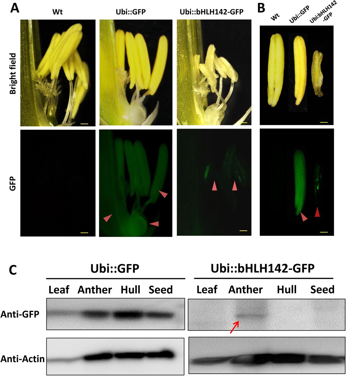
Previous RNA in situ hybridization (ISH) analysis indicated that bHLH142 is tissue specifically expressed in the anthers of Wt at S7 to S9 but not in the leaf (Fu et al., 2014;Ko et al., 2014). RNA ISH data indicated that bHLH142 transcript was localized specifically in the tapetum, middle layer, and meiocytes of the Wt (Fig. 3C). However, in OE142 transgenic line, bHLH142 transcript was detected in leaf tissue (Fig. 3E) and anther (Fig. 3B). Moreover, RNA ISH analysis further indicated that bHLH142 transcript is constitutively expressed in the hulls, anther walls, vascular bundle, and meiocytes of OE142 (Fig. 3D). Surprisingly, western blot analysis using anti-bHLH142 antibody showed that bHLH142 protein is only present in the anther but absent in the leaf of OE142 (Fig. 3E), suggesting post-transcriptional regulation of its expression. In addition, using Ubi::bHLH142-GFP transgenic plants generated in this study we demonstrated that GFP fluorescent signal was detected only in the anther but not in the hull of the transgenic line (Fig. 4). Consistently, western blot analysis of various tissues from Ubi::GFP and Ubi::bHLH142-GFP plants further demonstrated that GFP protein is only present in the anther but not in the leaf, hull or seed of Ubi::bHLH142-GFP transgenic line (Fig. 4C, right panel). Transgenic line overexpressing Ubi::GFP served as a good positive control showing constitutive expression of GFP protein in all tested organs (Fig. 4C, left panel). These results show that both Ubi::bHLH142 (OE142) and Ubi::bHLH142-GFP constructs drove the expression of bHLH142 protein specifically in the anther. Clearly, bHLH142 is expressed in an anther specific manner in OE142. Moreover, both Ubi::bHLH142 (OE142) and Ubi::bHLH142-GFP transgenic lines showed a similar male sterility phenotype, presumably due to the overexpression of bHLH142.

GFP signals observed under fluorescence microcopy for spikelet (A) and anthers (B). Upper panel shows bright field and lower panel shows GFP signals. Arrowheads show GFP signals in the spikelets and anthers of transgenic lines. Scale bars: 50 μm (A), 20 μm (B). (C) Western blotting using anti-eGFP antibody indicated bHLH142 protein is tissue specifically expressed in the anther of Ubi::bHLH142-GFP plant only. Transgenic rice overexpressing GFP as driven by ubiquitin promoter was included as a control. Anthers at vacuolated pollen stage were collected for protein isolation.
Overexpression of bHLH142 alters transcriptional regulation of several known genes related to pollen development
To understand whether the pollen development regulatory network in OE142 was altered, qRT-PCR analysis of several of the known regulatory TFs that are involved in pollen development was carried out. As the expression of bHLH142 in the OE142 lines was driven by the strong constitutive ubiquitin promoter, its mRNA expression in the OE142 lines was consistently upregulated throughout all stages of anther development (Fig. 5A). Interestingly, the expression of GAMYB, UDT1 (bHLH164), and MYB35 (TDF1) was also upregulated in the OE142 anthers (Fig. 5). TDR1 (bHLH5) was upregulated at the early stage but then downregulated at the meiosis stage (S8) in OE142 (Fig. 5E). Similarly, EAT1 was upregulated at the early meiosis stages from S6 to S8 but strongly suppressed after reaching the young microspore stage (S9) in the anthers of OE142 (Fig. 5F). In addition, the expression of MYB80 was found downregulated at meiosis onwards (Fig. 5G). Similar to MYB80, PTC1, a key regulator of tapetal PCD and pollen wall biosynthesis (Li et al., 2011), declined significantly to a negligible amount at S9 in the OE142 anthers (Fig. 5H). Clearly, constitutively overexpressing bHLH142 alters the expression of the key regulatory TFs associated with pollen development.

SC, sporogenous cell, S6; MMC, microspore mother cell, S7; Mei, meiosis, S8; YM, young microspore, S9. Error bars indicate the SD of mean from three replicates.
Overexpression of bHLH142 downregulates PCD related functional genes
EAT1 is known to trigger tapetal PCD by regulating the expression of two Aspartic Proteases (AP37, AP25) at the young microspore stage (Niu et al., 2013). In OE142 anthers, EAT1 was significantly downregulated at S9 (Fig. 5F). TUNEL assay indicated premature onset of tapetal PCD in OE142 anthers (Fig. 2), which was correlated with the upregulation of EAT1 before the meiosis stage (Fig. 5F). Slightly higher expression of AP37 and AP25 at the early stages of OE142 anther development was observed (Fig. 6). Normally, Wt rice exhibits the strongest expression of EAT1, AP37, AP25, and CP1 at S9 to turn on timely tapetal PCD (Fig. 5F, 6). Thus, the negligible expression levels of these marker genes associated with PCD at S9 in the anthers of OE142 further supports the notion that decreased expression of these tapetal functional genes might disrupt timely tapetal PCD (Fig. 2). Consistently, reduced expression of these two proteases (AP37 and AP25) coincided with the reduction of EAT1 mRNA in OE142 at S9 (Fig. 5F, 6). Thus, collectively these results further supported the previous finding that EAT1 regulates AP37 and AP25 (Niu et al., 2013).

(A) Gene expression of AP37. Red area is magnification. (B) Gene expression of AP25. (C) Gene expression of CP1. Abbreviations are as described in the legend for Fig. 5.
OE142 has altered ROS metabolism in the anther
Timely accumulation of ROS is essential to induce PCD during tapetum degeneration (Hu et al., 2011;Yi et al., 2016). The premature onset of tapetal PCD as shown in Fig. 2 prompted us to hypothesize that OE142 may have altered ROS metabolism in the anthers. Therefore, quantitative measurement of superoxide ion using WST-1 was performed in the anthers of the Wt and OE142 line at various developmental stages. Normally, ROS accumulates more at S8 to S9 to trigger tapetal PCD (Xie et al., 2014). Our results showed that the Wt anthers accumulated the highest level of superoxide anions during the meiosis stage; however, OE142 had significantly lower level of superoxide anions compared to the Wt. In addition, the anthers of OE142 accumulated more superoxide anions at the later stage of anther development (Fig. 7A) that might be toxic for OE142 anther development. A previous study suggested that tapetal PCD requires timely and precise control of ROS levels (Xie et al., 2014). We therefore compared the expression of rice ROS scavenging associate genes in OE142 at various stages of anther development. Our qRT-PCR analysis indicated that OE142 reduced the expression of MADS3 and MT2b (Fig. 7). Earlier studies suggest that MADS3 involved in ROS metabolism to trigger PCD (Hu et al., 2011). Our qRT-PCR results indicated that MADS3, and MT2b were significantly downregulated in the anthers of OE142 (Fig.7B, C), which is consistent with the higher ROS accumulation in OE142 as compared to the Wt (Fig. 7A). Taken together, these results suggest that decreased ROS scavenging activity in OE142 anthers affects ROS metabolism and initiation of synchronized PCD, resulting in defective pollen grains (Fig. 1E). Based on the fact that EAT1 expression is downregulated at the S9 stage in OE142 anthers, we contemplated whether EAT1 may play an important regulatory role in ROS metabolism. To verify the possible gene hierarchy in this regulatory process, qRT-PCR analyses of the expression of ROS marker genes in eat1 (Tos17 mutant) anthers were carried out. The results indicated that MADS3 and MT2b were downregulated in eat1 mutant (Fig. 7D, E), implies that MADS3 and MT2b genes might be located downstream of the EAT1 regulatory network. Taken together, these data suggest that overexpression of bHLH142 causes downregulation of EAT1 at the late stage of anther development, which in turn alters the expression of ROS scavenging genes with decreased scavenging activity and accumulation of ROS molecules, leading to defected male gametophyte development.

(A) Alter superoxide anion levels in the anthers of OE142. (B) Comparison of Wt and OE142 using qRT-PCR to analyze gene expression patterns of MADS3 and MT2b (C). (D-E) Mutagenesis analysis indicated gene hierarchy of MADS3 and MT2b locate downstream of EAT1. Wild type for eat1 is in Hitomebore background. Abbreviations are as described in the legend of Fig. 5.
Overexpression of bLHL142 impairs sporopollenin biosynthesis
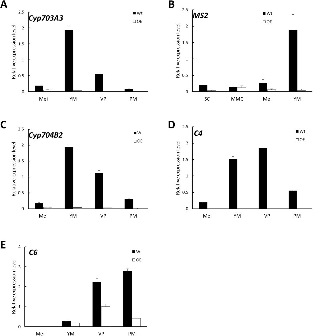
Lipidic exine synthesis is an important component of the pollen wall in rice and Arabidopsis (Yang et al., 2007). The anthers of OE142 were weakly stained by the lipid specific dye Sudan Black compared to that of Wt (Supplementary Fig. S2B). Moreover, the TF PTC1 was downregulated in OE142 (Fig. 5H). Several lipid transfer proteins were also downregulated in ptc1 anthers (Li et al., 2011). Therefore, the expression of these marker genes related to pollen sporopollenin biosynthesis was monitored by real time PCR during OE142 pollen development. The results demonstrated that overexpression of bHLH142 sharply reduced the expression of these genes related to sporopollenin and pollen wall biosynthesis. The transcripts of Cyp703A3, Cyp704B2, MS2, and C4 were almost not detectable in OE142. The expression of C6 was also down-regulated in the anthers of OE142 at the late stage of development (Fig. 8). Our analyses with rice tdr1 and eat1 mutants also indicated that MYB80 was downregulated at S9. Similarly, PTC1, a key regulator of sporopollenin biosynthesis, was significantly downregulated in the eat1 anther (Supplementary Fig. S5). Taken together, these results support the idea that both MYB80 and PTC1 regulate sporopollenin biosynthesis in both monocots and dicots. Thus, overexpressing bHLH142 caused downregulation of EAT1 at S9, which might severely inhibit MYB80 and PTC1 and reduce sporopollenin gene expression (Fig. 8) and interrupt normal sporopollenin biosynthesis with defected pollen wall in OE142 transgenic lines.

Comparison of Wt and OE142 using qRT-PCR to analyze gene expression patterns of Cyp703A3 (A), MS2 (B), Cyp704B2 (C), C4 (D), and C6 (E). Abbreviations are as described in the legend of Fig. 5.
OE142 anther exhibits parallel changes in EAT1 transcript and protein
In this study, we found that EAT1 was upregulated at stages S6 to S8 but then downregulated at S9 in the anthers of OE142 (Fig. 5F, 9A). To understand the spatial and temporal expression patterns of EAT1 in OE142, we carried out RNA ISH hybridized EAT1 Dig-labeling probe in the anthers of Wt vs. OE142 at S8a and S9. The results revealed that EAT1 mRNA was highly expressed in the tapetum, middle layer, meiocycte, microspore, vascular bundle, and hull of the Wt at S9 (Fig. 9B). However, ISH positive signal of EAT1 was strong in the anthers of OE142 at early meiosis (S8a), but significantly reduced to a negligible level at YM (S9). The ISH results support our EAT1 qRT-PCR data (Fig. 5F), providing a clear picture of the in vivo transcriptional map of EAT1.

(A) qRT-PCR revealed upregulation of EAT1 at (S7) and downregulation at YM (S9). (B) RNA ISH hybridization to EAT1-antisense probe in the Wt and OE142 anthers at stages S8 to S10. (C) Western blotting showed premature expression of EAT1 protein at MMC and downregulation at YM in OE142. The knockout mutant eat1 was used as a negative control. Abbreviations are as described in the legend of Fig. 5. Scale bars: 20 μm.

Overexpression of bHLH142 upregulates UDT1, GAMYB, TDR1, and EAT1 at an early stage of anther development. However, EAT1 is downregulated at the young microspore stage (YM, S9), which in turn reduces the expression of the downstream genes involved in ROS scavenging (MADS3, MT2b), leading to premature onset of tapetal PCD at meiosis-I (S8a). Moreoer, OE142 downregulated PCD (AP37, AP25, CP1), and sporopollenin biosynthesis (MYB80, PTC1, MS2, CYP704B2, C4, C6). Thus, defected tapetal PCD at YM stage (S9) and defected pollen wall development together leads to male sterility in the overexpression line. Genes marked in red or green denote upregulation or downregulation, respectively. Solid arrow lines indicate direct regulation, while dotted arrow lines indicate indirect regulation. Double arrows represent protein-protein interaction. SC, sporogenous cell; MMC, microspore mother cell; YM, young microspore stage (S9).
Western blot analysis further revealed that bHLH142 protein was specifically accumulated in the OE142 anthers (Fig. 3F). EAT1 protein was not detectable at S8a but accumulated at a high level at S9 in the Wt (S9). However, OE142 anthers precociously expressed EAT1 protein at S8a but its expression was reduced to negligible level at S9 (Fig. 9C). These data suggest that overexpression of bHLH142 prematurely upregulate EAT1 transcription (Fig. 5F, 9A) as well as its protein level (Fig. 9C) in the anthers of OE142. Thus, the decreased transcript abundance and protein amount of EAT1 at S9 might interrupt the normal anther development in the OE142 transgenic lines.
In our previous study, we demonstrated that bHLH142 interacts with TDR1 to co-modulate EAT1 transcriptional activity (Ko et al., 2014). Overexpression of bHLH142 increases bHLH142 protein level, which may in turn enhance bHLH142/TDR1 protein-protein interaction to increase EAT1 expression at the early stage of anther development. However, downregulation of TDR1 expression at S8 and onward (Fig. 5E) might decrease TDR1 protein translation and then hamper TDR1/bHLH12 protein-protein interaction, therefore significantly reduce EAT1 expression at later stages even though high bHLH142 protein is present in the anthers of OE142. In additions, a low EAT1 protein level at S9 (Fig. 9C) might further reduce TDR1/EAT1 interaction and subsequently influence the regulatory cascade of downstream target genes and result in defected pollen development in OE142 anthers.
DISCUSSION
Overexpression of bHLH142 causes male sterility by triggering premature PCD
In an effort to provide greater insight into the functionality of bHLH142 in rice pollen development, we generated transgenic lines constitutively overexpressing bHLH142. To our surprise, overexpression of bHLH142 also leads to male sterility in rice, similar to the knockout mutant reported previously (Ko et al., 2014). Except for the defect in pollen development, OE142 transgenic lines maintain wild-type like vegetative growth (Fig. 1; Supplementary Fig. S1, S2).
Our in-depth molecular characterization suggests that overexpression of bHLH142 significantly alters in vivo homeostasis of the known key pollen development-related regulatory TFs in the OE142 anthers (Fig. 5). For example, UDT1, GAMYB, MYB35, TDR1, and EAT1 were upregulated in OE142 at the early stages of anther development (Fig. 5). Presumably, this triggers a premature onset of tapetal PCD in OE142 anthers before the maturation of pollen grains, as shown in the TUNEL assay (Fig. 2). The reduced expression of EAT1 at the young microspore stage (S9) and onward in OE142 anthers (Fig. 5F) further impairs the normal development of pollen grains because of reduced expression of the downstream genes in the PCD pathway, such as AP37, AP25, and CP1 (Fig. 6) and pollen wall biosynthesis, such as MYB80, PTC1, MS2, Cyp704B2, C4, and C6 (Fig. 8). Obviously, interference with the timely expression of these pollen development associated genes leads to male sterility in OE142.
Tightly regulated bHLH TFs are essential for pollen development
Our study revealed that overexpression of bHLH142 causes significant changes in the expression of known regulatory genes associated with tapetal PCD, ROS metabolism, and pollen wall development (Fig. 6–8), thus leading to male sterility in OE142 transgenic plants. This may result from the interference in its protein interaction with TDR1 in activational transcription of EAT1 (Ko et al., 2014). This study advances our knowledge of the molecular mechanism underlying the bHLH142 and EAT1 transcriptional circuits controlling pollen development in rice and possibly in other plants as well. Timely expression and maintenance of proper expression levels of these bHLH TFs must be tightly regulated developmentally for normal pollen maturation. The hierarchy of several known regulatory network genes associated with pollen development is therefore clarified in this study.
Based on this and previous studies, we propose a mechanistic model of genic male sterility in rice as caused by overexpressing bHLH142 (Fig. 10). According to the model, overexpressing bHLH142 causes upregulation of UDT1, GAMYB, TDR1, and EAT1 at the early stage of anther development. This consequently leads to premature onset of tapetal PCD. However, EAT1 is downregulated at the young microspore stage (YM, S9) in OE142 anthers, which in turn further reduces the expression of the downstream functional genes involved in PCD (AP37, AP25, and CP1), ROS scavenging (MADS3 and MT2b), and pollen wall biosynthesis (MYB80, PTC1, Cyp704B2, MS2, and C4) and impairs normal pollen grain maturation. Thus, increased ROS accumulation, defect in timely tapetal PCD at the YM stage, and defect in pollen wall development, eventually lead to male sterility in the OE142 plants. The alterations in homeostasis of key TFs in pollen development or protein-protein interaction between bHLH142/TDR1 or TDR1/EAT1 may account for the decreased expression of downstream pollen development marker genes regulated by EAT1 (Ko et al., 2014).
Potential of establishing a male sterility line by overexpressing key TFs
Our finding that overexpression of bHLH142 (TIP2) causes male sterility by triggering premature PCD in rice is similar to previous results obtained by overexpressing several pollen development related transcription factors in other species. A total of 148 out of 196 Arabidopsis transformants overexpressing AMS (rice homolog of TDR1) produced sterile pollen, mimicking the ams mutant phenotype (Sorensen et al., 2003). It is claimed that the resulting male sterility might be due to co-suppression of AMS, but our results tend to suggest that altered homeostasis of the related TFs may be the major cause. Moreover, overexpressing MS1 as driven by the CaMV35S promoter also caused stunted plants with sterile pollen in Arabidopsis (Yang et al., 2007).
Recently, the ortholog of MS1 in barley (HvMS1) was cloned and its expression was altered to be either overexpressed or suppressed (Fernandez Gomez and Wilson, 2014). Both RNAi and overexpression of HvMS1 full-length cDNA under the control of the maize ubiquitin promoter caused male sterile phenotype in the transgenic barley plants. Also, knockout of AtCEP1, which encodes a papain-like cysteine protease involved in tapetal PCD, delayed tapetal PCD, while its overexpression caused premature tapetal PCD (Zhang et al., 2014). Thus, we hypothesize that overexpression of other key TFs in the pollen development regulatory network, such as GAMYB, UDT1, TDR1, or EAT1 (DTD1, bHLH141) may also cause male sterile phenotype in rice due to alteration in the homeostasis of the regulatory cascades in pollen development.
Advantages of using OE142 in hybrid seed production
In this study, overexpression of bHLH142, an anther-specific TF gene, by a strong constitutive promoter led to its ubiquitous transcription in leaves, hulls, as well as in the anther of OE142, as expected (Fig. 3). However, bHLH142 protein expression was not constitutively expressed. Rather, its expression was maintained in a tissue specific manner in the anthers (Fig. 3F and 4). The ubiquitin promoter is expected to drive ubiquitous gene expression. However, protein expression level is determined by the rate of transcription and by post-transcriptional processes that lead to changes in mRNA transport, stability, and translational efficiency. The nuclear localization sequence (NLS) mediates the transport of nuclear proteins into the nucleus. bHLH142 contains two NLS and targeted protein is localized in the nucleus (Ko et al., 2014). The basis for the specific expression of bHLH142 in anthers, as driven by the ubiquitous promoter, requires further study. Some unknown posttranscriptional factor(s) specifically in the anthers may be required for its translation. In fact, overexpressing target genes in an anther-specific manner is desirable from the perspective of GMO food biosafety because anther-specific expression will avoid any unintended expression in other tissues, especially in the edible part of the seed (Fig. 4). This may be more acceptable to consumers.
Future prospects
Cytoplasmic male sterility (CMS) is widely used in F1 seed production in rice. Wild abortive (WA) CMS gene encodes a tapetal mitochondrial protein, WA352, which interacts with the nucleus encoded mitochondrial protein COX11 to inhibit ROS-scavenging activity, thus triggering premature tapetal PCD and pollen abortion (Luo et al., 2013). Whereas CMS guarantees a high degree of purity of hybrid seeds it increases production cost. Therefore, there is a great demand for the generation of new male sterility lines for production of hybrid crops, such as rice, maize and wheat, etc. Here, we showed that overexpression of bHLH142 may provide a novel and simple way to generate genic male sterility lines in rice. Normally, genetic engineering using the overexpression approach is preferred to RNAi by the biotech industry. However, one major concern with this technology is the maintenance of the genic male sterility line as it fails to produce viable seeds as stock. A strategy to generate inducible male sterility and restorer hybrid seed production has been demonstrated in Arabidopsis (Li et al., 2007). In this system, a complete male sterility transgenic line was generated using AtMYB103EAR chimeric repressor construct under the control of the native AtMYB103 promoter, whereas the restorer containing the AtMYB103 gene under the control of a stronger anther-specific promoter (p39) was introduced into the pollen parent (Li et al., 2007). Fertility is restored when the restorer line is crossed with the male sterile plant. Another possible solution to alleviate this problem is to employ an inducible promoter to drive its expression. GM male sterility plants generated by overexpression of a MYC5-SRDX chimeric repressor under the control of an inducible promoter have been demonstrated in Arabidopsis (Figueroa and Browse, 2015). Other promoters inducible by chemicals, temperature or light can also be considered and tested.
Accession numbers
Sequence data from this article can be found in the GenBank/EMBL database under the following accession numbers: bHLH142 (Os01g0293100), protein (NP_001042795.1). Additional loci are presented in Supplementary Table S1.
Competing interests
The authors declare that they have no competing interests.
Acknowledgments
We thank the Tos17, Postech, and TRIM Mutant Libraries for providing rice mutant seeds. We appreciate the technical support of the Biotechnology Center in Southern Taiwan, Academia Sinica, GMO greenhouse core facility. This work was supported in part by the Biotechnology Center in Southern Taiwan, Academia Sinica, and a Ministry of Science and Technology of Taiwan grant to Dr. Swee-Suak Ko’s project (MOST104-2313- B-001 -003). We thank Ms. Miranda Loney for English editing.
Footnotes
E-mail addresses for authors: Swee-Suak Ko: sweesuak{at}gate.sinica.edu.tw, Min-Jeng Li: lmz8332{at}yahoo.com.tw, Yi-Jyun Lin: hbabyi{at}gate.sinica.edu.tw, Hong-Xian Hsing: sanny618{at}gmail.com, Ting-Ting Yang: tty0318{at}gmail.com Tien-Kuan Chen: s4life2010{at}gmail.com, Chung-Min Jhong: amo2175111{at}gmail.com, Maurice Sun-Ben Ku: mku{at}mail.ncyu.edu.tw
Authors’ contributions SSK designed the experiments, MJL performed most of the experiments, YJL, HXH, TTY, TKC, CMC, SSK conducted parts of the experiments, MJL and SSK analyzed the data, and SSK and MSBK wrote the manuscript.


