

Abstract
Antibiotic resistance continues to be a major global health risk with an increase in multi-drug resistant infections seen across nearly all bacterial diseases. This global burden is further pronounced by the misuse of antibiotics and the availability of only a handful of antibiotics in the development pipeline. Amongst the drug-resistant infections, mycobacterial infections such as Tuberculosis (TB) and Non-Tuberculosis infections have seen a significant increase in the incidence of multi-drug resistant and extensively drug-resistant infections. With this increase in drug-resistant Mycobacteria, mycobacteriophage therapy offers a promising alternative. However, a comprehensive study on the infection dynamics of mycobacteriophage against their host bacteria and the evolution of bacteriophage (phage) resistance in the bacteria remains elusive. We aim to study the infection dynamics of a mixture of phage against Mycobacteria under various pathophysiological conditions such as low pH, low growth rate and hypoxia. We show that mycobacteriophages are effective against M. smegmatis under various conditions and that the mixture of phages prevents the evolution of resistance for long durations. Mycobacteriophages are effective against antibiotic-resistant strains of Mycobacterium and show synergy with antibiotics. Finally, we also show that mycobacteriophages are infective and efficient against M. tuberculosis and prevent its growth for several weeks. These findings have important implications for developing phage therapy against Mycobacterium.
INTRODUCTION
Prolonged and unsupervised use of antibiotics results in an increase of antibiotic resistance across all bacterial species which is considered a major threat to human health (1, 2). The Mycobacterium genus with over 190 recognized species contains various pathogenic species including M. tuberculosis, M. leprae, M. abscessus, M. avium complex, etc. (3). These infections are often difficult to treat owing to their resilient cell walls which render them resistant to prolonged exposure to acidic and alkaline environments (3). This thick, hydrophobic cell wall rich in mycolic acids also provides resistance to various cell-wall synthesis disrupting antibiotics such as Penicillin (3–5). In addition, several mycobacterial species rapidly evolve resistance to antibiotics. A World Health Organization (WHO) surveillance network reported the emergence of rifampicin-resistant M. leprae leading to drug-resistant leprosy (6). In a total of 1143 relapse cases, 5.1% were found to be rifampicin-resistant and in a total of 789 new cases, rifampicin resistance was observed to be at 2% (6). Mycobacterium abscessus has been recognized as the most drug-resistant pathogen amongst the rapidly growing Non-Tuberculosis Mycobacteria (NTMs) and drug-resistant M. abscessus infections have posed a clinical challenge in a number of cases (7). Similarly, the Mycobacterium avium Complex (MAC) infections are noted to be on the rise, especially in AIDS and pulmonary disease patients (8) and MAC infections are often associated with an increase in mortality (8, 9).
Tuberculosis (TB), caused by Mycobacterium tuberculosis, is one of the most prevalent infection in modern times with an estimated 10 million new cases and 1.5 million TB-related deaths in 2018 (10). Conventional therapy of TB includes the use of antibiotics such as isoniazid, rifampicin, ethambutol and pyrazinamide for 6-12 months (11). However, the emergence of drug-resistant, multi-drug resistant and extensively drug-resistant TB poses a challenge in the treatment and eradication of the disease (5, 10). According to a recent estimate by the WHO (10), 4.84% of total TB cases in 2018 are rifampicin-resistant, of which 78% are multi-drug resistant. Across the multi-drug resistant TB, 6.2% are extensively drug-resistant TB. The antibiotic treatment regimen for TB is associated with the distinct and long-lasting changes to the gut flora, often resulting in the depletion of various species of commensal bacteria (12). Antibiotics are also associated with various adverse side-effects ranging from nausea, vomiting and fever to anaphylaxis, hypersensitivity and photodermatitis (13). In order to tackle this growing rate of antibiotic resistance and the side effects of antibiotic treatment regimens, various therapeutic strategies have been proposed, including phage therapy that has shown tremendous potential against various bacterial pathogens (14).
Phages are viruses that are host specific towards bacteria and are inherently non-pathogenic to humans. They are abundantly present in the natural microflora of the human body (15, 16). A recent non-comparative clinical trial showed that the intravenous delivery of anti-staphylococcal phages in patients with severe infection did not pose any challenges to tolerability and safety (17). Moreover, phages remain infective and lyse antibiotic-resistant bacteria (14). To circumvent the narrow-host range specificity and development of resistance (18), multiple phages are used in phage therapy. Successful compassionate use of phages against antibiotic-resistant bacteria on patients in the USA and Europe has been on the rise and shows their safety and translation potential (19, 20).
With over 10,000 mycobacteriophages isolated and more than 1500 sequenced (21), there is a large library of phages against Mycobacterium. Although the desired lytic phenotype of phages for phage therapy brings the number down, development of Bacteriophage Recombineering of Electroporated DNA (BRED) technique (22) allows rapid conversion of lysogenic phages to lytic phages for phage therapy. Recently, a three-phage mixture has been successfully used in a clinic for the treatment of a cystic fibrosis patient suffering from Mycobacterium abscessus infection (23). Prophylactic phage (D29) therapy against TB also showed some success in a preclinical animal model in reducing the burden of infection (24). A study employing phage therapy in treating subcutaneously injected M. tuberculosis (H37Rv) infection in guinea pigs showed that mycobacteriophages have a therapeutic effect (25).
Phage therapy, although is a strong candidate for treating mycobacterial infections, presents a few physiological hurdles that need to be taken into consideration. For instance, in TB infection, bacterial cells are known to reside and eventually replicate under nutrient starvation, acidic and hypoxic environments (26–28). For a phage to successfully eliminate these bacilli, it needs to be active and efficient under such low pH and hypoxic conditions. Since most bacteria enter a non-replicative stationary phase, the phage should also be able to infect and kill bacteria in the stationary phase where most of the current antibiotics fail. Designing a phage therapy that would be effective in such complex physiological environments would necessitate a better understanding of the bacteria-phage dynamics under various conditions. However, to our knowledge, no study has looked at the efficacy of phages over time in such different environments. Hence, there is a need to study the efficacy of phage mixtures in the physiological and pathophysiological conditions in which bacteria survive.
In this study, we have performed phage-bacteria kinetics under various pathophysiological conditions of mycobacterial diseases-low pH, hypoxia and stationary phase. We further study the emergence of phage resistance in M. smegmatis and the combinatory effect of antibiotics and phage therapy. We show that mycobacteriophage mixtures are effective against Mycobacterium under the low pH, hypoxic and stationary conditions and showed synergy with antibiotics. Finally, we also find that mycobacteriophages are able to prevent growth of Mycobacterium tuberculosis and encourages further exploration of phages for diseases caused by Mycobacterium such as TB.
MATERIALS AND METHODS
Chemicals and Reagents
Middlebrook 7H9 Broth, Middlebrook 7H10 Agar and DMSO were purchased from Sigma (St Louis, MO, USA). Middlebrook 7H11 agar was purchased from BD Difco (NJ, USA). ADC growth supplement, Ziehl-Neelsen stain, rifampicin, and isoniazid were purchased from HiMedia (India). Tween 80 and Glycerol were purchased from Fisher Scientific (MA, USA). Calcium Chloride, Magnesium Sulphate, and Sodium Chloride were purchased from Sri Tirumala Chemicals (India).
Bacterial cell culture and maintenance
Primary Mycobacterium smegmatis (mc2155) or Mycobacterium tuberculosis (H37Ra) cultures were grown in Middlebrook 7H9 broth supplemented with Glycerol, ADC and Tween-80 (0.1% v/v). A log phase primary culture was inoculated into a secondary culture without Tween-80 and supplemented with CaCl2 (2 mM) to promote an efficient infection of phages. Frozen stocks were prepared by adding 300 μL of a late log phase culture to 35% (v/v) final concentration of glycerol and stored at −80°C until future use. All cultures were maintained at 37°C with rotary shaking at 180 rpm.
Soft agar overlay and spot assays for phage enumeration
For phage titrations and phage confirmation, spot assays were carried out in which late-log phase M. smegmatis cultures were plated on a Middlebrook 7H10 media plate supplemented with CaCl2 (2 mM) and glycerol to form a bacterial lawn. Phage samples were serially diluted and spotted on to the bacterial lawn. The plates were incubated at 37°C for 24 h and the number of plaques formed at various dilutions was counted.
Soft agar overlay technique was also used to detect and quantify phages. Briefly, soft agar was prepared by adding agar (0.8%) to 7H9 media along with the required supplements. The suspension was autoclaved and allowed to cool down to 42°C. 1 mL of late log phase bacterial culture and/or 100 μL of phage dilutions was added to 5 mL of the soft agar and was poured onto a 7H10 plate supplemented with CaCl2. The soft agar was allowed to cool down and solidify. The plate was incubated at 37°C overnight.
Phage amplification and maintenance
Five phages (D29, TM4, Che7, PDRPv (29) and PDRPxv (30)) used in this study were amplified on M. smegmatis. Similarly, three phages (D29, TM4 and DS6A) were amplified on M. tuberculosis. For liquid amplification, 100 μL of phage sample was added to a log phase culture and incubated for 12-24 h at 37°C with rotary shaking at 180 rpm. The cultures were then centrifuged at 4000 g for 10 min and the supernatant was collected and filtered. The spot assay was used to determine the titers in filtered supernatant. For plate amplification, a soft agar overlay containing M. smegmatis was prepared and the phage sample was spotted on top of the soft agar. The plates were incubated for 12-24 h at 37°C following which, the soft agar was collected into a 50 mL falcon tube. An appropriate amount of Magnesium Sulphate-Tris Chloride-Sodium Chloride Buffer (SM Buffer) was added to the falcon tube to completely immerse the soft agar and the falcon tube was incubated for 2-4 h at 37°C or overnight at 4°C. The falcon tube was then subjected to centrifuge at 4000 g for 10 min at 4°C and the supernatant was collected and filtered using 0.22 μm syringe filters (Biofil, FPV203030-A). The spot assay or soft agar overlay was used to determine the phage titers.
Bacterial growth kinetics
In order to study the bacterial growth kinetics, 7H9 broth supplemented with ADC and CaCl2 was prepared and inoculated with 50 μL of log-phase secondary bacterial culture per 10 mL of the media. The cultures were then incubated at 37°C with rotary shaking at 180 rpm. Periodic measurements of bacterial optical density (OD) were taken using a spectrophotometer (Jenway 7205 UV/Visible Spectrophotometer) at 600 nm against a media blank. For phage treated samples, a 10:1 MOI (for M. smegmatis) or 0.0003:1 for (M. tuberculosis) of each phage in the respective phage solution was added at the appropriate time (either at the start of the experiment (T0) or at a specified interval of mid-log phase).
Development of phage resistant M. smegmatis strains
One mL stationary-phase M. smegmatis cultures were treated with 100 μL of respective phages and were transferred into prewarmed overlay agar. Soft agar overlay was carried out and the plates were incubated at 37°C and monitored for bacterial growth for 3-5 days. Single colonies were picked from the plates and Ziehl-Neelsen staining was performed to confirm the growth of M. smegmatis and lack of contaminants. These cultures were then expanded in 7H9 broth supplemented with ADC and CaCl2 and checked against all the phages for resistance through spot assay.
Bacterial growth kinetics with phage – pH, stationary phase and hypoxia
To study the effects of acidic pH on phage infection dynamics, phage kinetics assay was performed at varying the pH of the media. Briefly, the pH of the prepared media was reduced from 6.8 prior to the experiments using HCl to either a pH of 6.0 or pH of 5.5. The media was filtered and used for future growth kinetics experiments. Bacteria were inoculated into the media at pH 6.8, 6.0 and 5.5. For phage treated samples, a 10:1 MOI for each phage in the 5-phage mixture was added at an appropriate time (either at the start of the experiment (T0) or at a specified interval of mid-log phase. OD was measured at 600 nm.
For stationary phase experiments, M. smegmatis cultures were grown in 7H9 media supplemented with Glycerol, ADC and CaCl2 (2 mM) until an OD of over 10 was reached. The stationary phase cultures were then either treated with the 5-phage mixture at 10:1 MOI or left untreated. OD values were obtained at regular intervals at 600 nm.
For the studies involving hypoxia, the cultures were grown in plastic K2-EDTA Vacutainers (BD Cat No. 367525) to generate hypoxia. Briefly, K2-EDTA Vacutainers were obtained from BD and washed with autoclaved sterile water over a period of 24 h to remove the EDTA coating. The vacutainers were then washed with prepared media before experimentation. Upon inoculation, the vacutainers were sealed with the rubber septa caps and parafilm to prevent oxygen diffusion. Samples were collected through a syringe and 20G needle for spectrophotometer readings. Oxygen levels were monitored by adding Methylene Blue (0.01%) to the samples (31) and measuring the OD at 665 nm.
Ziehl-Neelsen staining
Ziehl-Neelsen staining was performed periodically to check for contamination and for the confirmation of Mycobacterium spp. as described by the manufacturer. Briefly, the bacterial suspension was spread evenly over the slide and air-dried for 30 min. The slide was then heat-fixed on a hot plate for 15 min at 60°C and then flooded with Carbol Fuchsin stain at 85°C. Care was taken not to overheat or boil the stain and the slide was frequently replenished with additional Carbol Fuchsin stain. The slide was allowed to remain on the hot plate for 10 min following which it was removed and allowed to incubate for an additional 5 min. The stain was washed away under running tap water and flooded with decolorizer for 2 min or until the smear was sufficiently decolorized. The slide was washed under running tap water and treated with Methylene Blue for 30 s. The slide was subjected to one final washing under running tap water to remove the Methylene Blue stain and was air-dried following which, it was observed under a 100X oil immersion objective using an upright fluorescence microscope (Olympus BX53F) for the presence of either pink rod-shaped acid-fast bacteria or blue stained contamination.
Antibiotic Minimum Inhibitory Concentration (MIC) determination
The MICs of both rifampicin and isoniazid were determined to establish the antimycobacterial activities of the respective antibiotics against M. smegmatis strain mc2 155. Antibiotic stock solutions were prepared at concentration of 10 mg/mL in sterile deionized H2O for isoniazid and in DMSO for rifampicin. Stocks were stored at −20°C until further usage. Several dilutions were prepared in a 96-well plate for each of the antibiotic stock solutions to obtain various concentrations ranging from 1.5 μg/mL to 100 μg/mL. A total of 2×10⁵ cells of M. smegmatis were added to each of the wells. The 96-well plate was placed in a rotary shaker incubator at 37° C for 48 h and was observed for growth against control be measuring OD at 600 nm. The lowest concentration of antibiotic without bacterial growth was termed as MIC for the respective antibiotic.
Antibiotic growth kinetics
In order to study the effect of antibiotics-isoniazid and rifampicin on M. smegmatis growth, 7H9 broth supplemented with ADC and CaCl2 was prepared and inoculated with 50 μL of log-phase secondary bacterial culture per 10 mL of the media. The cultures were then incubated at 37°C with rotary shaking at 180 rpm. Periodic measurements of bacterial OD at 600 nm against a media blank. For antibiotic-treated samples, a final concentration of rifampicin at 40 μg/mL and 100 μg/mL and isoniazid at 40 μg/mL and 100 μg/mL were added at the appropriate time (either at lag phase or at a specified interval of mid-log phase).
Phage kinetics against isoniazid resistant M. smegmatis
To study the effect of phage mixture on antibiotic-resistant strain, 7H9 broth supplemented with ADC, CaCl2 and isoniazid (40 μg/mL) was prepared and inoculated with 50 μL of isoniazid-resistant (mc2 4XR1) (32–34) log phase secondary bacterial culture per 10 mL of media. The cultures were then incubated at 37°C with rotary shaking at 180 rpm. Periodic measurements of bacterial OD were taken using a spectrophotometer at 600 nm against a media blank. For phage treated samples, the 5-phage mixture was added at 10:1 MOI at an appropriate time (either at T0 or at a specified interval of mid-log phase).
Statistical analysis
All experiments were performed on independent biological replicates. Statistical significance was determined for control and experimental groups using multiple t-test with Holm-Sidak method, with alpha = 0.05. Each time point was analysed individually, without assuming a consistent SD. For analysis of multiple groups, 2-way ANOVA was used with Sidak’s multiple comparisons test, with alpha = 0.05. Graphpad (Prism) was used for all statistical analysis.
RESULTS
Mycobacteriophages reduce bacterial growth in M. smegmatis
We first started with M. smegmatis as the model organism due to its fast replication and ease of handling. Several lytic Mycobacteriophages were procured and amplified to titers greater than 109 pfu/mL. In order to test their efficacy, bacterial cultures were infected with phages at the MOI of 10:1 either in the lag phase (termed at before growth) or in the mid-log phase (termed as after growth). The optical density of the cultures at 600 nm was monitored using a spectrophotometer at various time intervals. Cultures treated with phages D29, TM4, Che7, PDRPv and PDRPxv individually showed a significant reduction in the optical density when phages were added at log phase (time of addition shown by red arrows) (Fig 1). Similarly, when the phage infection was carried out at lag phase, no growth was observed for many hours. It is well known that bacteria evolve resistance to phages over a period of time. M. smegmatis cultures infected with phages were also monitored for phage resistance. Phage resistance was observed in all phage treated M. smegmatis cultures. While D29 and Che7 treated cultures showed phage resistance at around 40 h, TM4 treated cultures showed phage resistance at 60 h; PDRPv and PDRPxv treated cultures showed phage resistance at 83 h and 101 h respectively (Fig 1). Bacterial cultures infected with phages at an MOI of 30:1 showed no significant increase in the onset of phage resistance (Supplementary fig S1).

A) Photograph showing spot clearance of a lawn of M. smegmatis after treatment with D29. Growth curves of M. smegmatis in liquid culture in the presence of B) D29, C) Che7, D) TM4, E) PDRPv and F) PDRPxv phage. Red arrow represents the time point at which phage solution was added to the cultures. These experiments were repeated several times (≥2) and the representative graphs are shown here. Multiple t-test with Holm-Sidak method was used to estimate the statistical significance. Symbols, with starting time in brackets for respective panels, represent a significant difference (p<0.05) between control and phage (after growth): α (21 h), β (28 h), χ (21 h), δ (28 h) and ε (24 h).
Single phage resistant M. smegmatis is susceptible to other phages
To design an optimal phage mixture with broad host-range and varied resistance inducing mechanisms, it is desirable that all phages in a mixture target the bacteria through different mechanisms. To test this, we generated single phage resistant M. smegmatis strains for D29, TM4, Che7, PDRPv and PDRPxv by picking colonies growing in soft agar overlay after phage challenge. We checked for the phage titers (>109 against wild-type M. smegmatis) against the resistant M. smegmatis strains (Table 1). We observed several log order reductions in titer of the phage which was used to generate the mutants in the respective phage resistant M. smegmatis. However, comparatively, other phages still had high titers on the mutant bacteria, which suggested a possible alternative resistance mechanism for each of the 5 phages.
Phage mixtures show synergy in growth reduction
To test the effect of phage mixtures, M. smegmatis cultures infected with multiple phages (a mixture of 3 or a mixture of 5) at 10:1 MOI of each phage were monitored for OD at 600 nm (Fig 2). Phage mixture treatment significantly reduced the optical density of the bacterial cultures compared to controls. Phage resistance in mixture treated cultures emerged at around 105 h in case of 3 phage mixture (D29, TM4 and Che7) (Fig 2A) and at 322 h in case of 5 phage mixture (D29, TM4, Che7, PDRPv and PDRPxv) (Fig 2B). The corresponding Colony Forming Units (CFU)/mL readings were obtained and the viable cell count was observed to be correlating with OD readings (Supplementary fig S2). This shows that the mixture of phages can act synergistically to delay the evolution of resistance. The 5-phage mixture was selected and used for future experiments.
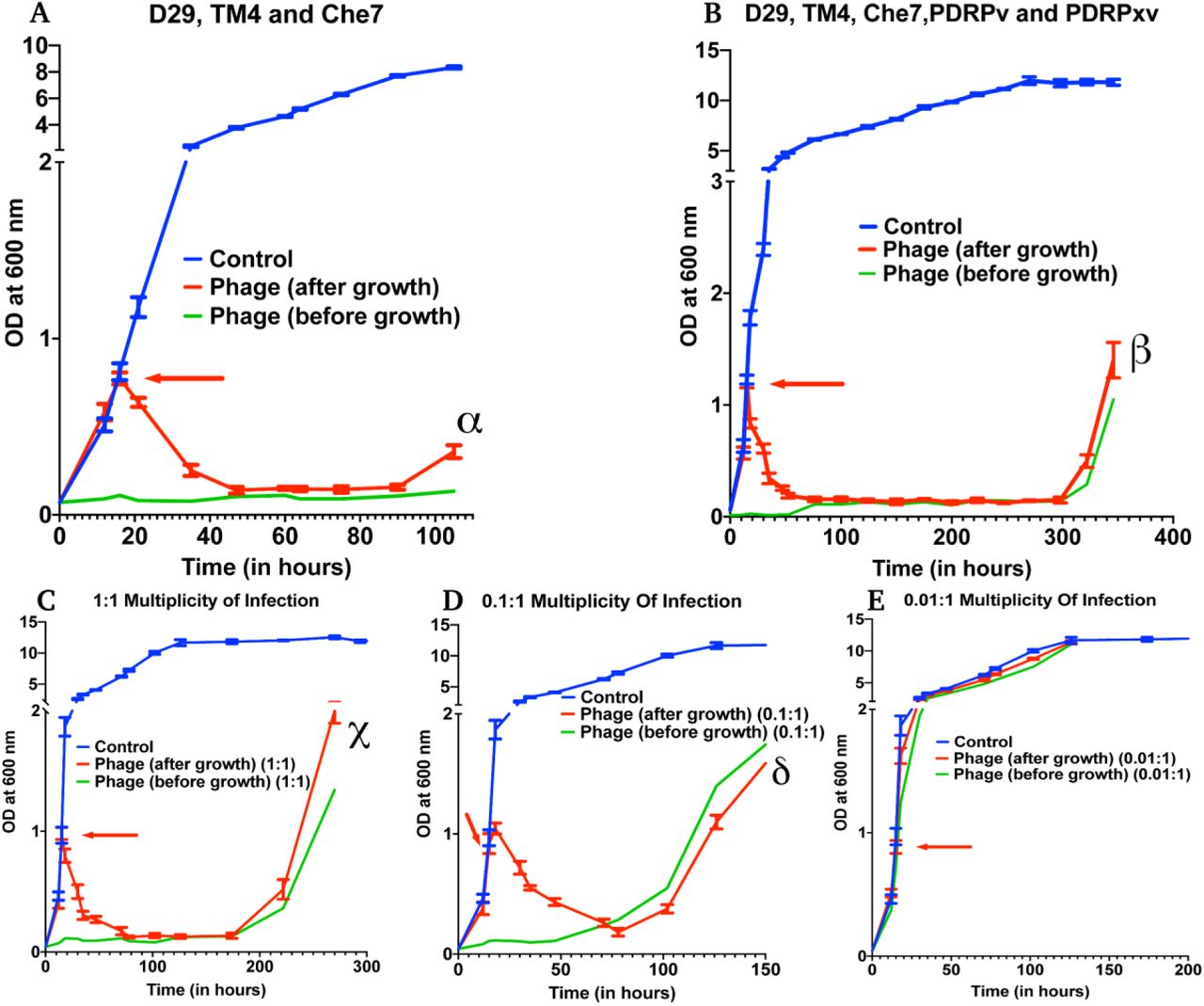
Growth kinetics of M. smegmatis cultures treated with A) 3-phage mixture (D29, Che7 and TM4) and B) 5-phage mixture (D29, TM4, Che7, PDRPv and PDRPxv) at MOI of 10:1 for each phage against a non-treated control. Growth kinetics of M. smegmatis cultures treated with a 5-phage mixture at the MOI of C) 1:1, D) 0.1:1 and E) 0.01:1 for each phage in the phage mixture. The experiments with MOI of 10:1 with 3- and 5-phage mixtures were repeated twice and the representative graphs are shown here. Red arrow represents the time point phage solution was added to the cultures. Multiple t-test with Holm-Sidak method was used to estimate the statistical significance (p<0.05). Symbols, with starting time in brackets for respective panels, represent a significant difference between control and phage (after growth): α (21 h), β (18 h), χ (18 h) and δ (18 h).
To test for the efficacy of mycobacteriophages at various Multiplicity of Infection (MOI), M. smegmatis cultures were infected with a MOI of 10:1, 1:1, 0.1:1 and 0.01:1 of each phage: bacteria in either the lag phase or the mid-log phase. A 10:1 MOI (Fig 2B) significantly reduced the growth of bacterial culture with phage resistance emerging at 322 h. A 1:1 MOI (Fig 2C) also resulted in a significant reduction of bacterial growth, however, phage resistance emerged at around 222 h. While a 0.1:1 multiplicity of infection (Fig 2D) resulted in a reduction in growth, resistance was observed at 102 h for mid-log phase treated cultures. A 0.01:1 MOI did not result in any significant reduction in the bacterial growth (Fig 2E). For all future experiments, we have used the MOI of 10:1 to evaluate the maximum lysing potential of phages against Mycobacterium.
Mycobacteriophages are effective in stationary phase of bacterial growth
Within the host environment, Mycobacterial species slow their growth rate and often enter a non-replicative state (27, 28, 35). This reduced growth state poses a significant hurdle in the treatment of infections with conventional antibiotics (36). For phage therapy to be efficient against mycobacterial infections, phages need to retain the infectivity against the slow growing bacteria. To test the effect of phage mixture against M. smegmatis in stationary phase, M. smegmatis cultures were allowed to reach an OD>10. The stationary phase cultures were then infected with the 5-phage mixture without any supplementation with nutrients and periodic OD measurements were taken at 600 nm. Significant reduction in OD and CFU were observed in phage treated samples demonstrating that the 5-phage mixture is effective in stationary phase conditions (Fig 3A, Supplementary fig S3A). Interestingly, even after 350 h of culture, no growth of bacteria was observed.

Growth kinetics of M. smegmatis cultures treated with a 5-phage mixture at A) stationary growth phase, B) pH conditions of 6.8, 6.0 and 5.5 and C) hypoxic conditions. For hypoxia experiment, solid lines represent bacterial OD at 600 nm and dashed lines are the respective Methylene Blue readings at 665 nm. Red arrow represents the time point phage solution was added to the cultures. These experiments were repeated twice and the representative graphs are shown here. For A and B, multiple t-test with Holm-Sidak method was used to estimate the statistical significance (p<0.05). Symbols, with starting time in brackets for respective panels, represent a significant difference between control and phage (after growth): α (24 h), β (36 h, for pH 6.8), χ (36 h, for pH 6.0) and δ (73 h for pH 5.5). For C, 2-way ANOVA with Sidak’s multiple comparisons test was used to determine statistical significance (p<0.05). ε represents a significant difference between control and phage (at high hypoxia) from 94 h and ϕ represents a significant difference between control and phage (at low hypoxia) from 45 h
Mycobacteriophages are effective in under low pH and hypoxia
Several species of Mycobacterium exists as intracellular pathogens in acidic and hypoxic environments (3, 35, 37–40). In order for phage therapy to be efficient against such mycobacterial infections, phages need to retain their activity and infectivity in these extreme environments. We, therefore, evaluated the efficiency of our 5-phage mixture under low pH conditions normally observed in infected phagosomes (ranges from pH 4-6.5) of mammalian cells. M. smegmatis cultures did not grow at pH 5.0 and below. Hence, we chose pH 5.5 and 6.0 to test the effectiveness of phages as these pH ranges are known to exist in early and late endosomes (41). Under low pH conditions of 6.0 and 5.5, the mycobacteriophages remain efficient in infecting and killing M. smegmatis and showed resistance around 300 h, similar to those observed at pH 6.8 (standard pH of 7H9 medium) (Fig 3B and Supplementary fig S3B). Efficacy of the phages under hypoxic conditions was checked by culturing bacteria in sealed vacutainers with septa. Since, there is no exchange of gases in this model, as the bacteria grows over time, the oxygen present in the tube depletes creating a hypoxic environment. This was confirmed by adding methylene blue to the media that turns colorless in the absence of oxygen (Supplementary fig S4) (42). We infected cultures with the 5-phage mixture, at either low hypoxia (intermediate methylene blue absorbance) or high hypoxia (at lowest methylene blue absorbance). We observed efficient lysis of bacteria by the 5-phage mixture in both the hypoxic conditions. (Fig 3C and Supplementary fig S3C). Similar to pH experiments, the emergence of phage resistance was observed, both in phage treated samples both at low and high hypoxia around 300 h.
Efficacy of isoniazid and rifampicin against M. smegmatis
Tuberculosis infections are conventionally treated by a combination of antibiotics, including the first-line drugs, isoniazid and rifampicin (10). To compare our phage kinetics with these antibiotics, we first determined the minimal inhibitory concentration of rifampicin (MIC: 25 μg/mL) and isoniazid (MIC: 15 μg/mL) on M. smegmatis (mc2155). We then treated the cultures with 40 μg/mL and 100 μg/mL of the respective antibiotic at either lag phase (before growth) or at mid-log phase (after growth). For 40 μg/mL antibiotic concentration, there was no significant reduction of bacterial growth at log phase (Supplementary fig S5). For 100 μg/mL, we observed that only a transient growth inhibition of M. smegmatis compared to the phage treatment. The cultures continue to grow even in the presence of high concentration (several times the MIC values) of antibiotics. This growth inhibition was much more pronounced in lag-phase cultures treated with antibiotics than log-phase cultures treated with antibiotics. (Fig 4A, 4B). These results are consistent with the previously observed results that isoniazid had reduced efficacy on exponentially growing M. smegmatis cultures (43).

Growth kinetics of M. smegmatis cultures treated with A) rifampicin (100 µg/mL), B) isoniazid (100 µg/mL) and C) 5-phage mixtures and rifampicin (20 µg/mL)-δ, and ε represent a significant difference between control, rifampicin + phage (after growth), phage (after growth) from 36 h respectively. χ, and ϕ represent phage (before growth), rifampicin + phage (before growth) from 15 h respectively. All sample groups were performed in triplicates and data was collated from two separate experiments. D) Growth kinetics of isoniazid-resistant M. smegmatis (4XR1) cultures treated with a 5-phage mixture. γ represents a significant difference between control and phage (after growth) from 40 h. Red arrow represents the time point antibiotic/phage solution was added to the cultures after bacterial growth. Multiple t-test with Holm-Sidak method was used to estimate the statistical significance (p<0.05). Symbols, with starting time in brackets for respective panels, represent a significant difference between control and antibiotic (after growth): α (21 h), and β (21 h).
Testing synergy between Mycobacteriophages and antibiotics
To check whether treatment with the 5-phage mixture enhances the efficacy of controlling bacterial growth in antibiotic-treated cultures, we added 20 μg/mL of rifampicin (∼MIC value) along with a 5-phage mixture solution at a MOI of 10:1 during the lag phase (before growth) or the mid-log phase (20 h of growth). We observed a significant growth reduction in phage treated samples compared to untreated samples and rifampicin only treated samples. Phage only and phage-rifampicin co-treated samples (at the log-phase) showed a significant reduction in growth compared to controls and the emergence of phage resistance at 338 h. However, lag-phase sample co-treated with both rifampicin and phage mixture showed no onset of growth emergence up to 750 h (Fig 4C), possibly owing to antibiotic-phage synergy previously observed in various other bacterial infections including P. aeruginosa infection treatment (44), S. aureus infection treatment (45), A. baumannii infection treatment (46). This shows that phage and antibiotics together show synergy in preventing bacterial growth when treated at lag-phase.
Mycobacteriophages are able to lyse antibiotic-resistant M. smegmatis
Antibiotic resistance is frequently observed in clinics (47). Isoniazid is one of the most commonly used antibiotics for the treatment of TB infections (10). Isoniazid-resistant tuberculosis infections are observed to be on the rise across the globe (10). We, therefore, looked at the efficacy of phage infection and mycobacteriophages against isoniazid-resistant strain of M. smegmatis (32–34). We observed that the 5-phage mixture remains effective in infecting antibiotic-resistant M. smegmatis (Fig 4D). We observed a sharp decrease in the OD of phage-treated log-phase cultures and an extended delay in the growth of lag-phase cultures. We also observed growth of phage treated bacterial cultures at 294 h, owing to the emergence of phage resistance similar to the previous experiments.
Mycobacteriophages are effective against M. tuberculosis
To check whether other slow-growing species of Mycobacteria also exhibit similar dynamics with Mycobacteriophages, we used a M. tuberculosis strain to test the phage mixture. In our 5-phage mixture, two phages (D29 and TM4) are known to infect Mtb strains. These two phages and another phage DS6A were amplified using M. tuberculosis (H37Ra) as a host. For infection experiments, a phage mixture was prepared using these three phages. Since our phage titers against Mtb strains were low (∼106 PFU/mL), we used a low MOI (0.003:1) of each phage to perform infection experiments. Cultures of Mtb (H37Ra strain) were treated with this three-phage mixture in the lag phase. Phage treatment was successful and efficient in preventing the growth of Mtb for several weeks (Fig 5). Emergence of phage resistance was observed after 1248 h (52 days).

Growth kinetics of M. tuberculosis (H37Ra) after treatment with a 3-phage mixture (D29, Che7 and DS6A). This experiment was repeated twice and the representative graph is shown here. Multiple t-test with Holm-Sidak method was used to estimate the statistical significance (p<0.05). α represents a significant difference between control and phage group from 240 h.
Discussion
Phage therapy has been proposed as an alternative to combat the rise in antibiotic resistance (14) and has successfully been used in the treatment of various bacterial infections including P. aeruginosa urinary tract infection (48), multidrug-resistant A. baumannii infection (19), P. aeruginosa aortic graft infection (49), and more recently, Mycobacterium abscessus infection of a 15-year old cystic fibrosis patient (23). Although phage therapy holds strong potential, there is little understanding of phage-host dynamics in disease environments. In this study, we studied the host-phage infection dynamics under various physiological and pathophysiological conditions the bacteria thrive in.
We chose mycobacterial species as our phage host, as persistent and multidrug-resistant mycobacterial infections have been increasing in incidence over the past decades and there is a need for new therapy against such infections (5–7, 47, 50). Further complicating the scenario is the existence at low pH (intracellularly) of various Mycobacterium species (M. tuberculosis (28), M. avium complex (37), M. bovis (38) and M. leprae (39)) which poses a challenge in efficacy of anti-bacterial. M. tuberculosis, usually residing within a granulomatous structure, is marked by a gradient of hypoxia, nutrient deprivation and acidity (51). These granulomatous structures however are not limited to M. tuberculosis. A recent study reports the granulomatous existence of M. avium complex (40) suggesting similar environments across the mycobacterial infection range. These necessitate the need to design new therapeutic strategies to combat such long-lasting infections.
For a phage therapy to be successful and efficient against TB and NTM infections, the phages need to retain their infectious activity in these extreme environments and infect non-replicative stationary phase bacteria. D29 is a well-known phage against M. tuberculosis known to produce superoxide radicals in oxygen-rich environments, which synergizes with phage mediated lysis to accelerate the killing of Mycobacterium (52). However, it is observed to be ineffective in hypoxic conditions (53). TM4, on the other hand, is shown to be efficient in infecting M. tuberculosis during stationary phases and hypoxic conditions (53). Biochemical analysis revealed that a peptidoglycan motif in the tape measure protein of TM4 phage confers an advantage in infecting bacteria in stationary phase (54). This complementary nature of phages suggests that a mixture of phages would be effective in the elimination of mycobacterial infections across a broad range of environments and growth states. Although research has been carried out on individual mycobacteriophages under specific environments, little information exists which solidifies our understanding of phage mixture infection kinetics and evolution of phage resistance under different pathophysiological conditions.
In this study, we show that a 5-phage mixture is efficient against M. smegmatis at various pH, low oxygen availability and different growth rate of bacteria. Furthermore, we found that a 3-phage mixture of D29, TM4 and DS6A was successful in preventing the growth of M. tuberculosis (H37Ra) for several weeks (Fig 5). Further studies are warranted in testing the efficiency of these phages on M. tuberculosis for longer durations and in acidic and hypoxic conditions.
We also found that two frontline TB antibiotics-isoniazid and rifampicin are limited in their ability to control the growth of M. smegmatis in the exponential phase of bacterial growth (Fig 4A-B). The more striking result from our data is the synergy of phages with rifampicin and effectiveness of phages on isoniazid-resistant M. smegmatis. This finding stands in line with observations of synergy during phage-antibiotic combination treatments for other bacterial systems (44–46, 55). Although phage therapy holds promise in treating bacterial infections and antibiotic resistance, bacteria are known to develop resistance against phages through various mechanisms (18). However, unlike antibiotics which have limited classes and require scrutinized drug design, phages are abundant in nature (56) and the mycobacteriophage repository alone is over 10,000 in number (21), providing a much larger repertoire of arsenal against bacterial infections. However, when designing a phage mixture, the phage resistance needs to be considered. The phage-bacterial dynamics and evolution of resistance described here are some the first studies performed on mycobacterial species and provide useful information for future exploration of phages for therapeutic use. Bacterial hosts are known to develop resistance against phages through various mechanisms including the blockage of the injection of phage DNA, cleavage of injected phage DNA, interference with phage assembly, abortive infection and other phage defense systems such as the R-M system, DISARM system or the CRISPR-Cas system (57). Whether Mycobacterium spp. utilize these mechanisms to overcome the phage challenge is not clear and requires further research. In our studies, we observed a strong correlation between the delay in the emergence of phage resistance and the number of phages used in our phage mixture.
Our phage infection at different MOI suggests that it is important to deliver a high number of phages at the site of infection for therapy to be successful. Several approaches have been developed to deliver active phages to the deep lung such as vibrating mesh nebulizer, soft mist inhalers and dry powder inhalers (58–62). Similarly, delivery to macrophages by direct phagocytosis of phages (63) or through nanoparticles/microparticles can result in targeting of the intracellular niche of the bacteria (60, 64, 65). However, the current understanding of the diffusion of phages across the granuloma remains limited and merits further studies in animal models. Another challenge for phage therapy is the human immunological response. Although phages have shown to elicit immune response (66), previous studies have shown that phages and host immune response can synergistically result in the elimination of acute and chronic bacterial infections (67–69). This is further supported by the successful treatment of several patients after phage administration in clinics.
Conclusions
We demonstrate the infection dynamics of five bacteriophages individually and in combination against Mycobacterium. The phages were found to be effective under various pH ranges (6.8, 6.0 and 5.5), stationary phase as well as under hypoxic conditions. One of the more important findings of the study is the efficacy of phages in infecting antibiotic-resistant bacteria. Co-treatment of lag-phase bacteria with both phage and antibiotics resulted in synergism and was able to delay the evolution of resistance. Importantly, we also found that a three-phage mixture was effective in preventing the growth of M. tuberculosis strain for over several weeks. Overall, this study provides evidence of phage efficacy over time under various physiological conditions and strengthens the idea that phage therapy could be employed in treating persistent, drug-resistant mycobacterial infections.
Acknowledgements
We acknowledge Dr Sujoy K. Das (Bose Institute, Kolkata), Dr Urmi Bajpai (Acharya Narendra Dev College, Delhi), Dr V.N. Azger Dusthackeer (National Institute for Research in Tuberculosis, Chennai) and Dr Graham Hatfull (University of Pittsburgh) for providing us with mycobacteriophages. We are grateful towards Dr Amit Singh (Indian Institute of Science, Bengaluru) for providing bacterial strains. We thank Dr Nagasuma Chandra for kindly providing us with the isoniazid-resistant M. smegmatis. We thank the Molecular Reproduction, Development and Genetics, and Materials Engineering common facility for instrument access. We are extremely grateful towards Ramanujan fellowship (SB/S2/RJN-036/2017, Department of Science and Technology, India), Bill & Melinda Gates Foundation (OPP1210498), Indian Institute of Science, Mr. Lakshmi Narayanan financial support and Department of Biotechnology (Pallavi) for funding the project.
References


