

ABSTRACT
While the biogenesis of microRNAs (miRNAs) in both animals and plants depends on Dicer, a conserved RNAse III enzyme, its helping partner proteins are considered distinct for each kingdom. Nevertheless, recent discovery of homologs of Hyponastic Leaves1 (HYL1), a “plant-specific” Dicer partner, in the metazoan phylum Cnidaria challenges the view that miRNAs evolved convergently in animals and plants. Here we show that the HYL1 homolog Hyl1-like a (Hyl1La) is crucial for proper development and miRNA biogenesis in the cnidarian model Nematostella vectensis. Inhibition of Hyl1La resulted in arresting of metamorphosis in Nematostella embryos. Moreover, most miRNAs are significantly downregulated in Hyl1La knockdown animals. These results support the participation of cnidarian HYL1 homologs in miRNA biogenesis and points towards the function of this pathway in cnidarian development. Further, it suggests that the last common ancestor of animals and plants carried a HYL1 homolog that took essential part in miRNA biogenesis.
INTRODUCTION
MicroRNAs (miRNAs) are 21-24 nucleotides long small RNAs that are known to be involved in post-transcriptional gene regulation and play important roles in both plant and animal development (Alvarez-Garcia and Miska, 2005; Bartel and Bartel, 2003; Voinnet, 2009). The miRNA is transcribed by RNA polymerase II into a long primary transcript, which is further processed into miRNA precursor and finally sliced into ~22 nucleotide miRNA/miRNA* duplex (Bartel, 2004, 2018; Voinnet, 2009). The processing of miRNA varies between plants and animals (Moran et al., 2013). In animals, the biogenesis of miRNAs is compartmentalized as the processing occurs in both nucleus and cytoplasm. Within the nucleus, the RNase type III Drosha and its partner Pasha (also called DGCR8) constitute a microprocessor complex (Kim et al., 2009). This complex acts on primary miRNA transcripts and process them into a precursor. The precursor is then transported by Exportin 5 into the cytoplasm where they get processed into the mature miRNA by the RNase type III Dicer with the help of other double-stranded RNA binding proteins such as Loquacious (Loqs), TRBP and PACT in the cytoplasm (Han et al., 2004a; Redfern et al., 2013; Saito et al., 2005). Contrastingly, in plants both primary and precursor transcripts are processed into mature miRNA by a single RNase type III, called DICER-LIKE1 (DCL1) assisted by its partner the double-stranded RNA binding motif (DSRM)-containing protein HYL1 within the nucleus (Han et al., 2004b; Voinnet, 2009). In both plants and animals, the miRNA duplex interacts with Argonaute proteins (AGOs) and forms the RNA-induced silencing complex (RISC) in the cytoplasm. The RISC complex commences miRNA guided cleavage and/or translational inhibition of complementary targets genes (Kim et al., 2009).
The metazoan lineages of Bilateria and its sister group Cnidaria separated more than 600 million years ago (MYA). While Bilateria include the vast majority of animals, Cnidaria include sea anemones, corals, hydroids and jellyfish. The phylogenetic position of cnidarians makes them an important comparative group for inferring animal evolution. In a previous study we identified different components of miRNA biogenesis machinery in Cnidaria and observed that most bilaterian components have cnidarian homologs. However, cnidarians lack homologs of classical bilaterian Dicer protein partners such as PACT, Loqs or TRBP (Moran et al., 2013). Interestingly two homologs of HYL1 called Hyl1-Like a (NveHyl1La) and Hyl1-Likeb (NveHyl1Lb) were identified in the sea anemone Nematostella vectensis (Moran et al., 2013). Apart from this it was also found that cnidarian miRNAs possess several interesting features that are common to their counterparts in plants: cnidarian miRNAs and their targets show perfect complementarity and frequently regulate their targets by mRNA cleavage (Moran et al., 2014). Recently, some other common features with plants that were identified in Nematostella included methylation of miRNAs by HEN1 (Modepalli et al., 2018), a feature rarely found in animals and the origin of miRNAs from inverted duplication of their target genes (Fridrich et al., 2020), a feature previously considered specific to plant miRNAs.
In addition to the presence of HYL1 homologs in Cnidaria, homologs are also present in other non-bilaterian animals such as sponges (Amphimedon queenslandica) and in ctenophores (M. leidyi) (Fig. 1; Ref. (Moran et al., 2013). However, we could not detect HYL1 homologs in Placozoa (T. adhaerens) (Figure 1a). Additionally, we also could not find any homologs in bilaterian animals and in unicellular organism like Fungi and Ichthyosporea. However, deep phylogenetic study of DSRM proteins showed that the protozoans and fungi are phylogenetically closer to the DSRM protein of plants (Dias et al., 2017). These results suggested that the HYL1-like proteins were already present in the common ancestor of plants and animals which during evolution has been lost in Bilateria and Ichthyosporea. These sequence-based observations led us to experimentally test the function of a HYL1 homolog of Nematostella, which could provide better insight into the evolution and origin of the miRNA biogenesis pathway.

(a) Phylogenetic tree representing the presence (green circles) and absence (open circles) of miRNA, Dicer and Dicer interacting protein in different plant and animal phyla. The names of often-studied organisms in different phyla are given in brackets. The names of Dicer interacting proteins are given near the green circles.
(b) Domain structure of different Dicer interacting proteins predicted by using the Pfam (https://pfam.xfam.org/). NCBI gene ID is shown in brackets.
RESULTS AND DISCUSSION
Hyl1La play an essential role in Nematostella development
Mutants of miRNA biogenesis pathway components exhibit severe developmental defects in both plants and animals (Alvarez-Garcia and Miska, 2005; Schauer et al., 2002). HYL1 protein has been known to play an essential role in growth and development of the model plant Arabidopsis thaliana by regulating miRNA biogenesis (Achkar et al., 2018; Han et al., 2004b). Similarly, in mice, TRBP mutants show multiple developmental abnormities and reduction in miRNA accumulation (Koscianska et al., 2011; Zhong et al., 1999). The Hyl1La gene of Nematostella contains 11 exons and 10 introns translating into protein containing three DSRM domains (Figure 1b and Figure 2a). Unlike its paralog Hyl1Lb that is specific to stinging cells and carries unique protein domains, Hyl1La is ubiquitously distributed throughout Nematostella tissues and shares its domain structure with other cnidarian HYL1 homologs (Moran et al., 2013); therefore, we decided to focus our analysis on this gene. To decipher the function of Hyl1La in Nematostella, we designed two different splicing morpholino (Hyl1La SI MO1 and Hyl1La SI MO2) to knockdown by mis-splicing the gene at two different intron-exons junctions. Additionally, the gene was also targeted for inhibition by using the translation-blocking morpholino (Hyl1La TB MO) which binds on the 5′ UTR and sterically blocks the translation (Figure 2a). We injected each of the three MOs in Nematostella zygotes in parallel with a control morpholino (Control MO) designed to bind no target in the sea anemone genome. The effect of SI MOs was validated by PCR followed by cloning and sequencing which revealed in both cases intron retention (Figure S1 and Table S1). All the injected animals were studied until nine days post-fertilization (dpf). We observed that more than 80% of the animals injected with control MO developed normally and metamorphosed into primary polyps. In contrast, the animals injected with any of the three Hyl1La MOs showed developmental abnormalities where more than 90% of the animals did not develop into primary polyp until nine dpf (Figure 2b-e). The developmental abnormalities observed here were grossly similar to those observed in morphants of other miRNA processing components such as HEN1, Dicer1 and AGO knockdown animals (Fridrich et al., 2020; Modepalli et al., 2018). These results indicated that Hyl1La plays an essential role in Nematostella development, possibly by regulating the processing and expression of miRNAs.

cDNAs were amplified by using two different primer sets for different morpholinos (Hy1La SI MO1 and Hyl1La SI MO2). The control morpholino lane showed the band of spliced Hyl1La while the Hyl1La SI morpholino lane showed the band of size equivalent for intron retention. The genomic DNA was amplified by using the same primer pairs to check the size and primer efficiency.

(a) Schematic representation of the Hyl1La gene showing the intron-exon junction as defined by comparing the transcript (NCBI Accession KF192067.1) to the Nematostella vectensis genome. The positions targeted by different morpholinos used in the study are shown by red symbols. The black arrows represent the position of primers designed for the validation of splicing morpholino.
(b-d) Images of 9 dpf animals showing similar developmental defects in different morphants.
(e) Bar chart representing percentage of developed and undeveloped animals for each morphants. More than 80% of Hyl1La depleted animals did not develop into the primary polyp stage after 9 dpf. Data was taken in triplicates, in each n=200, ***P<0.001 (Student’ t-test).
Hyl1La regulates the miRNA biogenesis
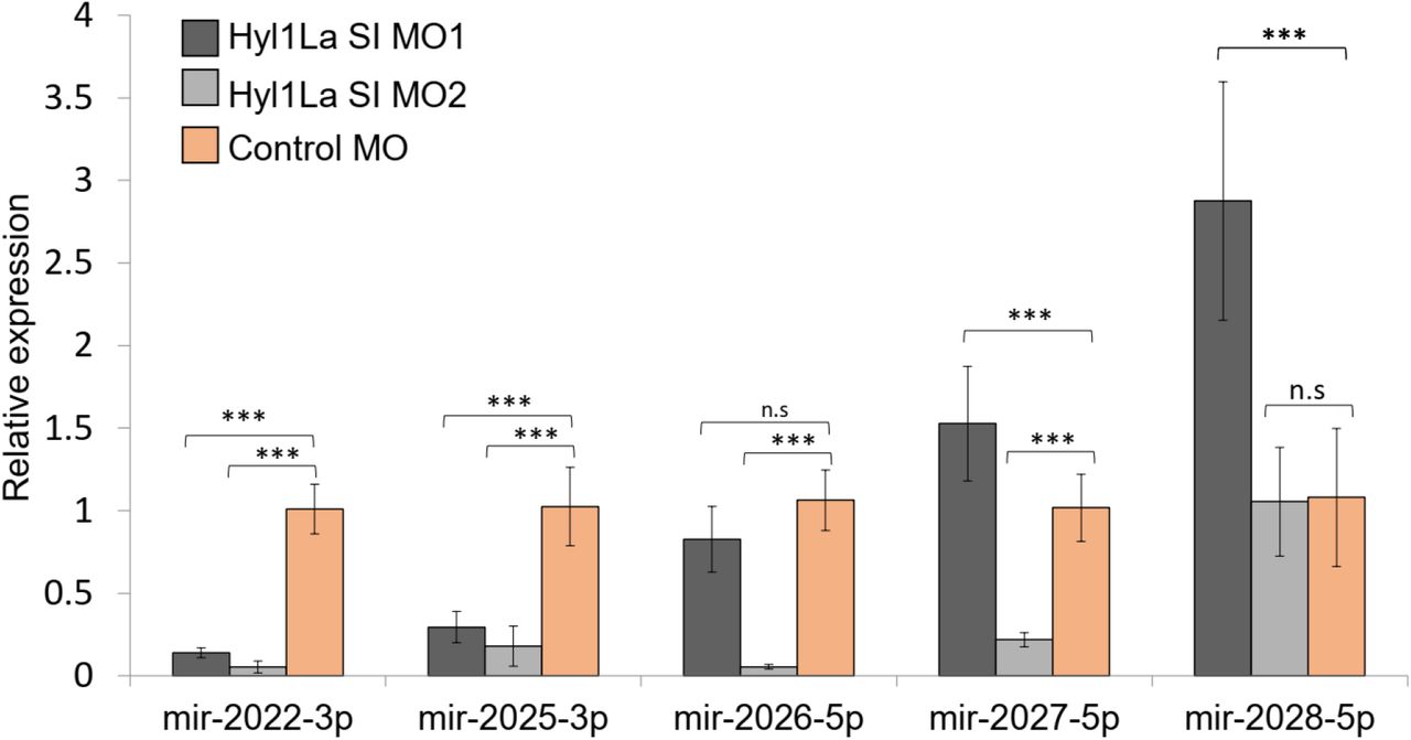
The above observed developmental defects suggested the possible involvement of Hyl1La in miRNA biogenesis, as mutants defective in their miRNA biogenesis exhibit abnormal development in both animals and plants (Achkar et al., 2018; Zhong et al., 1999). The HYL1 in Arabidopsis interacts with the stem region of miRNA precursor by using its DSRM domains and woks with DCL1 synergistically (Song et al., 2007). Although the Dicer alone is capable of processing the precursor into mature miRNA, the presence of HYL1 enhances the accuracy as well as efficiency of miRNA biogenesis in plants (Dong et al., 2008). To assay the possible role of Hyl1La on miRNA expression in Nematostella, we performed small RNA sequencing of animals, injected with Hyl1La SI MO1 and with control MO. The analysis of read length distribution showed that the small RNA reads that lied between the size of miRNA (20-24nt) were higher (P < 0.01) in control as compared to knockdown embryos. Interestingly, the small RNAs that lie in the range of piRNAs (26-30nt) were slightly higher (p = 0.0428) in Hyl1La MO (Figure 3a), indicating that knockdown of Hyl1La MO affects negatively the biogenesis of miRNAs but increases piRNA reads, as those small RNAs are produced by different machinery. We suggest this is probably due to more sequencing reads available for piRNAs following the decrease in miRNAs. Further, we analyzed the miRNA expression by using miRProf (Stocks et al., 2012) and normalized the miRNA reads in transcripts per million (TPM) (Table S2). For the miRNA quantification we used the most recent Nematostella miRNA datasets that were obtained by AGO-immunoprecipitation (Fridrich et al., 2020). The expression of normalized miRNA reads was compared between control and Hyl1La MO. About 54% of the total identified miRNAs showed downregulation of more than two-fold in Hyl1La MO injected animals as compared to the control (Figure 3b). A significant reduction in overall miRNA abundance was also observed in the knockdown mutants (P < 0.00001, Wilcoxon signed-rank test) (Figure 3c). The expression variation caused by the action of other two morpholinos (Hyl1La SI MO2 and Hyl1La TB) was also checked by quantitative stem-loop PCR of five miRNAs (Figure 3d and Figure S2). A significant downregulation of three miRNAs: miR2022-3p, miR2025-3p and miR2026-5p was found in all three MOs, which supported the small RNA sequencing results. In contrast, two miRNAs, miR2027-5p and miR2028-5p either showed upregulation or were not significantly affected by the Hyl1La knockdown. Previous studies have also shown that these two miRNAs respond similarly in HEN1 and Dicer knockdown morphants of Nematostella (Modepalli et al., 2018). The results of the present study, together with previous studies, establish the fact that these two miRNAs might be produced by some non-canonical miRNA biogenesis pathways. The presence of similar expression patterns for miRNA further strengthens our study and the involvement of Hyl1La in miRNA biogenesis. Further, we also checked for the processing accuracy of all the identified miRNA by mapping them onto their respective precursors. The analysis did not reveal aberrant processing. These results further suggested that like its homolog in plants, Hyl1La in Nematostella might be involved in enhancement of Dicer efficiency and is not involved in size selection.

The expression of miR-2022, miR-2025, miR-2026, miR-2027 and miR-2028 were checked by using the stem loop PCR between the Hyl1La SI MO1 vs. Control MO and Hyl1La SI MO2 vs. Control MO. The data represents the mean of four independent biological replicates ± SD. ***P<0.001, ** P ≤ 0.01. * P ≤ 0.05, (Student’s t-test), n.s (not significant).

(a) Average read length distribution of small RNA reads after adapter removal.
(b) Scatter plot representing normalized read counts of miRNAs in control and treated animals. Each dot represents the average expression of an individual miRNA. The miRNAs showing a depletion greater than two-fold are indicated in green. The axes are scaled to Log 10 of normalized read counts. The data represents the mean of three independent biological replicates.
(c) Box plot showing average of abundance of miRNA read counts in Hyl1La SI MO1 and control MO. A significant reduction of miRNA read counts is noted in Hyl1La SI MO1, (P < .0001, Wilcoxon signed-rank test). The data represents the mean of three independent biological replicates ± SD.
(d) Bar plot showing the expression of miR-2022, miR-2025, miR-2026, miR-2027 and miR-2028 as quantified using stem loop PCR in translation blocking (TB) and control morpholino. The data represents the mean of three independent biological replicates ± SD. ***P<0.001, ** P ≤ 0.01. * P ≤ 0.05, (Student’s t-test), n.s (not significant).
To further support our results, we attempted to knockdown this gene by using short-hairpin RNAs (shRNAs), a method previously established in Nematostella (Karabulut et al., 2019). We designed three different shRNAs from three different regions of Hyl1La gene (Hyl1La shRNA1, Hyl1La shRNA2 and Hyl1La shRNA3) (Figure 4 a-c) and injected them into Nematostella zygotes. In parallel we also used a control shRNA with no target in Nematostella genome that was previously used as control for similar experiments (He et al., 2018; Karabulut et al., 2019). To assess the effect of these shRNAs on Hyl1La expression, we performed qRT-PCR from three days-old injected animals. Unexpectedly, we did not find any difference in Hyl1La expression (Figure 4d). Additionally, we also assessed the phenotype, but we could not identify any phenotypic difference as well. Next, we employed stem-loop PCR to test whether small RNAs are generated from an injected shRNA and indeed the small RNAs were produced as expected (Figure 4e). Thus, the failure of the shRNAs to knockdown Hyl1La transcripts was probably not due to the lack of small RNA production from the shRNAs. The possible explanation of this contrasting result between MO and shRNA probably lies between the different mode of action of these two molecules. In contrast to MOs that do not use the cellular machinery, shRNA requires the miRNA/RNA interference machinery for their production as well as in target recognition and inhibition. Thus, our combined results suggest that Hyl1La might have an additional effect on biogenesis steps that are downstream to the cleavage by Dicer such as loading of small RNAs into AGO, the protein at the heart of the RNA-induced silencing complex (RISC) (Hutvagner and Simard, 2008). Under such a condition the shRNA derived small RNA would be unable to load onto RISC and hence could not cleave the Hyl1La, rendering its expression unaffected. Further, in such a scenario after injection with the shRNAs the system might reach a balance point that is very close to the normal Hyl1La levels. Alternatively, it is possible that the three shRNAs are ineffective due to lack of accessibility of the three distinct target sites on the Hyl1La transcript to the RISC loaded with the shRNA derived small RNAs for unknown reasons.

(a-c) Structure of different shRNAs designed from different positions of Hyl1La gene along with GC content and their position are shown. In the shRNA sequence, the red colour shows the nucleotides edited for mismatch and blue colour represents loop region. The red coloured nucleotides on precursor’s structure indicate the small RNA derived from the shRNAs.
(d) Real time quantification of Hyl1La from animals injected with different shRNAs relative to control. The data represents the mean of three independent biological replicates ± SD.
(e) Quantification of small RNA produced from Hyl1La shRNA1. The quantification was performed by using stem loop qRT-PCR.
Altogether the absence of animal-like Dicer partner proteins such as TRBP or PACT and presence of a functional homolog of HYL1 (Hyl1La) in Nematostella indicated that a Hyl1-like protein might have been present in the last common ancestor of plants and animals. Apart from Nematostella, the presence of HYL1 homologs in additional members of Cnidaria and other non-bilaterian metazoan groups such as sponges (Figure 1a) further strengthens the notion of common ancestry of the miRNA systems of plants and animals. Further, unlike the cleavage mode of action and nearly-perfect target binding that could have evolved convergently in plants and cnidarians (Moran et al., 2014) due to functional constraints, the involvement in miRNA biogenesis of the Hyl1L in Cnidaria and its plant homolog HYL1 is far less likely to be the result of parallel evolution as it will require the independent recruitment of the same protein into the same system. Recently, it was found in Chlamydomonas (a unicellular green algae) that DUS16, which is a DSRM protein, and DCL3 were efficient enough for miRNA processing (Yamasaki et al., 2016). Further, various fungal groups also exhibit the presence of Dicer and plant-like DSRM proteins and lack animal-like accessory proteins, such as Drosha and Pasha (Dang et al., 2011; Dias et al., 2017). Contrastingly, DCL3 of Chlamydomonas exhibits some structural features that are reminiscent of metazoan Drosha (Valli et al., 2016). These observations suggest that the common ancestor of all these groups might have harbored only a single Dicer/Drosha-like RNAse III enzyme assisted by a DSRM protein resembling the ones found in plants (HYL1-like).
Finally, here we report that Hyl1La play an important role in miRNA biogenesis in Nematostella, a representative of Cnidaria which is the sister group of Bilateria. However, the functional importance of Hyl1La in Nematostella identified here raises another interesting evolutionary question of why Hyl1La was replaced by other DSRM proteins like TRBP, Loqs or PACT in bilaterian animals during evolution (Figure 1a). Interestingly, both Loqs in flies and TRBP in mammals, enable processing of some miRNA precursors into different mature miRNAs and by this significantly increase their variability and targeted sequences (Fukunaga et al., 2012; Lee and Doudna, 2012). Such variability is currently unknown in plants or cnidarians. It is intriguing to consider the possibility that this ability of the bilaterian proteins to increase small RNA variability was advantageous over Hyl1-like proteins and led to the loss of the latter in bilaterian lineages.
SUPPLEMENTAL INFORMATION
Supplementary information includes two figures and three tables (Table S1, Table S2 and Table S3).
AUTHOR CONTRIBUTIONS
Conceptualization: A.M.T. and Y.M.; Methodology: A.M.T. and Y.M.; Validation: A.M.T.; Software: A.M.T. and A.F.; Formal Analysis: A.M.T., A.F. and M.L.; Investigation: A.M.T., A.F. and M.L.; Resources: Y.M.; Data Curation: A.M.T.; Writing –Original Draft: A.M.T. and Y.M.; Writing –Review & Editing: A.F. and M.L.; Visualization: A.M.T. and A.F.; Supervision: Y.M.; Project Administration: Y.M.; Funding Acquisition: Y.M.
DECLARATION OF INTERESTS
The Authors declare no competing interests.
METHODS
KEY RESOURCES TABLE
LEAD CONTACT AND MATERIALS AVAILABILITY
Further information and requests for resources and reagents should be directed to and will be fulfilled by the Lead Contact, Yehu Moran (yehu.moran{at}mail.huji.ac.il). These resources and reagents will be made available upon request.
EXPERIMENTAL MODEL AND SUBJECT DETAILS
Nematostella vectensis (a common lab strain originating from Rhode River, MD) were grown in lab in 16‰ artificial seawater under controlled conditions. The knockdown animals (morphants) were generated by using the three different morpholinos. For cloning of splicing product, we used the DH5α E. coli cells provided by New England Biolabs (USA).
METHOD DETAILS
Animal culture and microinjection
Nematostella were in grown in 16‰ sea water at 18°C in a dark culture room. The growing animals were fed with freshly hatched Artemia salina nauplii three times a week. Induction of spawning was performed as previously described (Genikhovich and Technau, 2009): the mature male and female animals were induced to produce eggs and sperms by placing them in an incubator for eight hours under constant blue light and heat (25°C). After induction, the tanks were further kept in 18°C (in the culture room) for two hours to allow the release of egg packages and sperm. Further, the egg packages were fertilized for 30 min by placing the packages inside the male tanks. The quality of egg packages was checked under the stereomicroscope and egg packages of round shape and homogenous size were processed further for dejellying using 4% of L-Cysteine in 16‰ sea water pH 7.2 (titrated with NaOH). The selected eggs packages were kept in the cysteine solution for 45 min on a table shaker. The eggs were washed using 16‰ sea water in petri plates. These clean eggs (zygotes) were further used for injection. For injection 1mM (1000ng) stock solutions of both morpholino and shRNA were prepared by dissolving them into nuclease free water. The toxicity of morpholinos as well as shRNA was optimized by injecting different concentrations into the animals along with the control injected animals. The toxicity of less than 30% was considered suitable for injection. The morpholinos used here were designed and synthesized by Gene Tools, LLC (USA).
We found that optimum concentration was 300 μM, 900 μM and 450 μM for the Hyl1La TB, Hyl1La SI MO1 and Hyl1La SI MO2, respectively. For all the three shRNAs, 600 ng/μl concertation was found to be suitable. Similar concentration of control morpholino was used for injection in parallel with Hyl1La morpholinos. In every shift we injected 600 zygotes (300 Control MO and 300 Hyl1La MO) by mixing the injected material with dextran Alexa Fluor 488 which was used as a fluorescent tracer while injection was carried under magnification by a TS-100F fluorescent microscope (Nikon, Japan). The injected zygotes were kept at 22°C for further growth. The morphology of the animals was observed up to nine days after which the number of settled and unsettled animals were counted and documented under SMZ-18 fluorescent stereomicroscope (Nikon). For RNA extraction injected zygotes were flash frozen in liquid nitrogen after three days of growth and stored at −80°C until RNA extraction. All the above experiments were performed in three independent biological replicates with three distinct animals’ batches.
Small-RNA sequencing and analysis
The RNA was isolated using Trizol (Thermo Fisher Scientific) from three days old animals. Small RNA sequencing was performed for only Hyl1La SI MO1 and control MO injected animals. The small RNA library was prepared using NEBNext Multiplex Small RNA Library Prep Illumina kit (New England Biolabs) with some modifications (Plotnikova et al., 2019). In brief, small RNA was isolated (18nt-30nt) from 1μg of total RNA using 15% urea-PAGE (Bio-Rad) followed by overnight precipitation using 0.3% NaCl. The size selected small RNA was further precipitated using ice-cold ethanol (2.5x volume) and 1μl of GlycoBlue (Thermo Fisher Scientific) by centrifugation. The pellet was dissolved in 7.5 μl nuclease free water and used further for adapter ligation. The ligated products were subjected to 14 cycles of PCR amplification using adapter specific primers. The PCR product was run on 2% agarose gel followed by staining with GelRed (Biotium, USA). The band size between 137nt–149nt was selected and purified using Gel Extraction Kit (Macherey-Nagel, Germany). The quality of purified product (sRNA-seq libraries) was checked by using TapeStation system (Agilent, USA). The libraries having quality in range of the size (137nt–149nt) were used for sequencing. One ng of each sample was run on NextSeq500 (Illumina) in single end mode.
The small RNA data was analysed using miRProf (Stocks et al., 2012) with the following parameters: two mismatches allowed, minimum abundance 1, allowed overhang and not grouping mature and star stand. For mapping onto the genome and miRNA precursor for identification of aberrant processing of small RNAs miRDeep2 (Friedländer et al., 2012) was used. The miRNA expression was normalized in TPM (transcripts per million) by using only the transcripts that mapped on the reference genome. For read length distribution and scattered plot we used the average of expression obtained from the three biological replicates.
Synthesis of shRNA
Potential shRNA precursors for Hyl1La gene were predicted using the shRNA prediction tools (https://www.invivogen.com/sirnawizard/index.php) (Karabulut et al., 2019). Three precursors from three different regions were further chosen manually having GC% of more than 35%. Further, we also added the sequence for T7 promotor and three different mismatches at nucleotide position 10, 13 and 16 to create the bulges in precursors (Figure 4a-c). All these modified precursors were reverse complemented and synthesized by Integrated DNA Technologies (USA). The templates and reverse primer were mixed (1:1) and denatured at 98 °C for 5 min and cooled to 24 °C. Further, this mixture was mixed with the components of in-vitro transcription kit (Epicentre, USA) and incubated for eight hours at room temperature. The in-vitro transcribed product was further purified using the cleaning kit (Zymo Research, USA). The quality and size of the precursor was checked on agarose gel and its concentration were measured using Qubit Normal Sensitivity RNA Kit (Thermo, USA). The concentration ranged from 1500 ng to 2000 ng/μl.
Reverse transcription-quantitative PCR
For the quantification of Hyl1La transcripts from shRNA injected animals and for checking the splicing inhibition (Hyl1La SI MO injected animals), cDNA was prepared from 500 ng of total RNA using iScript™ cDNA Synthesis Kit (Bio-Rad, USA). For the quantification of miRNAs and shRNA, we designed the stem loop primers for five different miRNAs and shRNA (Chen et al., 2005). For cDNA preparation, 100 ng of total RNA was reverse transcribed using the SuperScript™ III Reverse Transcriptase (Thermo Fisher Scientific). The specificity of the miRNA primers was determined by using end point PCR (Varkonyi-Gasic et al., 2007). For this, we used 2 μl of cDNA as template, miRNAs specific forward primer and stem-loop specific reverse primer and run the PCR at 94°C for 2 min, followed by 35 cycles of 94°C for 15 s and 60°C for 1 min. For analyzing differential expression, we ran qRT-PCR with 5sRNA as internal control. For all the real time experiments, we used Fast SYBR® Green Master Mix (Thermo Fisher Scientific) and samples were run on StepOnePlus Real-Time PCR System (Thermo Fisher Scientific). All the real time experiments were performed in three independent replicates and data was analyzed using 2- ΔΔCt method (Livak and Schmittgen, 2001). All the primers are listed in Table S3.
Cloning and sequencing of Hyl1La SI MO injected animals
To validate the effect of splicing morpholinos, we designed the primers pairs spanning the introns lying on the boundary of exons. PCR of the Hyl1La was done using Q5® High-Fidelity DNA Polymerase (New England Biolabs, USA). The PCR products were run on the gel and the expected-sized PCR product was purified with kit. Then the purified PCR products were ligated into the pJet2.1 vector (Thermo Fisher Scientific) and transformed into the E. coli DH5α strain of and outsourced for Sanger sequencing (HyLabs, Israel).
QUANTIFICATION AND STATISTICAL ANALYSIS
For statistical analysis of qRT-PCR data, Student’s t-test was performed on ΔCt values between different comparisons. For phenotypic analysis we performed Student’s t-test between the number of developed and undeveloped animals. To check overall significant difference between the miRNA expression levels, Wilcoxon signed-rank test was done. The Student’s t-test was conducted in excel while Wilcoxon signed-rank test was done using socscistatistics (https://www.socscistatistics.com/tests/signedranks/default.aspx).
DATA AND CODE AVAILABILITY
The small RNA sequencing data were submitted to NCBI-SRA under BioProject ID PRJNA630340.
ACKNOWLEDGMENTS
The authors would like to thank Dr. Michal Bronstein and Ms. Adi Turjeman of the Center for Genomic Technologies (The Hebrew University) for their help with sequencing. The authors would also like to thank Dr. Reuven Aharoni (The Hebrew University) for his technical and administrative assistance in this work. This work was supported by European Research Council Starting Grant 637456 CNIDARIAMICRORNA to YM.
REFERENCES


