

Abstract
Gender is a risk factor for several infections that, for many pathogens, has been linked to sex hormones impacting host immunity and directly affecting microbial virulence. Candida albicans is a commensal of the urogenital tract and the predominant cause of vulvovaginal candidiasis (VVC). Factors that increase circulatory oestrogen levels like pregnancy, the use of oral contraceptives, and hormone replacement therapy predispose women to VVC, but the reasons for this are largely unknown. Here, we investigate how adaptation of C. albicans to oestrogen impacts the fungal host-pathogen. Physiologically relevant concentrations of oestrogen promoted fungal virulence by enabling C. albicans to avoid the actions of the innate immune system. Oestrogen-induced innate immune evasion was mediated via inhibition of opsonophagocytosis through enhanced acquisition of Factor H on the fungal cell surface and was dependent on the C. albicans moonlighting protein Gpd2. Oestrogen dependent derepression of GPD2 was mediated via a non-canonical signalling pathway involving Ebp1 and Bcr1. Therefore, we propose that, in addition to affecting the antifungal potential of vaginal epithelial cells, elevated oestrogen levels predispose women to VVC by directly enhancing fungal pathogenicity. The discovery of this new hormone sensing pathway might pave the way in explaining gender biases associated with fungal infections and may provide an alternative approach to improving women’s health.
Introduction
Microbial infections exhibiting gender bias are common. This bias may predispose one sex to infection over the other, or result in one sex exhibiting severer infection outcomes [1–3]. Sex hormones like oestrogen, testosterone and progesterone regulate many functions of the immune system, and generally males are more prone to infection than females, as overall immune responses are lower in the male population [4–6]. Besides the impact of sex hormones on immune cell function, sex hormones also have a direct effect on microbial pathogenicity increasing microbial persistence, metabolism and virulence gene expression [7, 8].
The opportunistic fungal pathogen Candida albicans is the predominant fungal coloniser of the female reproductive tract and the major cause of genital thrush (vulvovaginal candidiasis, VVC). Approximately 75% of the female population will encounter at least one episode of VVC in their life-time, while up to 15% experience recurrent infection (RVVC) defined as four or more episodes in a twelve-month period [9]. Although not life threatening, mucosal infections are expensive to treat, impact quality of life, and increase population morbidity.
One of the major risk factors associated with the development of VVC is elevated levels of oestrogen which occur as a result of pregnancy, the use of high oestrogen-containing oral contraceptives and hormone replacement therapy [10, 11]. Therefore, oestrogen plays a key role in predisposing women to VVC, but the precise mechanism(s) for this are unknown. Oestrogen promotes glycogen production at the vaginal mucosa which might provide a nutrient rich environment to promote the expansion of C. albicans [10]. In mouse models of VVC, were pseudoestus is induced to maintain fungal colonisation, vaginal epithelial cells have a diminished ability to control the growth of C. albicans [12]. In addition, oestrogen decreases the infiltration of phagocytes and suppresses cell mediated immunity [13]. However, in rats, preincubation of C. albicans with oestrogen prior to vaginal infection enhances fungal survival [14], suggesting that in addition to affecting host immunity, oestrogen may directly affect the virulence of C. albicans. In agreement with this idea, oestrogen has been shown to promote hyphal morphogenesis of C. albicans [15], a key virulence factor of the fungus. Furthermore, an oestrogen binding protein (Ebp1) has been identified in C. albicans [16], although the importance of this protein in VVC is not known. Therefore, how C. albicans adapts to oestrogen is still unclear.
The C. albicans cell wall is a multi-layered structure consisting of an inner layer of chitin and beta-glucan, and an outer layer of heavily glycosylated proteins [17]. The fungal cell wall is a highly dynamic structure providing rigidity, strength and protection from the environment. In addition, many components of the cell wall act as pathogen associated molecular patterns (PAMPs) and are recognised by the innate immune system [17, 18]. Recently, remodelling of the cell wall in response to adaptation to host environments has been shown to regulate the host-pathogen interaction [19–30]. In addition to direct recognition of cell wall PAMPs mediating phagocytosis, C. albicans also activates the alternative complement system, resulting in the deposition of complement proteins (i.e. C3) on the fungal cell surface, resulting in opsonophagocytosis [31]. However, C. albicans can evade opsonophagocytosis through the binding of complement regulatory proteins to its cell surface that inactivate the complement cascade [32]. Here we show that C. albicans does adapt to oestrogen, and that this adaptation perturbs the host-pathogen interaction, inhibiting phagocytosis of the fungal pathogen. Avoidance of the innate immune system was mediated via the fungal cell surface protein, Gpd2, recruiting the human complement regulatory protein, Factor H, to the fungal cell surface inactivating the alternative complement system.
Results
Adaptation of C. albicans to oestrogen promotes immune evasion
Pregnant women and women taking high oestrogen-containing oral contraceptives are more prone to vulvovaginal candidiasis (VVC) [33], while VVC occurs less frequently in postmenopausal women, indicating that oestrogen may play a role in promoting the virulence of C. albicans. There are four main forms of oestrogen, estrone (E1), 17β-estradiol (E2), estriol (E3) and 17α-ethynylestradiol (EE2). Estrone is the weakest form of oestrogen produced by the ovaries and adipose tissue, and is only found in menopausal women, while 17β-estradiol is the strongest form of oestrogen produced by the ovaries and has been associated with many gynaecological disorders. Estriol is a by-product from the metabolism of estradiol, and as such is found in high concentrations during pregnancy. Finally, 17α-ethynylestradiol is a synthetic oestrogen used in oral contraceptive pills. To ascertain whether adaptation of C. albicans to oestrogen affects the host-pathogen interaction, C. albicans cells were grown in the presence of physiological (0.0001 μM) and super-physiological (0.1 μM, 10 μM) concentrations of 17β-estradiol, estriol, or 17α-ethynylestradiol, and phagocytosis rates quantified. Physiological and super-physiological concentrations of all three forms of oestrogen significantly inhibited macrophage and neutrophil phagocytosis of C. albicans, resulting in a 50% drop in phagocytosis compared to the ethanol vehicle control (Fig 1A-C, S1 Fig A, B). The reduced rates of C. albicans phagocytosis were independent of any impact of oestrogen on fungal growth (S1 Fig C) or morphology (S1 Fig D). Given that all tested forms of oestrogen elicited similar results, all subsequent experiments were performed only with 17β-estradiol.

C. albicans cells were grown in YPD with or without oestrogen supplementation. Cells were harvested, washed in PBS and co-incubated with (A) J774A.1 cells (B) human macrophages (C), human neutrophils. (D) J774A.1 phagocytosis rates of C. albicans clinical isolates after exposure to 10 μM 17β-estradiol. (E) J774A.1 phagocytosis rates of C. albicans clinical isolates after exposure to progesterone (F) J774A.1 phagocytosis rates of C. albicans clinical isolates after exposure to progesterone and oestrogen. Cells were fixed with 4% PFA, imaged by microscopy, scored using ImageJ software and phagocytic index determined. All data represent the mean ± SEM from at least three independent experiments. (*p < 0.05, **p < 0.01, ****p < 0.0001).
To determine whether the observed inhibition of phagocytosis is truly mediated by adaptation of C. albicans to oestrogen, and not a result of residual oestrogen coating the surface of C. albicans inhibiting phagocyte function, macrophages were pre-treated with oestrogen prior to the addition of C. albicans or latex beads. Macrophages pre-incubated with oestrogen phagocytosed C. albicans at rates comparable to non-treated cells (S1 Fig E), suggesting that under the tested conditions oestrogen does not directly impact the ability of macrophages to phagocytose target particles. Pre-incubating inert particles like latex beads with oestrogen had no effect on the ability of macrophages to phagocytose the particles (S1 Fig F), suggesting that any residual oestrogen on the surface of particles does not interfere with the ability of macrophages to phagocytose them. In agreement with this, incubation of C. albicans with oestrogen on ice, which inhibits fungal growth and metabolism, did not affect phagocytosis rates (S1 Fig G). Furthermore, re-inoculation of oestrogen-adapted C. albicans cells into fresh YPD media restored C. albicans phagocytosis rates (S1 Fig H). Therefore, in response to oestrogen, C. albicans undergoes some form of adaptation that alters the host-pathogen interaction.
Finally, we tested several vaginal C. albicans isolates and observed the all isolates displayed reduced phagocytosis after adaptation to oestrogen, confirming that oestrogen-induced immune evasion is a general trait of clinically relevant C. albicans (Fig 1D). Taking this data together we conclude that the reduction in C. albicans phagocytosis is due to the fungus reversibly adapting to the oestrogen, and that this adaptation interferes with the host mechanism of fungal clearance.
Hormone induced innate immune evasion is specific to Oestrogen
Progesterone been shown to affect phagocytosis rates [34], and is structurally quite similar to oestrogen. Therefore, we hypothesised that C. albicans may adapt to progesterone in a similar way to oestrogen to evade the innate immune system. C. albicans cells pre-exposed to physiological and super-physiological concentrations of progesterone exhibited comparable phagocytosis rates as C. albicans cells grown in YPD (Fig 1E), while C. albicans treated with both oestrogen and progesterone still exhibited innate immune evasion (Fig 1F). Therefore, the promotion of innate immune evasion appears to be a specific and dominant attribute of oestrogen.
Altered phagocytosis rates does not affect pro-inflammatory cytokine production
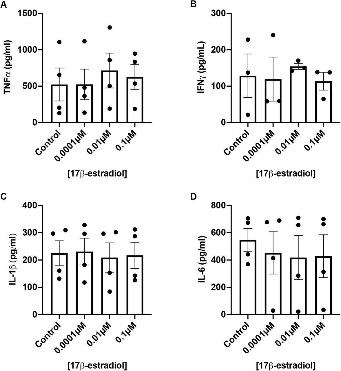
Previous studies have shown that oestrogen can interfere with NF-кB signalling to attenuate inflammatory responses [35, 36]. Therefore, we investigated whether adaptation of C. albicans to oestrogen dampens the pro-inflammatory innate immune response. Human peripheral blood monocytes (PBMCs) were exposed to oestrogen-adapted C. albicans cells and secretion of TNFα, IFN-γ, IL-6 and IL-1β was evaluated. Adaptation to oestrogen did not affect the cytokine response (Fig 2). Therefore, although significantly fewer oestrogen-adapted cells are phagocytosed by the innate immune system, the extracellular C. albicans cells still stimulate significant cytokine production, likely through non-phagocytic receptors like TLR4.

C. albicans cells were grown in YPD with or without 10 μM 17β-estradiol for 4 h. Cells were washed, fixed with 4% PFA and co-incubated with PBMCs for 24h. Secretion of (A) TNFα, (B) IFNγ (C) IL-1β and (D) IL-16 by PBMCs was quantified by ELISA. Data represent the mean ± SEM from four independent experiments using different donors.
Oestrogen dependent innate immune evasion is not dependent on fungal carbohydrate cell wall remodelling
Recently, modulation of the C. albicans cell wall carbohydrates has been shown to influence the host-pathogen interaction [19–30]. Therefore, the ability of oestrogen to promote remodelling of the fungal cell wall was quantified by flow cytometry. However, initial studies confirmed that adaptation of C. albicans to oestrogen does not result in increased incorporation of mannan, β-glucan or chitin into the cell wall (Fig 3A-C).

C. albicans cells were grown in YPD with or without 10 μM 17β-estradiol. Cells were harvested, washed in PBS, fixed with 4% PFA and stained for (A) total mannan (B) total glucan (C) total chitin. (D) To assess the role of Dectin-1 in C. albicans phagocytosis, Dectin-1 was expressed in NIH 3T3 fibroblasts. C. albicans cells were stained for (E) exposed β1,3-glucan and (F) exposed chitin levels. Staining was quantified by flow cytometry and median fluorescence intensities (MFI) determined. All data represent the mean ± SEM from at least three independent experiments.
Dectin-1 is a phagocytic receptor important for controlling fungal infections through the recognition of surface exposed beta-glucan [37]. Host environmental cues that reduce C. albicans phagocytosis do so through masking surface exposed beta-glucan inhibiting Dectin-1 dependent recognition [21–23, 30]. To ascertain whether the observed reduction in phagocytosis was dependent on Dectin-1, the attachment of oestrogen-adapted C. albicans cells to Dectin-1 expressing fibroblasts was quantified. Dectin-1 recognised and bound oestrogen-adapted cells at rates comparable to control cells, suggesting that the altered phagocytosis rates are not due to reduced Dectin-1 recognition (Fig 3D). In agreement with this, adaptation of C. albicans to oestrogen did not affect the surface exposure of either β-glucan or chitin (Fig 3E, F). Taken together, these data indicate that oestrogen does not affect the overall structure of the C. albicans cell wall, and that the oestrogen-dependent reduction in phagocytosis is not dependent on fungal carbohydrate cell wall remodelling.
Oestrogen-induced innate immune evasion is mediated via inhibition of opsonophagocytosis
In addition to direct detection of cell wall PAMPs leading to phagocytosis, C. albicans can be opsonised. C. albicans activates the alternative complement cascade resulting in the deposition of complement (C3) on its surface inducing opsonophagocytosis via the complement receptors CR1 and CR3. To confirm whether oestrogen-induce innate immune evasion was mediated via inhibition of complement, we assessed phagocytosis rates in heat-inactivated serum, where the major complement proteins (i.e. C3) are denatured. Overall, the phagocytosis rates of C. albicans in heat-inactivated serum were lower than in live serum (Fig 4A), confirming the importance of opsonophagocytosis in the recognition of C. albicans. However, adaptation of C. albicans to oestrogen did not result in further evasion of phagocytosis in heat-inactivated serum (Fig 4A), suggesting that pre-exposure of C. albicans to oestrogen impacts complement activation. Supplementation of heat-inactivated serum with purified C3 restored oestrogen-induced immune evasion (Fig 4B), confirming that oestrogen-induced immune evasion likely occurs through the avoidance of opsonophagocytosis.

A) J774A.1 macrophages were maintained in either complete serum or heat-inactivated (HI) serum and infected with C. albicans pre-exposed to YPD, 0.3% ethanol or 10 μM 17β-estradiol at a MOI of 5. B) J774A.1 macrophages were maintained in either complete serum, heat-inactivated (HI) serum, or heat-inactivated serum supplemented with purified C3 and infected with C. albicans pre-exposed to YPD, 0.3% ethanol or 10 μM 17β-estradiol at a MOI of 5. C) C. albicans was grown in YPD with or without 10 μM 17β-estradiol, incubated in human serum for 20 minutes, fixed with PFA and C3 and C3b binding quantified by FACS. (D). C. albicans was grown in YPD with or without 10 μM 17β-estradiol, incubated in human serum for 20 minutes, fixed with PFA and Factor H binding quantified by FACS. Data represent the mean ± SEM from at least three independent experiments. (*p < 0.05, **p < 0.01, ***p < 0.001, ****p < 0.0001).
Next, we tested whether adaptation of C. albicans to oestrogen affected the deposition of C3 and C3b on the fungal cell surface. Oestrogen-adapted cells exhibited enhanced C3 and C3b deposition compared to non-adapted cells (Fig 4C), suggesting that adaptation to oestrogen promotes C3 and C3b binding on the C. albicans cell surface. Increased deposition of complement is normally associated with increased phagocytosis. However, processing of C3 can be inhibited by the recruitment of host regulatory proteins like Factor H, and several pathogens are known to avoid opsonophagocytosis through enhanced recruitment of Factor H [38, 39]. Therefore, the ability of oestrogen adapted C. albicans cells to bind Factor H was quantified. Oestrogen-adapted cells bound significantly more Factor H, compared to the solvent control (Fig 4D), suggesting that oestrogen-adapted C. albicans evade the innate immune system through enhanced Factor H recruitment and decreased opsonophagocytosis.
Oestrogen-induced innate immune evasion is mediated through Gpd2
GPD2 is predicted to encode a cytosolic protein with glycerol-3-phosphate dehydrogenase activity [40]. However, more recently, Gpd2 has been shown to localise to the fungal cell surface in response to serum [41], and has been classed as a moonlighting protein (a protein that has a biological function not predicted by its amino acid sequence). The moonlighting activities of Gpd2 include binding Factor H, Factor H like protein-1 (FHL-1) and plasminogen [40], key components of the alternative complement system. Binding of these host complement regulatory proteins to microbial surfaces has been shown to promote immune evasion via inhibition of complement-mediated phagocytosis [42]. Thus, we hypothesised that adaptation of C. albicans to oestrogen promotes binding of these complement regulatory proteins to Gpd2 thereby inhibiting opsonophagocytosis. To test this hypothesis, GPD2 was deleted in C. albicans and phagocytosis rates in the presence and absence of oestrogen determined. Deletion of GPD2 prevented oestrogen-dependent inhibition of macrophage phagocytosis rates (Fig 5A), confirming that GPD2 is required for oestrogen dependent innate immune evasion.

A) J77A.1 macrophages were infected with C. albicans strains grown in YPD with or without 10 μM 17β-estradiol and phagocytosis rates quantified. B) J774A.1 macrophages were infected with CAI4-pSM2 and CAI4-pSM2-GPD2 (GPD2 over-expression strain) grown in YPD with or without 10 μM 17β-estradiol and phagocytosis rates quantified. C) C. albicans strains were grown in YPD with or without 10 μM 17β-estradiol, incubated in human serum for 20 minutes, fixed with PFA and Factor H binding quantified by FACS. All data represent the mean ± SEM from at least three independent experiments. (*p < 0.05, **p < 0.01, ***p < 0.001).
To determine whether increased expression of GPD2 is sufficient to promote innate immune evasion, we over-expressed GPD2 in C. albicans. Over-expression of GPD2 (2.75-fold increased mRNA expression compared to wild type cells) resulted in reduced phagocytosis rates irrespective of oestrogen treatment (Fig 5B), thereby confirming that enhanced expression of GPD2 is sufficient to promote C. albicans innate immune evasion. To determine whether the expression levels of GPD2 correlate with Factor H binding, we quantified Factor H binding in the gpd2Δ mutant. As predicted, the gpd2Δ mutant did not bind more Factor H than wild type cells in the presence of oestrogen (Fig 5C). Therefore, the expression of Gpd2 plays a key role in regulating the host-pathogen interaction.
Oestrogen elicits a mild transcriptional response in C. albicans
To elucidate how quickly C. albicans adapts to oestrogen the fungus was grown in the presence of 10 μM 17β-estradiol for varying lengths of time before macrophage phagocytosis rates were quantified. This time course analysis confirmed that phagocytosis rates gradually reduced after 60 minutes of growth in the presence of oestrogen (S2 Fig A), suggesting that C. albicans undergoes some form of transcriptional or translational response to oestrogen. To identify how oestrogen affects the global transcriptional profile of C. albicans, RNA-Seq was performed on C. albicans cells that had been adapted to 10 μM 17β-estradiol for 4 hours. Despite having a strong impact on the host-pathogen interaction, oestrogen only had a mild effect on the transcriptome of C. albicans, (S2 Fig B, Table 1) with 5 genes being significantly up-regulated and 23 genes being significantly down-regulated in the presence of oestrogen (fold change > 2, FDR < 0.01). Genes that were upregulated were largely involved in cellular redox or efflux, suggesting that oestrogen imposes oxidative stress on C. albicans. The most significantly down regulated genes were FRE7, SAP2, HWP1 and ECE1. FRE7 is a cupric reductase that has previously been confirmed to be repressed by oestrogen [43]. ECE1 and HWP1 are strongly associated with hyphal formation, suggesting that in YPD oestrogen represses hyphal formation. As phagocytosis occurs through interactions with the fungal cell surface we further analysed the expression of cell surface proteins in response to oestrogen stimulation to see if other surface proteins contributed to the observed innate immune evasion. In addition to GPD2, transcripts corresponding to CDR1, EBP1, SOD6, ALS2, UTR2 and FGR23 were moderately up regulated in the presence of oestrogen (Table 2). Oestrogen is a substrate of Cdr1, and it is thought that CDR1 is upregulated in the presence of oestrogen to increase the efflux of oestrogen thereby reducing toxicity [43]. To determine whether Cdr1 is involved in oestrogen-dependent innate immune evasion, phagocytosis rates of the cdr1Δ and cdr1Δ/cdr2Δ mutants were quantified. Deletion of CDR1 alone or in combination with CDR2 did not affect oestrogen dependent inhibition of C. albicans phagocytosis (S2 Fig C).
Ebp1 is a negative regulator of innate immune evasion
The most significantly differentially regulated cell surface gene was oestrogen binding protein 1 (Ebp1). Ebp1 is homologous to old yellow enzyme 2 (OYE2) of S. cerevisiae and is an NADPH oxidoreductase [16]. Oestrogen inhibits the NADPH oxidoreductase activity of recombinant Ebp1, yet the function of this enzyme in the C. albicans oestrogen response is unknown. To determine whether Ebp1 plays a role in oestrogen induced innate immune evasion, EBP1 was deleted in C. albicans and phagocytosis rates of the mutant in the presence and absence of oestrogen quantified. Deletion of EBP1 resulted in consistently low phagocytosis even in the absence of hormone stimulation (Fig 6A). To determine whether this enhanced innate immune evasion was linked to GPD2, the expression of GPD2 in the ebp1Δ mutant was quantified by RT-PCR. Deletion of EBP1 resulted in 2.74-fold higher expression of GPD2 (comparable levels of GPD2 expression to the over-expression strain) compared to wild type cells (Fig 6B), suggesting Ebp1 is a negative regulator of GPD2 expression and therefore innate immune evasion.
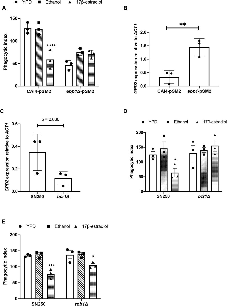
A) The ebp1Δ mutant and the parental control strain were grown with or without 10 μM 17β-estradiol for 4 hrs and then exposed to J774A.1 macrophages for 45 minutes and phagocytosis rates quantified. B) The ebp1Δ mutant and the parental control strain were grown in YPD to mid-log phase, total RNA extracted and GPD2 gene expression quantified by RT-PCR relative to ACT1. C) The bcr1Δ mutant and the parental control strain were grown in YPD to mid-log phase, total RNA extracted and GPD2 gene expression quantified by RT-PCR relative to ACT1. D) The bcr1Δ mutant and parental control strain were grown with or without 10 μM 17β-estradiol for 4 hrs and then exposed to J774A.1 macrophages for 45 minutes and phagocytosis rates quantified. E) The rob1Δ mutant and the parental control strain were grown in YPD with or without 10 μM 17β-estradiol for 4 h and then co-incubated with J774A.1 macrophages for 45 min and phagocytosis rates quantified. All data represent the mean and SEM from three biological replicates (*p < 0.05, **p < 0.01, ***p < 0.001, ****p < 0.0001).
Oestrogen dependent innate immune evasion requires a threshold level of GPD2 expression
To identify transcription factors involved in the expression of GPD2, we analysed the promoter region of GPD2. The Pathoyeastract program [44] identified 17 transcription factors associated with the promoter region of GPD2. Of these 17 transcription factors, 10 have been deleted in C. albicans and are available in the transcription factor knockout library [45]. To investigate the role of these 10 transcription factors in the regulation of GPD2, the expression of GPD2 was quantified by qPCR. Of the 10 transcription factor mutants tested, deletion of BCR1 and ROB1 had a significant impact on the expression of GPD2 (Fig 6C, S3 Fig), reducing the expression of GPD2 to 25% of the wildtype control. To assess whether the reduced expression levels of GPD2 in the bcr1Δ and rob1Δ mutants are sufficient to perturb the response to oestrogen, the phagocytosis rates of the bcr1Δ and rob1Δ mutants in the presence or absence of oestrogen were quantified. Deletion of BCR1 resulted in a loss of oestrogen-mediated innate immune evasion (Fig 6D), while deletion of ROB1 partially blocked innate immune evasion (Fig 6E), confirming that a threshold level of GPD2 expression is required for innate immune evasion.
Oestrogen-induced immune evasion plays a key role inC. albicans pathogenicity
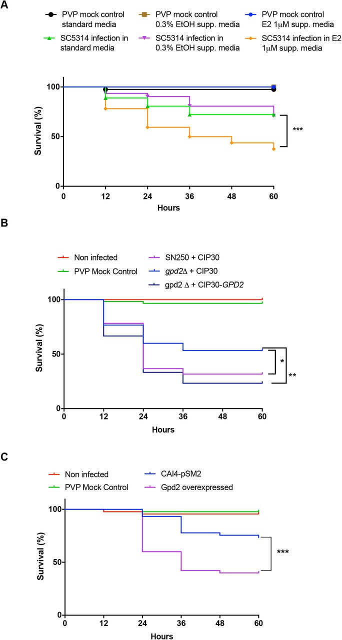
Having established that adaptation of C. albicans to oestrogen inhibits phagocytosis, we explored whether this phenomenon could influence virulence in vivo using a zebrafish larval model for disseminated disease. Previously, it was shown that exposing zebrafish larvae (3 hours post fertilisation) to media containing 1 μM oestrogen results in an in vivo oestrogen concentration of 0.0057 μM, equivalent to physiological levels during pregnancy in humans [46, 47]. Taking advantage of this observation, zebrafish larvae were infected with C. albicans SC5314 and maintained in E3 media supplemented with 1 μM oestrogen and survival rate monitored for up to 5 days post fertilisation. Compared to zebrafish incubated in E3 media alone, or E3 media supplemented with ethanol, supplementation of E3 media with 1 μM oestrogen enhanced the virulence of C. albicans, resulting in an 63% reduction in zebrafish survival 60 hrs post infection (Fig 7A).

A) C. albicans (SC5314) were microinjected into the hindbrain ventricle of zebrafish larvae in the Prim25 stage. Infected larvae were maintained in E3 media, or E3 media supplemented with 0.3% ethanol, or 1 μM 17β-estradiol and larval survival monitored every 24 h until day 5 post fertilisation. B) SN250-CIP30, gpd2Δ-CIP30 and gpd2Δ-CIP30-GPD2 were microinjected into the hindbrain ventricle of zebrafish larvae in the Prim25 stage dpf. Larvae were maintained in E3 media and larval survival monitored every 24 h until day 5 post fertilisation. C) CAI4-pSM2 or CAI4-pSM2-GPD2 were microinjected into the hindbrain ventricle of zebrafish larvae in the Prim25 stage dpf. Larvae were maintained in E3 media and larval survival monitored every 24 h until day 5 post fertilisation. The survival curves represent data pooled from three independent experiments. Statistically significant differences were determined by Log-rank (Mantel-Cox) test. (*p < 0.05, **p < 0.01; ***p < 0.001).
To investigate whether Gpd2 is required for virulence in this model, zebrafish were infected with the parental control strain, the gpd2Δ mutant, or reconstituted control strains of C. albicans. Deletion of GPD2 led to attenuation of C. albicans virulence, which was restored to parental control levels via complementation with a single copy of GPD2 (Fig 7B). To assess whether increased expression of Gpd2 is sufficient to drive fungal virulence, GPD2 was over expressed in the CAI4 background. Infection of zebrafish larvae with C. albicans over-expressing GPD2 enhanced C. albicans virulence in the absence of oestrogen compared to the respective control strain (Fig 7C). Therefore, Gpd2 plays a key role in promoting C. albicans virulence in vivo.
Discussion
Vulvovaginal candidiasis (VVC) is a mucosal infection affecting 75% of the female population of reproductive age [11]. Oestrogen is known to govern susceptibility to VVC infections, with women with low circulatory oestrogen levels (i.e. postmenopausal women) having a low risk of developing VVC, and women with high oestrogen levels (i.e. during pregnancy, or women taking high oestrogen containing oral contracepts) having a high risk of VVC. Elevated oestrogen levels increase glycogen at the vaginal mucosa, reduce leukocyte infiltration, and reduce antifungal activity of epithelial cells, promoting infection [10, 12, 13]. However, here we show that, in addition to these effects on the host, oestrogen promotes adaptation responses in C. albicans which induce evasion of the innate immune system through Gpd2-dependent inhibition of complement mediated opsonophagocytosis. Unlike the induction of fungal morphogenesis, which appears to be limited to 17β-estradiol [43], all tested forms of oestrogen promoted innate immune evasion, suggesting that C. albicans has evolved at least two signalling pathways that are responsive to oestrogen.
Complement evasion is a successful mechanism employed by viruses, bacteria, parasites and fungi to escape the innate immune system [48]. Evasion of the complement system can be mediated through the secretion of degradative proteins, or by recruitment of host regulatory proteins [49]. One of the most common methods pathogens use to evade complement is through enhanced recruitment of Factor H, a key regulatory protein in the complement system, to the microbial surface. Bound Factor H prevents further activation of the alternative complement system through both the destabilisation of the C3 convertase and enhancement of Factor I mediated degradation of C3b to iC3b, reducing opsonisation and inhibiting the formation of the membrane attack complex [49]. C. albicans has been shown by others to recruit Factor H to its surface through the expression of moonlighting proteins. Moonlighting proteins are proteins that perform a biological function not predicted from the amino acid sequence. So far, four moonlighting proteins (Phr1, Gpd2, Hgt1 and Gpm1) have been identified in C. albicans [40, 50–52]. Although Gpd2 is predicted to be a cytoplasmic protein involved in cellular redox, Gpd2 has also been identified in cell wall proteomic studies [41], suggesting that Gpd2 functions in the cell wall. Interestingly, Gpd2 was only identified in the cell wall proteome of C. albicans grown in complete serum, and not heat-inactivated serum where complement proteins have been inactivated [41]. Therefore, deposition of complement on the fungal cell surface may drive localisation of Gpd2 to the cell surface.
Purified Gpd2 binds all three complement regulatory proteins [40], although the biological significance of these interactions in infections is not known. Factor H and FHL1 bound to Gpd2 remain active, cleaving C3b and thereby inhibiting the alternative complement system [40]. In addition, Gpd2 can also bind plasminogen, which is then processed into plasmin, contributing to inactivation of the alternative complement system [40]. FACS analysis of the deposition of complement proteins on the surface of C. albicans in response to oestrogen suggested that oestrogen-adapted cells bound more Factor H, indicative of oestrogen promoting complement evasion. However, oestrogen-adapted C. albicans also bound more C3 and C3b, which is surprising as binding of Factor H should prevent C3 deposition. Factor H is a glycoprotein that is made up of 20 complement control proteins (CCP). Microbes bind Factor H at various positions, with Neisseria species binding Factor H at CCP6-8 [53]. However, multiple microbes bind Factor H at CCP20, and this has been termed the Common Microbial Binding site [54]. Binding of Factor H to some of these microbial cell surface proteins at CCP20 results in increased affinity of Factor H for C3b [54]. The enhanced affinity of Factor H for C3b might explain why we observe elevated binding of both C3b and Factor H, and would suggest that Gpd2 binds to CCP20. The formation of this stable tri-partite complex (microbial protein, Factor H, C3b) results in enhanced activity of Factor H and therefore rapid inactivation of the alternative complement cascade [54], which would explain why we observe reduced levels of phagocytosis.
Innate immune evasion was only observed after C. albicans had been grown in the presence of oestrogen for 60 minutes, suggesting that the response required either transcriptional or post-transcriptional regulation. However, in line with previous studies [43], global transcriptional analysis confirmed that only a small proportion of C. albicans genes were differentially regulated by oestrogen. In comparison to the work of Cheng et al., where most of the differentially regulated genes are involved in fungal morphogenesis [43], our analysis mainly identified genes involved in metabolism and oxidation-reduction processes. This lack of overlap is likely reflective of the growth conditions used, as Cheng et al., used RPMI where oestrogen promotes hyphal formation [13, 43], while under our experimental conditions (YPD) oestrogen did not promote hyphal formation. Although innate immune evasion was dependent on the presence of Gpd2, GPD2 mRNA levels were only moderately increased in response to oestrogen, suggesting that other factors (i.e. post-translational modifications, protein localisation) may be involved. Given that Gpd2 has only been identified as a cell wall protein under specific conditions [41], it might be that in response to oestrogen a higher level of Gpd2 is recruited to the cell wall, enabling the fungus to bind more Factor H. In S. cerevisiae, the activity of both Gpd1 and Gpd2 is regulated via post-translational modification [55]. Snf1-dependent phosphorylation of ScGpd1 (orthologue of CaGPD2) results in decreased enzyme activity [55]. Although CaGpd2 has several predicted phosphorylation sites, these are located in different regions of the protein compared to ScGpd1, and therefore it is possible that phosphorylation/dephosphorylation at any of these sites in CaGpd2 could affect the localisation, or potential to bind Factor H.
Ebp1, despite showing no homology to mammalian hormone binding proteins, has been described as an oestrogen binding protein. Ebp1 is homologous to old yellow enzyme 2 (OYE2) of S. cerevisiae, an NADPH oxidoreductase. Biochemical characterisation of recombinant Ebp1 confirms that Ebp1 is an NADPH oxidoreductase, and that oestrogen inhibits this enzymatic activity [16, 56]. The importance of Ebp1 enzymatic activity in C. albicans pathogenicity is not known, but here we demonstrate that Ebp1 may serve as a negative regulator of innate immune evasion through the regulation of GPD2. Interaction of oestrogen with Ebp1, through an as yet unidentified pathway likely involving Bcr1, results in elevated expression of GPD2 and, potentially in association with other post-translational modifications, promotes innate immune evasion and pathogenicity (Fig 8).
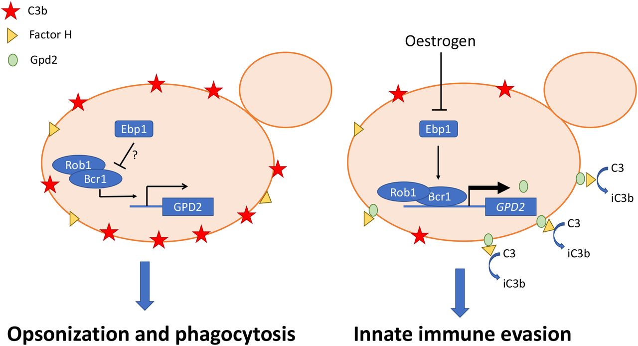
Under standard laboratory conditions the C3 is deposited on the surface of C. albicans resulting in effective phagocytosis. However, in the presence of oestrogen the NADPH activity of Ebp1 is reduced resulting in derepression (through an as yet to be identified signalling pathway) and surface localisation of Gpd2. Gpd2 recruits Factor H to the fungal cell surface, preventing the formation of the C3 convertase and disposition of C3 on the fungal cell surface. Reduced opsonisation results in innate immune evasion and insufficient clearance of C. albicans, promoting fungal pathogenicity.
In recent years zebrafish larvae have become an excellent model for studying pathogenicity mechanisms. Zebrafish have a complement system that is structurally and functionally similar to the mammalian complement system [57], and an orthologue of Factor H has been identified and cloned [58]. The presence of oestrogen in the zebrafish larval model of disseminated C. albicans infection, confirmed that oestrogen promotes the virulence of C. albicans, suggesting that oestrogen dependent inactivation of the alternative complement system promotes the virulence of C. albicans in vivo. In support of this, deletion of GPD2 attenuated fungal virulence, while over expression of GPD2 enhanced virulence in the zebrafish model of infection, confirming that Gpd2 is an important fungal virulence factor. Given the strong association of elevated oestrogen levels with the development of VVC, it is important to understand the role of Gpd2 in promoting innate immune evasion, and may lead to the development of alternative treatment options for VVC and RVVC.
Materials and Methods
Ethics
Protocols for human blood collection and isolation of neutrophils and peripheral blood mononuclear cells (PBMCs) were approved by ethical review board of the School of Biosciences at the University of Birmingham. Blood was collected anonymously and on voluntary basis after getting written informed consent.
Zebrafish care and experiment protocols were performed in accordance with the Home Office project license 40/3681 and personal license IE905E215 as per Animal Scientific Procedures Act 1986.
Strains and media
Unless indicated otherwise, all media and chemicals were purchased from Sigma-Aldrich UK. All strains used in the study are listed in S1 Table. Candida strains were routinely maintained on YPD agar (1% yeast extract, 1% peptone, 2% glucose and 2% agar). For broth cultures all strains were cultured in YPD (1% yeast extract, 1% bacto-peptone, 2% glucose) buffered to pH6 with 3.57% HEPES. Oestrogen was diluted in 10% ethanol to a stock concentration of 100 μg/ml and diluted into YPD at the required concentrations, maintaining the final ethanol concentration at 0.3%.
Phagocytosis experiments
Phagocytosis assay was performed as previously described [59]. Briefly, overnight cultures of C. albicans were sub-cultured 1:100 in fresh YPD media, media supplemented with 0.3% ethanol, or media supplemented with oestrogen (0.0001 μM, 0.01 μM or 10 μM) and incubated at 37⁰C, 200 rpm for 4 h. C. albicans cells were washed three times in PBS and 1 ×10⁵ J774A.1 macrophages (Sigma, UK) were infected with 5 × 10⁵ yeast cells (multiplicity of infection [MOI] of 5) for 45 min at 37°C, 5% CO2. Cells were washed with PBS to remove non-phagocytosed yeast cells, and phagocytosis stopped by fixing with 4% paraformaldehyde (PFA) for 20 minutes. Samples were stained for 30 minutes with 50 μg/ml ConA-FITC to stain non-phagocytosed fungal cells, washed and imaged. Phagocytosis events were scored from multiple fields of view using imageJ. When required, J774A.1 cells were maintained in DMEM supplemented with heat inactivated serum and the assay was complemented with 1 μg/mL C3 (Sigma, C2910) or C3b (Sigma, 204860). All experiments were performed in technical duplicates, with a minimum of three independent biological repeats. Data were analysed by Kruskal-Wallis test followed by a post-hoc Dunn’s multiple comparisons test at 95% confidence.
Human macrophages and neutrophils
PBMCs and neutrophils were isolated as previously described [20]. Neutrophils were seeded at 2 × 10⁵ cells/mL in 24-well plates in serum free RPMI supplemented with 100 mM L-glutamine, incubated for 1 h at 37°C, 5% CO2 and then co-incubated with 1 × 10⁶ C. albicans cells (MOI = 5) for 45 min at 37°C, 5% CO2. Cells were immediately fixed with 4% PFA and stained with 50 μg/ml ConA-FITC for 30 minutes to stain non-phagocytosed fungal cells, washed and imaged. Phagocytosis events were scored from multiple fields of view using imageJ. To assess primary macrophage phagocytosis rates PBMCs (0.5 ×10⁶) were seeded into 24 well plates in differentiation media (RPMI 1640 supplemented with 100 mM L-glutamine, 10% human AB serum, 100 mM Pen/Strep and 20 ng/ml M-CSF) for 7 days replacing the media every 2-3 days, and then phagocytosis rates determined as described above. To assess cytokine production 2.5 ×10⁴ PBMCs were stimulated with 5 ×10⁴ PFA fixed C. albicans for 24 hours, supernatants collected and stored at −20°C and cytokine concentrations quantified by ELISA. All experiments were performed in technical duplicates, with a minimum of three independent biological repeats using different donors. Data were analysed by Kruskal-Wallis test followed by a post-hoc Dunn’s multiple comparisons test at 95% confidence.
Genetic manipulation of C. albicans
All primers used in genetic manipulation of C. albicans are listed in S2 Table. To reintroduce GPD2 into C. albicans gpd2Δ mutant, the GPD2 locus was PCR-amplified from C. albicans SC5314 genomic DNA using primers GPD2-SacI-F and GPD2-NotI-R. The PCR product was cloned into CIp30 plasmid[60] restricted with SacI and NotI using T4 DNA ligase. The generated CIp30-GPD2 was linearized with StuI and transformed into C. albicans gpd2Δ mutant by standard heat-shock transformation [61].
To generate a C. albicans strain that over expresses GPD2, the open reading frame of GPD2 was cloned into pSM2[62] using primers GPD2-OE-F and GPD2-OE-R under the control of the TEF2 promoter. The resulting plasmid was linearized with PacI, and integrated into CAI4 at the URA3 locus by standard heat-shock transformation.
To generate ebp1Δ, 500 bp of the 5’ and 3’ UTR were amplified from genomic DNA using primers EBP1-5F, EBP1-5R, and EBP1-3F and EBP1-3R. The resulting PCR products were purified, digested with EcoRV and SacI or HindIII with KpnI and cloned into the mini URA blaster cassette pDDB57 [63]. The resulting disruption cassette was digested with EcoRV and KpnI and transformed in CAI4 by standard heat-shock transformation. Resulting colonies were screened by PCR and positive colonies were plated onto YNB supplemented with 5-fluoroorotic acid (5-FOA) and uridine to select for spontaneous homologous recombination and loss of URA3. Resulting colonies were then re-transformed with the EBP1 knockout cassette, and loss of both alleles was confirmed by PCR, and lack of expression was confirmed by qPCR. The URA baster cassette was recycled and URA3 replaced at its native locus by transforming the strain with pSM2.
Immunofluorescent staining of C. albicans cell wall components
C. albicans cells were stained as previously described [20]. Briefly, C. albicans cells from overnight culture were sub-cultured in YPD broth with or without oestrogen supplementation and incubated at 37°C, 200 rpm for 4 h. Cells were harvested by centrifugation, washed in PBS and fixed with 4% PFA. To quantify total mannan, glucan and chitin levels in the cell wall, cells were stained with 50 μg/ml TRITC-conjugated concanavalin A (Molecular Probes, Life Technologies), 33.3 μg/ml Aniline Blue fluorochrome (Bioscience supplies) and 3 μg/ml Calcofluor White for 30 minutes. To quantify surface exposure of β1,3-glucan and chitin, cells were stained with 3 μg/ml Fc-Dectin-1 (a gift from G. Brown, University of Aberdeen) and 50 μg/ml TRITC-conjugated wheat germ agglutinin (Molecular Probes, Life Technologies). Cells were analysed by flow cytometry and median fluorescence intensity (MFI) quantified. FACS data were analysed by Kruskal-Wallis test followed by a post-hoc Dunn’s multiple comparisons test at 95% confidence.
RNA sequencing
C. albicans cells were grown for 4 h in YPD broth with or without 10 μM 17β-estradiol at 37⁰C, 200 rpm. Cells were harvested by centrifugation, washed three times in PBS, and snapped frozen in liquid nitrogen. Total RNA was extracted as per manufacturer’s instructions using the RNeasy Plus mini kit (Qiagen). Total RNA was quantified using NanoDrop 8000 spectrophotometer (ND-8000-GL; Thermo Fisher). RNA samples were assessed for genomic DNA contamination by PCR and agarose gel electrophoresis. Samples were then processed as previously reported [59]. Sequencing reads are available at the Gene Expression Omnibus (GEO) database (http://www.ncbi.nlm.nih.gov/geo/) at the following accession number GSE145240.
Reads were analysed following a previous published method[59] and using CLC Genomic workbench 11.0.1 software (Qiagen). In summary, adapter and quality trimming was performed before reads were mapped to C. albicans reference genome (Assembly 21, version s02-m09-r10). Transcript Per Kilobase Million (TPM) were reported for each open reading frame (ORFs). Statistical analysis was performed after addition to all values of the lowest TPM measurement, then data were log10 transformed and differential expression between condition were considered significant if the absolute value Fold Change >2 and FDR < 0.01. CGD GO term finder [64] was used to perform Gene ontology (GO) analysis with P-values corresponding to Bonferroni-corrected hypergeometric test P-values.
RT-PCR
C. albicans cells were grown for 4 h in YPD broth with or without 10 μM 17β-estradiol at 37⁰C, 200 rpm. Cells were harvested by centrifugation, and snapped frozen in liquid nitrogen. RNA was extracted using the RNeasy Plus mini kit (Qiagen) as per manufacturer’s instructions. RNA quality and quantity were checked by electrophoresis and spectroscopy. The qRT-PCR was performed using the 2x qPCRBIO SyGreen mix kit (PCRbiosystem) according to manufacturer’s recommendations with 50 ng of total RNA (primers shown in S2 Table). Relative quantification of gene expression was determined by the Delta Delta Ct method with ACT1 as an endogenous control. mRNA expression was performed in technical triplicate, and data represent the mean and SEM from three independent biological repeats, and were analysed using a paired T-test with 95% confidence.
Complement binding
C. albicans cells were grown for 4 h in YPD broth with or without 10 μM 17β-estradiol at 37⁰C, 200 rpm. Cells were harvested, washed three times in PBS, fixed on ice for 30 min with 4% PFA. About 2 × 10⁶ yeast cells were labelled with 400 μL 10% normal human serum for 20 min at 37⁰C, 200 rpm. Cells were washed thrice in PBS and incubated on ice with 100 μL of either 10 μg/mL Anti-Factor H Goat pAb (Sigma, 341276-1ML) or 1 μg/mL Goat anti Chicken IgY (H+L) diluted in 1% BSA/PBS. Cells were washed thrice in PBS and incubated in dark with 100 μL of either Rabbit anti Goat IgG (H+L) Secondary Antibody, Alexa Fluor 488 (Invitrogen, A11078) or Goat anti Chicken IgY (H+L) Secondary Antibody, Alexa Fluor 594, (Invitrogen, A11042) diluted 1:200 in 1% BSA/PBS. Cells were analysed by flow cytometry and median fluorescence intensity determined. FACS data were analysed by Kruskal-Wallis test followed by a post-hoc Dunn’s multiple comparisons test at 95% confidence.
Zebrafish infection
Wild type (AB) Danio rerio zebrafish used in the study were housed in a recirculating system of gallon tanks at the University of Birmingham Zebrafish Facility. To obtain embryos, 4 male and 5 female fish were transferred into a breeding tank and maintained at 28°C, 14 h light/10 h dark cycle. Embryos were collected the following day, sorted and incubated at 30°C for 24 h in E3 media (5 mM NaCl, 0.17 mM KCl, 0.33 mM CaCl2, 0.33 mM MgSO4, 0.00003% methylene blue, pH 7). Embryos were maintained at a density of 100 per 14.5 cm dish containing 150 mL E3 media supplemented with 0.02 mg/mL Phenylthiourea.
Hind brain infections were performed as previously described [65]. Briefly, zebrafish at the prim25 stage were manually dechorionated, and anesthetized in 160 μg/mL Tricaine. Approximately 5 nL of injection buffer (10% PVP-40 in PBS, 0.05% phenol red) or C. albicans suspension at 5 × 10⁷ cells/mL in injection buffer was microinjected into the hindbrain ventricle via the otic vesicle to achieve a dose of 20-50 yeast/larva. Within 1 h of infection, larvae were screened by microscopy to remove fish noticeably traumatised from microinjection and to ascertain correct injection site and inoculum size. At least 15 larvae per condition were transferred into a 6-well plate, incubated at 28°C in E3 media either with or without 1 μM oestrogen and observed for survival every 24 h until day 5 post fertilisation. Data from three independent biological replicates were pooled together to determine percent survival. Data were analysed by Log-rank Mantel-Cox test and Gehan-Breslow-Wilcoxon test (extra weight for early time points).
Statistics
Unless indicated otherwise, data were analysed in Prism (version 8) using a Kruskal-Wallis test followed with a post-hoc Dunn’s multiple comparisons test at 95% confidence. Data represent the mean +/− SEM from at least three independent biological experiments.
Supporting information

A) J774A.1 phagocytosis rates of C. albicans (SC5314) grown in the presence of estriol. B) J774A.1 phagocytosis rates of C. albicans grown in the presence of 17α-ethynylestradiol. C) C. albicans cells were grown in 96-welled plate in YPD broth supplemented with 10 μM 17β-estradiol, 10 μM 17α-ethynylestradiol or 10 μM estriol. Optical densities (OD) of the cultures were recorded every 30 min for 18 h. D) C. albicans was grown in YPD broth supplemented with 17β-estradiol 10 μM for 4 h. Cells were washed with PBS, stained with concanavalin A, imaged by microscopy and analysed for shape. E) J774A.1 macrophages preincubated with 10 μM 17β-estradiol were infected with C. albicans cells grown in YPD and phagocytosis rates quantified. F) Beads were incubated at room temperature in PBS with or without 10 μM 17β-estradiol. Beads were washed and co-incubated with J774A.1 macrophages and phagocytosis rates quantified. G) C. albicans cells were incubated in PBS at 4°C for 4 h with or without 10 μM 17β-estradiol. Cells were washed and co-incubated with J774A.1 macrophages and phagocytosis rates quantified H) C. albicans cells previously grown in YPD with or without 10 μM 17β-estradiol were harvested, washed and re-incubated in fresh YPD for 24 h. Cells were co-incubated with J774A.1 macrophages and phagocytosis rates quantified. All data represent the mean ± SEM from at least three independent experiments. (**p<0.01, ***p<0.001, ****p<0.0001).

A) C. albicans (SC5314) was grown with or without 10 μM 17β-estradiol for the indicated time and then exposed to J774A.1 macrophages for 45 minutes and phagocytosis rates quantified. B) PCA of RNA Seq data. C) The cdr1Δ, cdr1Δ/cdr2Δ mutants and the parental control strain were grown with or without 10 μM 17β-estradiol for 4 hr, and then exposed to J774A.1 macrophages for 45 minutes and phagocytosis rates quantified. All data represent the mean ± SEM from at least three independent experiments. (*p < 0.05, **p < 0.01, ***p < 0.001, ****p < 0.0001).

Transcription factor mutants and the parental control strain were grown in YPD to mid-log phase, total RNA extracted and GPD2 gene expression quantified by RT-PCR relative to ACT1.
Acknowledgements
We would like to thank the fungal community for the donation of fungal strains, Prof G. Brown (University of Exeter) for FC-Dectin-1, Prof G. Ramage for clinical isolates, the Environmental Omics facility at the University of Birmingham for help with the RNA sequencing, and Elizabeth Ballou (University of Birmingham) for critical reading of the manuscript. PK is funded by the Wellcome trust strategic award for medical mycology (097377/Z/11/Z), FC and RAH are funded by the MRC (MR/L00903X/1) and BBSRC (BB/R00966X/1).
References


