

Abstract
Multispecies microbial adherent communities are widespread in nature and organisms but the principles of their assembly and development remain unclear. Yet, the demand to understand and predict the responses of such living communities to environmental changes is increasing, calling for new approaches. Here, we test the possibility to establish a simplified but relevant model of multispecies biofilm in a laboratory setup enabling in situ real-time monitoring of the community development and control of the environmental parameters in order to decipher the mechanisms underlying the formation of the community. Using video-microscopy and species combinatorial approach, we assess the global and individual species spatiotemporal development in millifluidic channels under constant flow of nutrients. Based on quantitative measurements of expansion kinetics, local dynamics and spatial distribution, we demonstrate that the four chosen species (Bacillus thuringiensis, Pseudomonas fluorescens, Kocuria varians and Rhodocyclus sp.) form a dynamical community that deterministically reaches its equilibrium after about 30 hours of growth. We evidence the emergence of complexity in this simplified community as reported by spatial heterogeneity rise and non-monotonic developmental kinetics. We find interspecies interactions consisting in competition for resources — in particular oxygen — and both direct and indirect physical interactions but no positive feedback. Thereby, we introduce a model of multispecies adherent community where effective couplings result from individual species quest for fitness optimization in a moving and heterogenous environment. This control and the understanding of this simplified experimental model shall open new avenues to apprehend adherent bacterial communities behavior in a context of rapid global change.
Introduction
Microbial communities are essential for global geochemical cycles and ecological equilibria e.g. (1). In nature, these communities typically comprise hundreds of distinct taxa with an enormous potential of direct and indirect interactions (2). These ecosystems arouse increasing scientific interest in the perspective of predicting and mitigating their response to global environmental change (3). High-throughput sequencing and meta-omics approaches coupled with computational models provide invaluable insights in the microbial taxonomic and functional structure of the complex communities harvested from their natural habitats — soils or oceans for instance (4–6). They make the case that multiple examples of ecological interactions such as syntrophy (7), catabolic parasitism (8) or competition for nutrient resources (9) occur in these microbial associations. Yet, the mechanistic understanding of the dynamics driving the community formation remains limited from these approaches due to the high complexity of a natural system. In addition, the inference of predictive networks in natural bacterial communities faces serious hurdles. Environment spatial heterogeneity is essentially overlooked while it is expected to play a significant role in the community structure and functioning (10). The appropriate time scale is also hard to capture. In particular, whether the community experiences a transient phase or a stationary state at the considered time points is usually unknown. Eventually, the environmental factors are intrinsically impossible to control.
Conversely, experimental models of microbial communities, highly simplified and limited in size, offer the advantage of enabling micro-organism population manipulation, traits and environmental parameters control (3, 11) as well as the completion of experimental replicates. As already argued (11, 12), these laboratory microcosms do not intend to reproduce natural communities but rather to uncover general principles operating in field communities as exemplified in the pioneer work of G.F. Gause (13) who, based on yeasts or paramecia co-cultures, described and conceptualized the interspecies interaction mechanisms that led to his theory of competitive exclusion.
Bacterial communities principally live attached to surfaces in nature, forming cell assemblages embedded into an extracellular polymer matrix that ensures their confinement and supports their structure, with major consequences for their development and properties (14–18). These adherent communities, so-called biofilms, are heterogeneous, highly concentrated and the site of multiple physico-chemical gradients (19). The resulting spatial organization (10, 20–24), is completely lost when samples are harvested from their initial development site to be analyzed in the laboratory. Recent advances in microscopy imaging (25, 26) and microfluidics (27–29) now facilitate biofilm in situ explorations and spatial features investigations. Recently, an increasing interest for multispecies systems has emerged. Experimental approaches dedicated to multispecies biofilms also begin to report a whole range of social interactions and spatial structures in specific models (30–36). However, due to the difficulty to implement accurate real-time imaging in composite systems, in most of these studies, biofilms are imaged at given time points using FISH and adaptations of the technique on fixed samples (22, 33, 37–44), which misses or blurs kinetic information. On the other hand, individual-based or continuum modeling approaches help to formalize mechanisms potentially involved in biofilm structure (45–49). Yet, the complexity of an adherent community development engages many processes combining cell biological traits and environmental physicochemical properties impossible to fully integrate in the model.
Hence, we reason that a kinetic analysis of the formation of a multispecies biofilm should provide more insights into the mechanistic bases underpinning community establishment and dynamics. For this work, we build an experimental model composed of four species of bacteria derived from a natural biofilm which accidentally developed in the industrial context of a pasteurization line (50). Our assembly which included Bacillus thuringiensis (Bt), Pseudomonas fluorescens (Pf), Kocuria varians (Kv) and Rhodocyclus sp. (Rh) aimed at a trade-off between simplification, preservation of a sufficient diversity and cultivability. The four species are a priori only related by their ability to co-exist spontaneously, which opens a large spectrum of mechanisms potentially behind the formation of the community. Our setup places the four species in a millifluidic channel under flow (51), realistic to a whole range of living or abiotic environments such as veins or streams (2). Made of PDMS and glass, the device provides regulated hydrodynamics and medium supply. In the presence of a growing biofilm, physical and chemical gradients are generated, offering a controlled habitat, complex enough to create environmental diversity. The spatiotemporal development of the composite adherent community is monitored using optical video-microscopy. We also implement a species combinatorial approach which implies building all the different combinations of the 4 species to decipher the full community specific traits.
Thus, we demonstrate that the four chosen species form a dynamic adherent community that deterministically reaches its equilibrium after about 30 hours of growth. We provide evidences for the emergence of spatial heterogeneity and non-monotonic developmental kinetics attesting for the rise of complexity in this simplified model based on competitive and physical interspecies interactions. We discuss the community mode of organization of this experimental model in the perspective of further investigations on the response of this system to perturbations.
Materials and Methods
Bacterial strains and culture conditions
Bacillus thuringiensis
(Bt) is a 407 Cry− strain (52). The fluorescent variants — Bt-FAST and Bt-GFP — were genetically engineered to express either the protein FAST (53) or the protein GFP under the control of the constitutive promoter Psar. Pseudomonas fluorescens (Pf) was an mCherry-expressing strain (WCS365 containing pMP7605)(54), a gift from E.L. Lagendijk from Leiden University (The Netherlands). Kocuria varians (CCL56) and Rhodocyclus sp. (CCL5) were isolated from a biofilm formed on a gasket in a milk pasteurization line (50). The strains were routinely cultivated at 30°C on M1 medium (see Supplementary information).
Millifludidic device
We micro-fabricated millifluidic channels 30 mm in length, 1 mm in width and height. A polydimethylsiloxane (PDMS) mixture (RTV615A+B from Momentive) was poured at ambient temperature in a polyvinyl chloride home-micromachined mold and left to cure at least 3 hours in an oven set at 65°C. Then, the recovered templates were drilled for further plugging of adapted connectors and tubings. PDMS templates and glass coverslips were then cleaned using an oxygen plasma cleaner (Harrick) and immediately bound together to seal the channels. The last step consisted in adapting connections: we used stainless steel connectors (0.013” ID and 0.025” OD) and microbore Tygon tubing (0.020” ID and 0.06” OD) supplied by Phymep (France). The thin metallic connectors accommodate a bottleneck on the flow circuit which prevented upstream colonization. The medium was pushed into the channels at a controlled rate using syringe pumps for the 36 to 40 hours of the experiment. Up to 12 channels could be run and monitored in parallel. The whole experiment was thermostated at 30°C.
Biofilm formation
Initiation
The same number of cells of each species — approx. 1.2×10⁵ cells — of exponentially growing cultures were mixed and immediately injected in the channels directly in the PDMS channels using a syringe with 22G needle before connecting the tubings. Then, the cells were allowed to settle down for 1h30 before starting medium flow. Nutrient flow triggering was taken at time t=0. For biofilm growth, we used MB medium, an adaptation of M1 medium. Additional details are given in Supplementary information.
Attached community development
The flow rate of 1 mL/h imposed a mean advection characteristic time τa of 2 min while in the stokes-Einstein approximation the characteristic sedimentation time τs is in the order of the hour and the diffusion characteristic time τd of the order of the hour for motile bacteria to tens of hours for non-motile cells (see details in Supplementary Information). This implies that, on average, bacteria suspended in the channel will be continuously quickly washed-out by the flow, so that essentially attached cells reside and divide in the channel together with a minority of freshly detached cells. Microscope time-lapse imaging of the channel bottom surface was started a few minutes before the triggering of the flow at time t=0.
Microscope imaging
Microscope
We used an inverted NIKON TE300 microscope equipped with motorized x, y, z displacements and shutters. Images were collected using a 20 × S plan Fluor objective, NA 0.45 WD 8.2-6.9. Bright field images were collected in direct illumination (no phase). Fluorescence acquisitions were performed using either the green channel filters for GFP and FAST:HBR-2,5-DM (Ex. 482/35, DM 506 Em. FF01-536/40) or the red one for m-Cherry (Ex 562/40nm DM 593 Em. 641/75). Excitation was performed using a LEDs box (CoolLed pE 4000).
Epifluorescence
We collected the fluorescence signals focusing the image on the bottom surface. Owing to the small numerical aperture (0.45) of the objective (20x) and the 1 mm-height of the channel, we collected the signal from the whole channel height (Fig. S1)
Confocal microscopy
For specific purpose, we also collected confocal images using a spinning disk Crest X light V2 module (Gataca, France distribution) exhibiting an axial resolution of 5.8 µm.
Image acquisition
We used a Hamamatsu ORCA-R2 EMCCD camera for time-lapse acquisitions of 1344×1024 pixels images with 12 bits grey level depth (4096 grey levels) and captured an xy field of view of 330 µm × 430 µm. Bright field and fluorescence images were usually collected for 36 hours at the frequency of 6 frames per hour. Excitation LEDs were set at a 50% power level and exposure times were 50 ms or 500 ms for the green emissions — GFP and FAST respectively — and 800 ms for the red emissions.
Image analysis
Image intensities
Timelapse images were analysed to derive the kinetics of biomass accumulation in the channel based on microscopic optical density (µOD) measured from transmitted light images according to µOD = ln (I0/I), where I0 is the intensity recorded on a channel filled with medium only and I the intensity recorded on a channel containing a growing biofilm (51). Image intensity per pixel averaged on the whole image or on defined regions of interest (ROIs) was collected using the NIKON proprietary software NIS. The data sheets edited by NIS were next exported to Matlab for further analysis of the biofilm development kinetics and growth parameters determination. Bt and Pf expansion kinetics was measured from timelapse images fluorescence intensities in their respective optical channel. Background was subtracted using the contribution to the fluorescence intensity of a channel of medium in the absence of bacteria. All curves are averaged over at least three independent replicates.
Kv delineation
Kv cells and clusters in the community were delineated using transmitted light image thresholding and morphological descriptor filtering from using NIS smart thresholding tool (see Fig. S2 for more details).
Pearson’s correlation coefficient
Fluorescent species dynamics was evaluated calculating rc, the Pearson’s correlation coefficient from consecutive frames of the growing-biofilm movies using ImageJ 1.52k correlator (55). This coefficient is equal to 1 for perfectly identical images and close to 0 for completely distinct ones (Fig. S3).
Results
The 4 species assemble according to a robust temporal sequence and form a community ultimately reaching dynamic equilibrium
Community formation
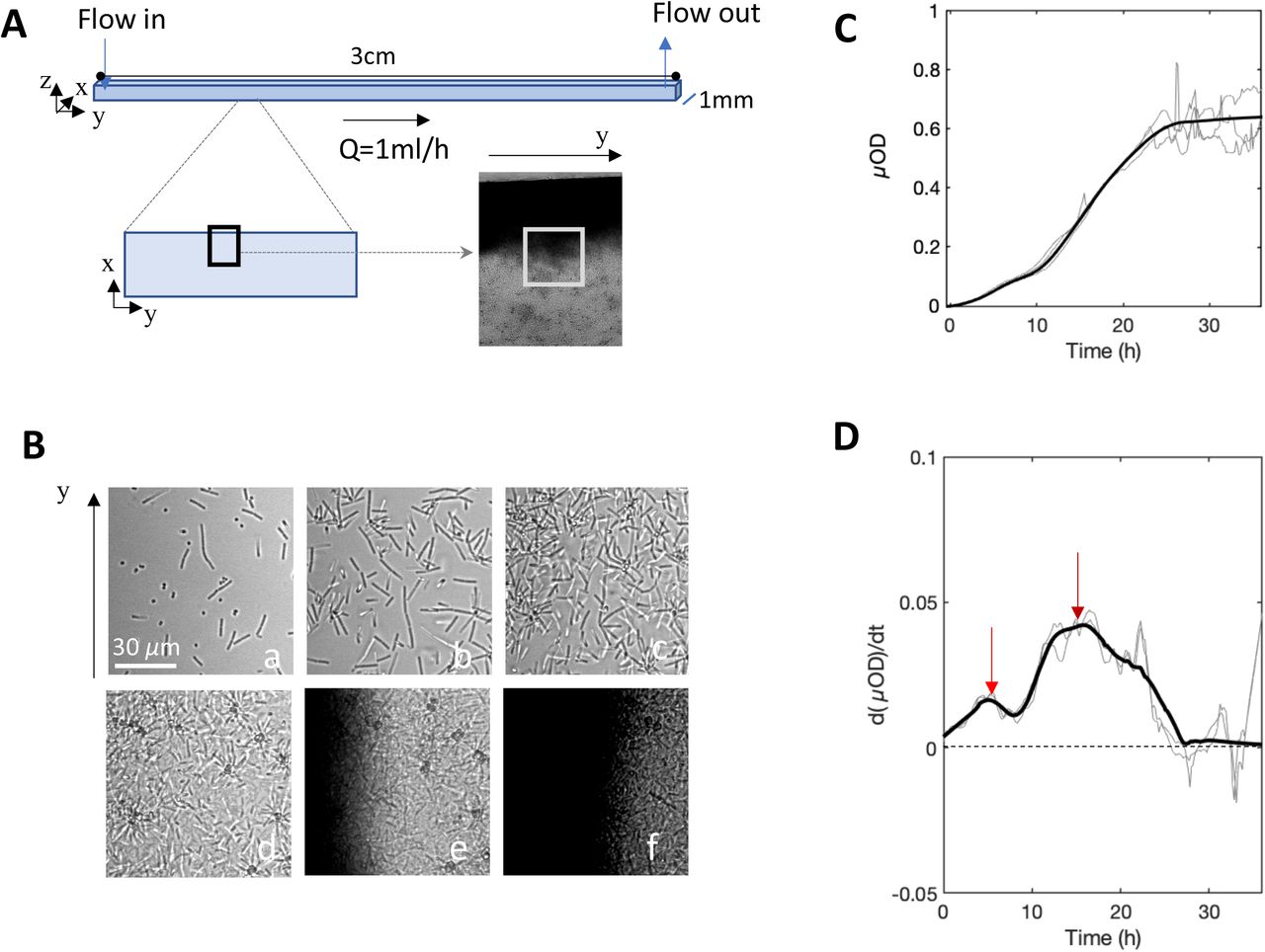
To assess the global development of the attached community under constant flow of growth medium in the millifluidic channel (Fig. 1A), we acquired time-lapse images (Fig. 1B) and measured the temporal variations of the biomass as reported by the microscopic optical density (Fig. 1C). The kinetics shows a biomass increase that levels off after approx. 25 hours. In addition, several inflections, robust across positions in the same channel, channels and biological replicates, attested for the succession of 2 characteristic temporal phases with climaxes sharply delineated by the derivative curve which measures biofilm expansion rate (Fig. 1D). A first biofilm initiation phase spread over the first 8 hours and included an accelerated growth phase (increasing expansion rate) followed by a damping phase (decreasing expansion rate) with a climax at t = 5 hours. Next, a second expansion phase arose exhibiting a climax around t = 16 hours followed by a kinetic steady-state. Remarkably, the stabilization of the average level of biomass coincided with a marked increase of the noise in biomass (Fig. 1C). Movies built from the image stacks clearly show that the noise came from the frequent passage of biofilm flocs detached upstream of the observed position (Movie S1). Locally, detachment events can be observed creating gaps refilled in a few tens of minutes (Movie S2). Thus, the kinetic steady-state signal likely results from the balance between biofilm growth and biofilm detachment, consistently with a dynamic equilibrium.

(A) A square channel of 1mm side and 30 mm long continuously fed with growth medium at 1 mL/h is settled on the stage of a microscope thermostated at 30°C. Images of 0.15 µm2 size, covering near half of the width of the channel from the edge to the middle (black rectangle) are taken every 10 mins. (B) Zooming in on center of these images (grey square in panel A) at time t = 10 min (a); 2h30 (b); 5h (c); 18h (d); 30h (e); 35h (f). (C) µOD — calculated from transmitted light images, i.e. ln(I0/I), and proportional to biomass — as a function of time. The intensity I is averaged over all the pixels of the 0.15 µm2 image (black frame in panel A). (D) Derivative of µOD signal with respect to time. The red arrows show maxima of the expansion rate. Light curves are from three independent biological replicates, the bold curve is the smoothed average.
The images (Fig. 1B) revealed the evolution of the spatial distribution of the biofilm in the channel. Initially, the colonization occurred and progressed uniformly on the bottom surface. Then, in the second expansion phase, an accumulation at the channel edges emerged while the central zone remained less densely populated.
Steady-state assessment
In order to assess the stability of the community steady-state, we evaluated the ability of the community to recover after a perturbation. To this purpose, at time t = 38 hours, we applied a major physical disturbance by injecting a 200 µl air bubble which detached approx. half of the attached biomass, indeed µOD was reduced by a factor of 2 bringing back the biofilm to the level it reached 20 hours earlier. The results in Fig. 2 show that the community biomass returned to its previous steady-state (before perturbation) in approx. 20 hours, suggesting the formation of a stable community (56, 57). Yet, the highest signal-to-noise ratio of the recovery part of the curve which corresponds to small floc detachment might reveal a slightly higher physical fragility of the material re-formed after the physical perturbation.

Evolution of the biomass (as reported by µOD) of the 4S biofilm grown in the 1 mm-side square channel at 1 mL/h for 38 hours at 30°C, then partially wrecked by the injection of a 200 µL-bubble of air (red arrow). The bold curve shows the average of 3 distinct positions in the channel (light grey curves).
Species-specific signal delineates individual kinetics and suggests interspecies coupling
To decipher the features of the 4-species (4S) biofilm development, we scrutinized the discrete contribution of each species to the community formation. To this purpose, we exploited Bt and Pf fluorescence signals (Fig. 3 A and B), Kv high contrast in transmitted-light mode (Fig. 3C) and we counted Rh cells at final time points after removing the biofilm from the channel (Fig. 3D). Bt and Pf fluorescence kinetics exhibited multiphasic curves composed of an initial oscillation followed by an intensive growth phase tending to a plateau as already detected on the global transmitted light signal. The derivatization of the kinetics featured the succession of the distinct phases and accentuated the main climaxes (Fig. 3 A and B) enabling extraction of three characteristic times for each species: t1, the maximum of the expansion rate (beginning of the dampening); t2, the end of the initial oscillation taken as the point where the derivative becomes positive again and t3, the maximum of the recovery corresponding to the inflexion preceding the plateau (table 1). Although Bt and Pf development followed similar kinetics, we observed a few hours shift in the characteristic times related to each species — intriguingly Pf exhibited delayed t1 and t2 but earlier plateau compared with Bt. The correspondence of the Bt and Pf kinetics suggested the existence of a coupling although climaxes were not fully synchronized.
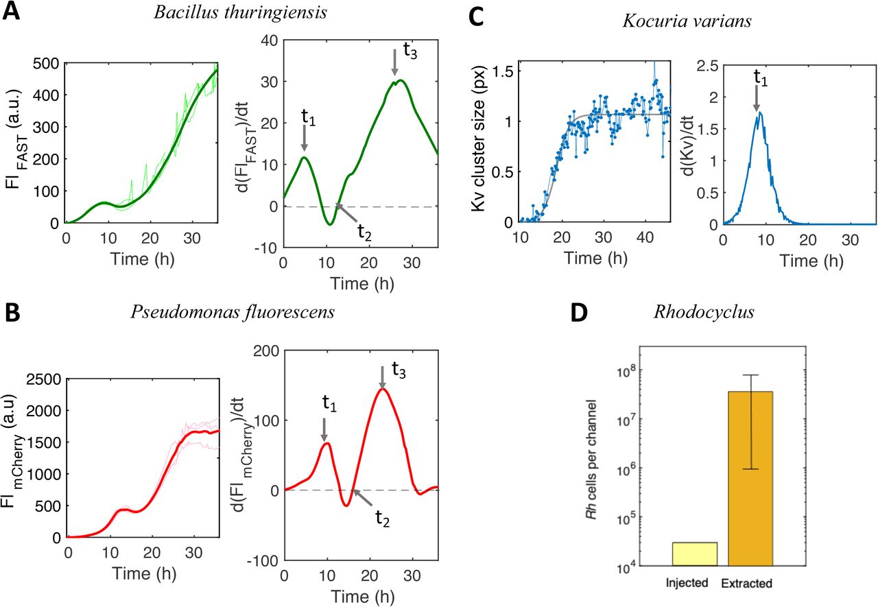
(A, B) Kinetics of the fluorescence intensity, Fl, from Bt-FAST (panel A, left side) and Pf-mCherry (panel B, left side) in the 4S biofilm with the corresponding Fl curve derivative (panels A and B, right side). Fl is the intensity per pixel averaged over the whole image. The arrows on the derivative curves mark the characteristic times t1, t2, t3 reported in table 1. (C) Kv area detected on transmitted light images as a function of time (left panel, blue line) and corresponding logistic adjustment (grey line) together with its derivative (right panel) (details in SI, Fig S2). In A, B and C, signals are collected from 0.15 µm2 images located as in Fig. 1A (black frame). Light curves are from 3 independent experiments, the bold curve is the smoothed average. (D) Bar graph of Rh cells injected in the channel (light yellow bar) and recovered after 36 hours of growth (dark yellow bar). Experimental conditions as in Fig. 1.
In parallel experiments, we monitored the fluorescence signal of Bt-GFP in the 4S community and compared it to Bt-FAST kinetics in order to get an indirect evaluation of O2 depletion along the biofilm formation. Indeed, as shown in a previous work (58), the proportionality between FAST and GFP signals is lost when environmental O2 level decreases below the threshold enabling GFP final maturation and fluorescence. We observed that the Bt-GFP fluorescence curve, initially superimposable with that of Bt-FAST, diverged at time t = 5h (Fig. 4), which corresponded to t1, the first Bt growth climax.

Kinetics of the fluorescence intensity, Fl, from Bt-FAST and Bt-GFP growing in parallel channels in the 4S biofilm. The curves represent the average of at least three independent measurements. The insert shows a zoom in of the first 8 hours of the biofilm formation. Experimental conditions as in Fig. 3A.
In the meantime, the Kv population developed from initially attached single cells under the form of small clusters, the size of which was taken as a growth index to plot Kv expansion kinetics in the community (Fig. 3C and SI). The obtained curve was formally adjusted to a logistic growth with only one characteristic time t1 reporting a single growth phase (table 1). Due to the small cell size, high motility and absence of labeling, the temporal evolution of the Rh population could not be accurately determined. Hence, the contribution of Rh was evaluated by enumerating the cells after biofilm extraction at the final stage which showed that the Rh population successfully thrived in the community, having multiplied by 3 logs along the biofilm development (Fig. 3D). In comparison, Bt and Pf were multiplied by approx. 3 and 4 logs respectively (Fig. S4).
The results suggest the existence of a coupling between Bt and Pf populations which both exhibit two successive growth phases, similarly to the global biomass kinetics. By contrast, Kv population exhibits an only one-phase expansion. The individual kinetics show that the four species were present all along the biofilm development. An oxygen depletion was deduced in correlation with the first climax of the community.
The 4S community displays species-specific spatial distribution
To document spatial heterogeneity emergence in the community, we examined the evolution of the species spatial distribution along the community expansion.
We first investigated the species vertical distribution. To this purpose, we run a series of confocal microscopy acquisitions to selectively image bottom and top surface of the channel whereas epifluorescence recordings captured the signal from the whole channel height. We found that Bt strictly dwelled on the bottom surface of the channel while Pf colonized both bottom and top surfaces (Fig. 5). Pf bottom and top surface populations displayed similar kinetic profiles except the first oscillation was delayed by 5 hours on top surface in comparison with the bottom surface. These acquisitions also showed that Bt and Pf exhibited synchronous kinetics on the bottom surface (see table 1 and table 2). Besides, transmitted light observations showed that Kv locations were also limited to bottom surface (Fig. S5). Thus, Pf appeared to share the bottom surface with the other species but also to colonize a specific niche on the top surface.

Confocal fluorescence intensity of (A) Bt and (B) Pf recorded with a focus on the bottom surface (light green (A) and light red (B)) and on the top surface (dark green (A) and dark red (B)). Axial resolutions are 5 µm and 5.8 µm for FAST and mCherry signals, respectively. (C) Confocal images recorded at time t=3h on the bottom surface (upper panels) and on the top surface (lower panels) for Bt (left panels) and Pf (right panels). Experimental conditions as in Fig. 1.
Next, we examined the lateral spatial distribution of the individual species, having in mind the spatial distribution shift of the global biomass reported above (Fig. 1B). During the first 5 hours of the colonization, both Bt and Pf developed a uniform distribution along the x and y axis on the bottom surface before priming a progressive shift towards the edges of the channel (Fig. 6A and B). In order to quantify this shift, we defined two 20µm-wide regions of interest, ROIEdge and ROICenter, located at 20µm and 360µm of the channel wall, respectively. Then, we calculated the ratio of their mean fluorescence intensities, FLEdge/ FLCenter which measured the intensity of the spatial shift. The evolution of this ratio along the formation of the biofilm together with representative kymographs are shown in Fig. 6C. They show a first 5 hours even distribution, then a trend to preferably populate the edge of the channel emerging between 5 and 10 hours and a clear-cut spatial distribution transition toward the channel edge arising shortly after the onset of the second growth phase.

Fluorescence images of Bt (A) and Pf (B) populations in the 4S biofilm, taken in the initial colonization phase (left image) and after the spatial transition (right image). The small bottom images are zoomed-in details showing local heterogeneities of the globally uniform distribution in the initial colonization phase. Graph of the ratio as a function of time of the fluorescence intensity at the channel edge (Fledge) to that at the channel center averaged on ROIs (20µm-wide) located at 20µm and 360µm of the channel wall, respectively; Bt in green and Pf in red (C upper panel). Kymographs of Bt (green blue) and Pf (Red) spatial distribution (C bottom panel). Detection of the Kv cells and clusters in the 4S biofilm using the NIS dark-spot detection tool (details in SI). Images correspond to time t=14h. In the bottom panel, detection results are overlaid on the original image shown in the upper panel (D). Kv spots are then counted in 30µm-wide ROIs from the edge to the center of the channel for time t= 10 mins (□); 20 mins (•); 7h30 (x); 16h30 (◻); 32h (+); error bars report the error of 10% made on spot counts as evaluated comparing automatic and visual detection.
Kv spatial distribution was analyzed collecting on the images the (x,y) coordinates of the Kv cells and clusters attached on the channel bottom surface (Fig. 6D and E). We found that the Kv discrete locations were evenly distributed over the bottom surface all along the biofilm development — despite short length scale heterogeneities — indicating that Kv did not participate to the spatial transition supported by Bt and Pf consistently with its kinetic profile also clearly distinct from that of Pf and Bt. The synchronized Pf and Bt spatial transition reinforces the hypothesis of a coupling of these two populations in the community on the bottom surface. In addition, the temporal correspondence of this spatial transition with the second growth phase indicates that this apparent translocation rather corresponds to a geographically-restricted growth. All the species coexisted on the bottom surface but Pf was in addition able to accommodate a private niche on the top surface.
Local dynamics analysis reveals both static and highly mobile species
To better characterize the initiation phase of the biofilm, we examined the four species local dynamics within the first 6 to 7 hours of the biofilm installation, when the colonized fraction of the surface is small enough to enable individual objects delineation — a period of time corresponding to the first growth phase.
The dynamics of the fluorescent species Bt and Pf was extracted from stacks of fluorescent images calculating the correlation coefficient, rc, between consecutive frames (Fig. 7A). Bt installation exhibited a correlation coefficient rc <0.5 (Fig. 7A), attesting for a loose attachment on the surface corroborated by the colocalization maps of successive images (Fig. 7B). After about 2 hours, the correlation coefficient increased, which coincided with the formation of fluorescent asters resulting from the progressive aggregation of Bt around Kv cells which physically stabilized the connected Bt cells while the others remained highly mobile (Movie S3).

(A) Consecutive frames correlation coefficient calculated from time-lapse recordings of Bt-FAST (green curve) and Pf-mCherry (red curve) in the 4S biofilm community. (B, C) Colocalization maps of Bt (B) and Pf (C) cells showing spatial self-overlap between time t and t’=t+10 min taken at t=1h (upper panels) and t=3h (lower panels). Light grey pixels correspond to non-moving cells (pixels unchanged between t and t’), blue pixels correspond to newly appeared cells (pixels dark at t and lighted at t’) and magenta pixels correspond to removed cells (pixels lighted at t and dark at t’). (D) Confocal images of Pf-mCherry in the 4S community taken at t= a) 6h40; b)7h; c)7h30 d)8h showing full detachment of the cell groups; see also colocalization maps in Fig. S6. (E) Transmitted light images of the 4S biofilm taken at time t = 1h (a) ; 12h (b) ; 37h (c) showing Kv (indicated by the yellow arrows) fixed localization all along the biofilm growth. (F) Successive frames (a,b,c) of the 4S biofilm in transmitted light and red fluorescence taken from t=1h (a) with a 10 mins interval. Upper right frame is a zoom in of (a). Yellow squares spot the Rh cells illustrating their high dynamics with different positions on each frame.
Pf cells exhibited a strong surface anchoring characterized by correlation coefficients comprised between 0.8 and 0.9 after 2 hours installation (Fig. 7A). The colocalization maps show that most of the initial attachments generated microcolonies (Fig. 7C). At time t≈5h, corresponding to the first climax of the development of Pf population on the bottom surface, rc decreased while Pf cells simultaneously started to detach, shifting from firmly attached to essentially detached cells in less than two hours (Fig.7D and Fig. S6). Noticeably, a similar detachment occurred on the top surface but delayed by about 6 hours consistently with the shift of the climaxes mentioned above.
Kv dynamics was evaluated from the localization maps established to evaluate its spatial distribution. Stable attachment of isolated cells randomly dispersed over the whole bottom surface of the channel was observed from the first minutes of the biofilm initiation. As biofilm grew, compact clusters of a few cells formed essentially at the same location than the initial attachment (Fig. 7E). At least 90% of the Kv clusters came from single cell initial anchors. The correlation coefficient calculated from the position map was found equal to 0.94 (Fig. S7).
Rh was difficult to follow, exhibiting high dynamics. On the first images, a few cells could be visually detected. Their surface-dwelling time was found below the 10 min of the frame period, meaning Rh cells were never found at the same location on consecutive frames (Fig. 7F). Therefore, the correlation coefficient for Rh within the installation period was assigned to 0 as a principle.
The 4 species installation dynamics ranges from highly dynamical with Rh (rc=0) to completely fixed with Kv (rc =0.95). The rc values which measure the bacterial residence on the surface in the presence of the flow show the significantly distinct capacities of the 4 species to adhere on the surface. The correlation profile of Bt which rc value increases upon its association with Kv also shows how physical interspecies interaction can improve the colonizing potential of poorly adhesive species.
The 4-species combinatorics highlights community-specific traits
Next, we raised the question of which are the community-specific traits. To this purpose, we designed a combinatorial approach consisting in injecting all the possible sub-combinations of the 4 species in different parallel channels (14 in total). Then, we collected the optical signals of all the channels along the first 36 hours following channel seeding. We exhaustively show the mixes recordings in supplementary material (Fig. S8) but focus in the following on the features the most significant for the interpretation of the community traits.
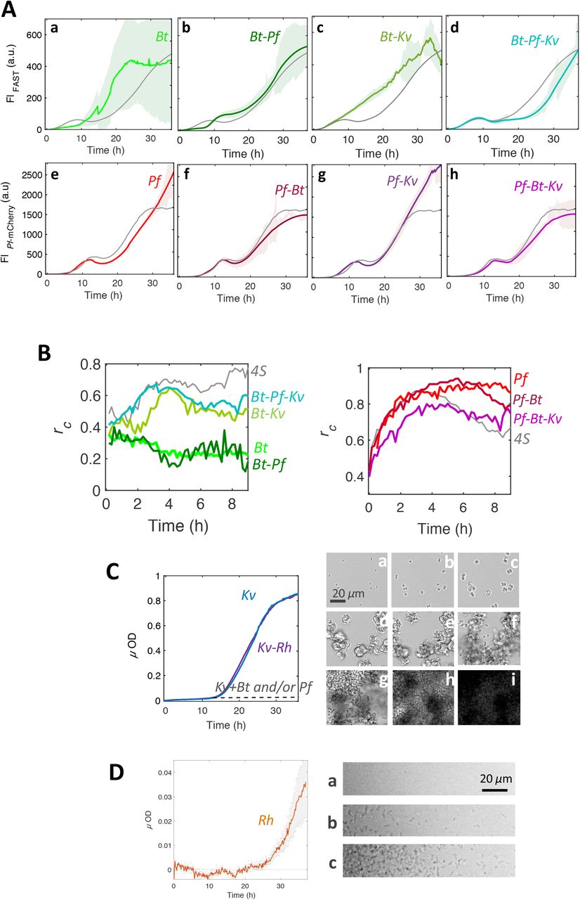
Bt improves its installation and physical stability in the 4S
The biofilm built by Bt alone exhibited several characteristics distinct from the 4-species biofilm. In particular the initial population oscillation was absent, replaced by a 10 to 12 hours-long lag phase where the cell amount at the surface remained low (Fig. 8A panel a). We also observed that local dynamics were characterized by correlation coefficient values below 0.4 (Fig. 8B) which indicated that Bt cells essentially did not attach on the surface in the absence of the other species. After 10 hours, the fluorescence signal significantly increased reporting a colonization phase similar to the second phase displayed by Bt in the 4S biofilm but affected by a strong instability. The standard deviation was of the order of the signal itself due to massive random detachment events which resulted in important variations of the signal (Movie S4). In addition, the single-species Bt-FAST and Bt-GFP biofilms exhibited fully superimposable fluorescence signals which indicates no O2 depletion occurred in this situation (58)(Fig. S9). In the presence of Pf, Bt development profiles did not drastically changed compared to that of Bt alone, except the colonization phase appeared to be paused for a few hours compared to the expansion of Bt alone (Fig. 8A panel b). The standard deviation of the data sets was also 3 times less than for Bt alone, suggesting a physically more stable biofilm had formed. The biofilm built by the Bt-Kv pair (Fig. 8A panel c) confirmed the specific Bt-Kv physical interaction which led to the formation of Bt asters around Kv (Fig. 6A and Movie S3) and to the physical grip effect that promoted Bt early installation as showed by the increase of the fluorescence intensity and of the consecutive frames correlation coefficient (Fig. 8B). In the 3-species Bt-Pf-Kv community, the Bt development profile combined sequentially (i) the traits of the Bt-Kv pair: the initial promoted Bt growth, and (ii) the traits of the Bt-Pf pair: the second phase induced latency (Fig. 8A panel d). This 3-species community closely resembles the 4S community with the initial oscillation and a second growth phase. Yet, it should be mentioned that, in comparison with to Bt-Pf and 4S, the 3-species Bt-Pf-Kv community exhibited an extended Bt second phase latency.
(A) Fluorescence signals of Bt (a-d) and Pf (e-h) in various sub-combinations of the 4 species : single-species (a,e), pairs (b,c,f,g) and triplets (d,h). (B) Consecutive frames correlation coefficient for Bt (left panel) and Pf (right panel) for the different combinations mentioned on the figure. Experimental conditions other than the biofilm composition are as in Fig. 3. (C) Micro-optical density of Kv single-species biofilm (in blue) and Kv-Rh pair (in purple) (graph on the left); the dashed line shows the level of Kv development in the presence of Bt, alone, Pf alone and Pf and Bt together, deduced from Kv area determination and mean µOD of a Kv cluster. Images on the right are snapshots of the single-species biofilm taken at time t = 10 min(a); 3h30(b); 9h(c); 15h(d); 16h30(e); 18h30(f); 23h30(g); 27h30(h); 30h(i). (D) Micro-optical density of Rh single-species biofilm (graph on the left) and corresponding snapshots taken at time t = 10h(a); 25h(b); 37h(c). The curve of the studied combination in each panel (color bold line) is the average of at least 3 independent samples and appears shaded with the standard deviation of the data set. The curve of the corresponding species in the 4S community is plotted in grey for the sake of comparison.
Pf undergoes a Bt-imposed dynamic equilibrium
Pf in the various combinations exhibited the same main traits evidenced in the 4S biofilm except the rise of the dynamic equilibrium which was absent in all the combinations devoid of Bt (Fig. 8A panel e-h). Pf appears as the less impacted species in the various combinations, simply forced to equilibrium by the presence of Bt after 30h of combined growth.
Kv development is inhibited in the 4S community
Injected alone in the channel, Kv population formed a profuse single-species biofilm. The cells immediately adhered on the bottom glass slide and multiplied in situ forming compact cubic clusters over the first 10 hours of the biofilm formation as described above in the 4S community (Fig. 8C). Then, whereas the Kv clusters were halted in this stage in the 4S community, they went on growing exponentially in the single-species mode, forming larger cohesive clusters of tens of cells before bursting again into single cells which were in part removed by the flow, and in part readdressed to the surface, efficiently forming a thick biofilm (Fig. 8C and Movie S5). We found that the Kv population was inhibited by the presence of Bt alone, Pf alone and both Bt and Pf as quantified on the images (Fig. 8C and Fig. S10) exactly the same way it happened in the 4S (Fig. S5). Noticeably, Kv cells extracted from two-species and three-species biofilms with Bt and/or Pf were recovered by plating on agar forming colonies, which indicated they were still viable.
Rh, the community neutral element
Rh alone exhibited poor colonization abilities. A thin layer of attached cells concentrated at the edge of the channel emerged after about 25 hours (Fig. 8D). Its contribution in mixed communities was always optically overwhelmed by that of the other species and could not be characterized in real time. Although their presence was attested by channel extraction counting, Rh cells did not alter any characteristics of the other species in multispecies communities (Fig. S8).
Discussion
We report here a real-time analysis of a 4-species bacterial assembly, composed from a more complex natural community (50) and grown under constant flow of growth medium in a laboratory millifluidic device. The simple and defined structure of the setup provided a controlled environment, the heterogeneity of which was sufficient to allow behavioral complexity to emerge.
We show that the 4S assembly forms a surface-attached community displaying a dynamic equilibrium, with a biomass robust to physical perturbation after about thirty hours seeding. We find that the 4S community settles according to a deterministic mechanism involving timed spatiotemporal phases of expansion and recession. However, two of them, Pf and Bt clearly dominate the developmental program. Remarkably, while the four species coexisted on the bottom surface, a specialist niche emerged on the top surface, accommodating exclusively Pf, who escaped gravity and bottom surface assignation. Considering the channel dimensions (section of 1 mm2) and the flow rate (1 ml/h), characteristic times of 2 mins for advection and of 10 mins to a few hours for molecular diffusion in the height of the channel can be evaluated from the Stokes-Einstein equation (59). Thereby, bottom and top surfaces habitats can be regarded as independent as well as their respective biomes, reminding — although at a much smaller scale and limited range — the biogeographical sorting that occurs in nature (60).
On the bottom surface, Pf and Bt appeared closely coupled in time and space. Our results support two distinct mechanisms of coupling. The first one, governing the first 25 hours of the biofilm formation, mediated by the environment and interpreted as a coupled response to the depletion in O2 of the environment principally resulting from the Pf population growth. This hypothesis is supported by the comparison of Bt-GFP and Bt-FAST fluorescence signals in the 4S and the single-species communities. Consistently, Pf development kinetics exhibited very similar profiles in a 4S and in a single-species biofilm within the first 25 hours of the formation, suggesting a prevalent imprint of Pf on the environment. In this scenario, the O2 depletion of the environment is also assumed to drive Pf detachment and spatial transition to the channel edges. Indeed, we know from previous experiments that, due to PDMS permeability to O2, renewal of O2 supplies occurs principally at the channel edges in our device (51, 58), explaining the selective growth of the aerobes in the regions of higher O2 level where they compete for the same resource. Here, we highlight a mechanism in which the spatial distribution is governed by the shaping of the environment by one of the species. Such a resource-driven spatial organization might be a key principle of the emergence of spatial structure in complex biofilms. It has also been previously described in colonies of mixed genotypes of Pseudomonas aeruginosa (61). Yet, reciprocal influence of spatial distribution on social interactions has also been described (40), confirming no unequivocal link between social behavior and spatial structure can be established (21). Moreover, we bring evidence that the spatial structure evolves along the biofilm development. The expression of a Bt-Pf interaction is also found in the characteristics of the dynamic equilibrium established after thirty hours of 4S biofilm growth. From the combinatorial assembly of multispecies communities, it appears clearly that Bt forces Pf to a plateau resulting from balanced growth and detachment by contrast to the continuous expansion Pf exhibits in its pure biofilm. The presence of Pf also provides a better physical stability to the co-existing population of Bt, as shown by the significant decrease of the standard deviation for Bt signal in the presence of Pf, and the reduction of the number of spikes due to detachment events.
These changes occurring in the stability of the biofilm point to the question of the assembly of the extracellular matrix in the multispecies context which just starts to be investigated (62). Indeed, biofilm stability is closely related to extracellular matrix properties such as its degree of cross-linking (63). The larger spectrum of components brought together in multispecies environment might offer novel opportunities to form cross-links. In this hypothesis, the matrix assembly is a community activity which occurs extracellularly as shown in a previous study on Candida albicans biofilms (64). This provides an illustration of how spatial reorganization leads to the emergence of novel properties, reinforcing the view of the critical role of the spatial distribution on the biofilm properties (20, 22, 24, 65, 66).
Bt and Kv displayed a specific physical interaction enabling several Bt cells to attach to a Kv cluster and to take advantage from the strong affinity of Kv cells for the bottom surface of the channel, in particular in the initial phase of the colonization where it hardly forms transient ties with the surface. This interspecies physical attachment has been mainly overlooked in the analyses of multispecies community structures compared to social interactions mediated by diffusible factors such as quorum sensing or metabolite tradeoffs (46, 67–69) except in the special case of the oral biofilms (70). A better understanding of the impact of these specific bindings on the biological functions as well as additional knowledge about their diversity and their evolutionary profile might open new perspectives in the comprehension of multispecies adherent communities.
Besides, Kv population experiences a major limitation of its development in the mixed communities containing Bt alone, Pf alone and both Bt and Pf. As both Bt and Pf inhibit Kv biofilm development without killing it, it is tempting to postulate an environmental effect driven by the development of neighbors rather than a specific interspecies interaction. However, at this stage, we have no cues for the involved parameter, in particular the inhibition did not show any positional dependence ruling out a link to O2 supply as assumed to explain Bt and Pf behaviors.
Rh appears as the freewheeling partner of the community. This highly motile cell, proved to develop poor attachment when grown as a single-species biofilm, still multiplies and persists in the 4S community in an apparently fully neutral mode with no detectable interaction with the other species. It should be mentioned that we gained poor information about it since it could not be tracked in the multispecies communities. However, we decided to conserve it in the consortium as an example of coexistence without interactions, anticipating a possible role of this neutral element in disturbed environmental conditions.
We conclude that our composite biofilm reaches its dynamical equilibrium based on local cell concentration, environmental physico-chemical properties constantly reshaped by the community development itself, and a deep correction of the species fitness by surface attachment. From a conceptual perspective, this community could be considered as individualistic to the sense of Gleason (71) but also as a limiting case of an organismic continuum as theoretically proposed by Liautaud et al. (72).
This multispecies biofilm emerges as the result of chance (a given species in a given environment) and necessity (individual species adaptation) deterministically leading to a unique community. The emergent properties could not be predicted from individual traits, involving a form of biofilm sociobiology which does not require that cells communicate with one another using specialized signaling molecules (73).
The operational experimental model and assembly principles we present here should prove useful to investigate multispecies bacterial community response to perturbations or elaborate new strategies (65) to manipulate biofilm functions in several fields including health, agriculture and environment.
Acknowledgments
Authors would like to thank Pierre Nicolas and Cyprien Guérin for fruitful discussions and Carounagarane Doré for technical assistance in the devices elaboration.
The work was supported by a grant from French Agence Nationale pour la Recherche (ANR-15-CE02-0001-01 ACToP) and a MESRI fellowship to WBY.
Footnotes
Competing InterestsThe authors declare there are no competing interests
References


