

Abstract
Understanding how a pathogen responds to the host stimuli and succeeds in causing disease is crucial for developing a novel treatment approach against the pathogen. Transcriptomic analysis facilitated by RNA-Seq technologies is used to examine bacterial responses at the global level. However, the ability to understand pathogen behavior inside the host tissues is hindered by much lower pathogen biomass than host tissue. Recently, we succeeded in establishing a method to enrich Staphylococcus aureus cells from infected organs. In this research, we analyzed the small non-coding RNA (sRNA) transcriptome of S. aureus inside the host and found that rsaC was among the highly expressed sRNAs. Furthermore, by gene disruption and complementation, we demonstrated that rsaC was required for full pathogenicity of S. aureus in a murine model. Besides, we found that ΔrsaC showed a difference in gene expression depending on the oxygen and host stress. The findings of this study suggest rsaC acts as a novel virulence factor in S. aureus and might facilitate the adaptation of staphylococci within the host.
Importance Drug-resistant Staphylococcus aureus is among the pathogen for which new treatment options are urgently needed. However, limited understanding of S. aureus pathogenesis in the host has hindered unearthing potential strategies to treat the infections. Here, based on the in vivo transcriptomic analysis, we present the identification of a small non-coding RNA (sRNA) rsaC as a novel virulence factor of S. aureus. Furthermore, we performed transcriptomic analysis of the rsaC disrupted mutant and identified different pathways, possibly controlled by rsaC, during aerobic, anaerobic, and in vivo conditions. These findings contribute to reveal the role of sRNA rsaC and broadens our understanding of the adaptation of S. aureus to host environments.
Text
The growth and behavior of a pathogenic microorganism differ between the host environment and in the in vitro culture. Understanding how a pathogen responds to the host stimuli and succeeds in causing disease is crucial for developing novel drugs against the pathogen. Comprehensive transcriptomic analysis facilitated by RNA-Seq technologies using next-generation sequencers has been widely used to understand pathogen response at the global level (1, 2). Several studies have used in vitro host-like or in vivo host environments to understand the infection process and alterations in the pathogen and the host (3, 4). Such studies aimed to elucidate the functional role of protein-coding genes, while the small non-coding RNAs (sRNAs) remain largely unattended. Studies that performed the comparative analysis of pathogen response in the host to the in vitro growth allowed us to evaluate how a pathogen behaves during infection situations and how the host responds to pathogen invasion (4–7). The current understanding of bacterial pathogenesis allowed us to identify key virulence determinants that could potentially be exploited as a drug target for antivirulence drugs. Whereas it can be expected that pathogens lacking these virulence factors are easy to be killed or cleared from the host, their behavior in the host has not been analyzed at the global level. Furthermore, a detailed evaluation of pathogenesis requires an understanding of pathogen behavior during actual infection conditions.
sRNAs have been identified in living organisms, including the bacterial kingdom. Although they do not encode functional proteins, they can accomplish a wide range of biological functions, including regulation of gene expression at the levels of transcription, RNA processing, mRNA stability, and translation (8). In bacteria, sRNAs have the potential to regulate the gene expression pattern of the bacteria by interacting with protein or mRNAs by cis- or trans-acting mechanisms (8), thus affecting multiple cellular processes such as pathogenesis and bacterial physiology in response to environmental cues, facilitating survival under unfavorable environments. In Staphylococcus aureus, a pathogen of global concern, several sRNAs have been identified (9–11), while the functional characterization of this class of RNAs has long been forsaken. One of the functionally characterized and extensively studied Staphylococcal sRNA, RNAIII, regulates the expression of several virulence factors both positively and negatively (12–14). rsaC, a part of polycistronic operon mntABC, is another staphylococcal sRNA known to be expressed in infected host (15) and modulate oxidative stress during manganese starvation (16). Whereas these studies provided a hint towards its role in S. aureus virulence, a cell-based study showed that the ΔrsaC strain was more persistent in macrophages and resistant to opsonophagocytosis than the wild-type strain (16) indicating its obscure role in pathogenesis.
We recently successfully enriched pathogen RNA from an infected animal and performed an in vivo RNA-Seq analysis of Streptococcus pyogenes (5) and S. aureus (6) using a two-step cell crush method. In this manuscript, we performed the comprehensive analysis of S. aureus sRNAs expressed within the host. Using RNA-Seq analysis of the rsaC disrupted mutant, we showed a clear difference between the gene expression patterns during in vivo and in vitro (aerobic and anaerobic) growth conditions. Furthermore, we found that the transcription of fermentation and oxidoreductase-related genes, virulence and toxin-related genes, and host evasion-related genes were affected during aerobic, anaerobic, and in vivo growth, respectively. Our results highlight the importance of in vivo transcriptomic analyses of two strains with the same genetic background to allow a direct comparison, revealing changes in different environments.
Materials and Methods
Ethics statement
All mouse experiments were performed at the University of Tokyo and Teikyo University, following the animal care and use regulations approved by the Animal Use Committee (approval numbers: P27-4 and 16-014 at respective institutes).
Bacterial strains and primers used in the study
Bacterial strains and primers used in this study are listed in Table 1. S. aureus and Escherichia coli were routinely grown in LB or TSB medium, respectively, at 37°C with shaking. The media were supplemented with antibiotics, as required.
Real-time RT-PCR
According to the manufacturer’s recommendations, one microgram of RNA was used to prepare cDNA using a High-Capacity RNA-to-cDNA™ Kit (Applied Biosystems; Foster City, CA, USA). From this, 15 ng of the cDNA was used as a template for RT-PCR using Fast SYBR Green Master Mix (Thermo Fisher Scientific) on a 7500 Fast (Applied Biosystems) machine with 40 cycles of denaturing at 95 °C followed by annealing/extension at 60 °C.
Construction of ΔrsaC strain and complementation
rsaC was disrupted using a double cross-over recombination method as previously described (24). Briefly, the genome regions up-and down-stream of rsaC were amplified by PCR using the listed primers. Then, overlap extension-PCR was performed using these two DNA fragments together with the aph gene amplified from the pSF151 vector using primers KmF and KmR. The PCR product was cloned into the pKOR3a vector (24) and introduced into the RN4220 strain (18) by electroporation. Integration of the mutant cassette in the genome was confirmed by PCR and further transformed into S. aureus Newman (17) by phage transduction using phage 80α as previously described (23, 25).
To prepare the overexpression strain, the rsaC coding region was amplified by the primer pair rsaC_bam_F and rsaC_sal_R (Table 1) and ligated to BamHI-SalI digested pND50-PfbaA (19, 20). The resulting plasmid, pND50-PfbaA-rsaC, was then transformed into S. aureus RN4220 by electroporation and then to S. aureus Newman wild and ΔrsaC strains by phage transduction.
Proteolytic and hemolytic assays
Bacteria were grown overnight in TSB medium, supplemented with antibiotics, as required, at 37°C with shaking. From this, 2 μl aliquots were spotted on TSB containing 3.3% skim milk or sheep blood agar plates (E-MR93; Eiken Chemical, Tokyo, Japan) to determine proteolysis and hemolysis, respectively. The plates were incubated at 37°C overnight. Sealed container with Anaero Pack (Mitsubishi Gas Chemical, Tokyo, Japan) were used to determine phenotypes under anaerobic conditions. The proteolytic and hemolytic activities were determined by the appearance of a clear zone surrounding the bacterial growth.
Mouse survival assay
S. aureus Newman wild-type and ΔrsaC strains were grown overnight on TSB medium supplemented with antibiotics, as required, on a rotary shaker maintained at 37 °C to obtain full growth. The full growth was diluted 100-fold with TSB and cultured overnight on the same shaker, and then the cells were centrifuged and resuspended in phosphate-buffered saline (PBS, pH 7.2) to have an optical density (OD600) of 0.7. An aliquot (200 μl) of the prepared cell suspension was then injected intravenously into C57BL/6J mouse, and mouse survival was observed. Survival analysis was performed using GraphPad Prism ver 8.0 (GraphPad Software), and statistical analysis was performed using the Log-rank (Mantel-Cox) test.
S. aureus infection, organ isolation and RNA isolation
Staphylococci were grown overnight on TSB medium at 37°C with shaking. The full growth was diluted 100-fold with 5 ml TSB and regrown for 16h under the same conditions. The cells were centrifuged and suspended in PBS (pH 7.2). The cells (OD600 = 0.7) were injected into C57BL/6J mice via the tail vein. On day 1, mice were killed to harvest organs. Organs were immediately placed either in ice to calculate viable cell numbers in each organ or liquid nitrogen and maintained at −80 °C for RNA extraction. Each experiment was conducted with three animals, and data are represented as an average. Mouse organs were homogenized, and total RNA was extracted, as explained previously (6). RNA extraction from in vitro culture was performed as explained (19). For anaerobic RNA-Seq, staphylococci were first grown aerobically to reach OD600 of 1.0 and then transferred to anaerobic growth for 30 minutes.
Library preparation and RNA-sequencing
Total RNA was subjected to rRNA depletion using a MICROBExpress™ Kit (Thermo Fisher Scientific, Waltham, MA) and used for library preparation for RNA-Seq using an Ion Total RNA-Seq Kit v2 following the manufacturer’s instructions. After confirming the size distribution and yield of the amplified library using a bioanalyzer, the libraries were enriched in an Ion PI Chip v2 using the Ion Chef (Thermo Fisher Scientific) and sequenced using Ion Proton System (Thermo Fisher Scientific). These data have been deposited in the NCBI Sequence Read Archive under accession number ######.
Differential gene expression analysis
All data were analyzed using CLC Genomics Workbench software, version 21.0.4 (CLC Bio, Aarhus, Denmark). Reads were aligned to the Newman genome annotated with ncRNA genes allowing a minimum length fraction of 0.95 and a minimum similarity fraction of 0.95. Differential gene expression analysis was performed using the default setting. Genes with a false discovery rate (FDR) p<0.05 were classified as having significantly different expressions.
Results
Comprehensive analysis of sRNAs expression in the host environment
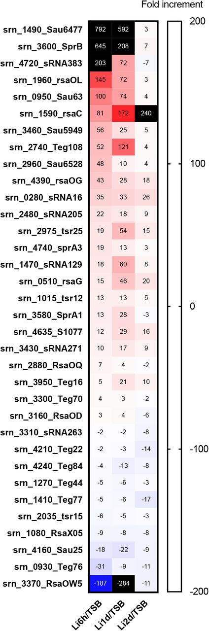
To quantitatively analyze the expression of S. aureus sRNAs within the host, we first annotated the sRNAs present in the Newman strain based on the report of Sassai et al. (11). Next, we performed an RNA-Seq analysis based on the reads obtained from our two-step cell crush method (6). A comparative expression analysis of RNA isolated from host liver at 6- hr, 1- day and 2- day post-infection to the RNA isolated from in-vitro culture was performed. We found that among 559 sRNAs present, 34 sRNAs were differentially expressed at all the three-time points compared to in vitro (Figure 1). Upregulation or downregulation of these sRNAs at all three time points suggested these sRNAs’ possible role to respond to host circumstances.

Differential expression of Newman sRNAs in mouse liver compared to TSB medium culture condition. sRNAs with common differential expression and FDR p < 0.05 on all the three-time points are shown.
Identification of RsaC as a virulence factor
Given that many sRNAs were differentially expressed under the host circumstances, we were interested in whether these sRNAs can modulate S. aureus pathogenesis. Among sRNAs, we focused on rsaC, as it falls among one of the studied staphylococcal sRNAs. Previously, rsaC was shown to have an increased expression in the infected host (15) and modulate oxidative stress during manganese starvation (16). However, its role in the regulation of pathogenesis has remained elusive. First, we confirmed the expression of rsaC in mouse organs using a real-time reverse transcription-polymerase chain reaction (RT-PCR). A higher expression of rsaC was observed in both the kidney and liver, as compared to TSB medium culture condition (Figure 2A). The gene organization of rsaC in the Newman genome (Figure 2B) showed that rsaC did not overlap with other genes, allowing us to construct a single gene disruption mutant. We constructed the rsaC disrupted mutant and examined its role in pathogenicity using a mouse infection model. We found that ΔrsaC had reduced ability to colonize in mouse heart 24h post-infection whereas the ability to colonize in kidney, liver, lungs and spleen were indistinguishable from that of the wild-type (Figure 2C). To further confirm the role of rsaC in virulence, we checked the survival of wild-type or ΔrsaC- infected mice. The results indicated that the ΔrsaC strain had reduced virulence (Figure 2D), evident by prolonged survival of the ΔrsaC infected mice. To eliminate the possibilities of polar effects associated with this disruption on pathogenicity, we complemented the wild-type and the ΔrsaC strains by reintroducing rsaC under the control of a constitutive promoter- PfbaA. Restored pathogenicity of the complemented strain unequivocally explained the involvement of rsaC in pathogenicity and established it as a virulence factor of S. aureus.

(A) Upregulation of rsaC in host organs compared with that in TSB medium was confirmed by RT-PCR. The data were standardized by the abundance of 16s rRNA in each sample, and statistical analysis was performed using ANOVA. (B) Position of the rsaC in S. aureus Newman chromosome. (C) The number of S. aureus cells in each organ after infection with (7.6×10⁷ CFU and 7.5×10⁷ CFU) S. aureus Newman and ΔrsaC strains, respectively. Statistical analysis was performed using 2way ANOVA with Sidak’s multiple comparisons in GraphPad Prism 8.4.3. (D) Survival of mice after the infection with a deletion mutant of the rsaC gene. Wild-type, ΔrsaC, wild-type/prsaC, or ΔrsaC/prsaC were injected intravenously at a dose of 5.0×10⁷, 5.9×10⁷, 4.8×10⁷ and 5.0×10⁷ CFU, respectively. Asterisk indicates a significant difference compared with the survival curve following wild-type injection (p<0.05) by the Log-rank (Mantel-Cox) test. The experiment was performed two times, and essentially the same results were obtained.
Next, we performed the phenotypic evaluation of the ΔrsaC mutant in vitro. We found that the growth of ΔrsaC strain was indistinguishable from that of the wild-type during both aerobic (Figure 3A) and anaerobic (Figure 3B) growth. In addition, ΔrsaC had unaltered proteolytic and hemolytic abilities compared to that of the wild-type during both aerobic and anaerobic culture conditions (Figure 3C, D). These results suggested no difference between the wild-type and mutant in terms of virulence-related in vitro phenotypes and necessitated an in vivo infection system to investigate the role of rsaC in pathogenesis.

(A) Growth curve of ΔrsaC and wild-type strains during aerobic culture. (B) Colony Forming Units (CFUs) of ΔrsaC and wild-type strains after growing anaerobically. Statistical analysis was performed using an unpaired t-test. (C) Proteolytic- and (D) hemolytic- ability of ΔrsaC and wild-type strains during aerobic and anaerobic conditions.
Role of rsaC in S. aureus transcriptome
Given that in vitro phenotypic analysis could not elucidate the possible link between the rsaC disruption and pathogenicity, we aimed to examine the transcriptome at the global level. We compared the gene expression pattern by performing an RNA-Seq analysis of the ΔrsaC and the wild-type strains under multiple growth conditions- aerobic, anaerobic, and in vivo (Figure 4A). Since we found higher colonization of S. aureus in the heart compared to other organs, and the ΔrsaC strain tended to colonize less in the heart compared to the wild-type (Figure 2D), we selected heart for in vivo RNA-Seq. The gene expression patterns of the ΔrsaC strain was compared to that of the wild-type strain in different growth conditions revealing the differential expression of diverse genes in the three tested conditions. Whereas 29, 254, and 47 genes were differentially expressed on RNA obtained from aerobic-growth, anaerobic-growth, and mouse heart, respectively, no commonly affected genes were observed (Figure 4B). This indicated a difference in the gene expression pattern depending on growth conditions. Fermentation-related genes, most of which are also associated with the oxidoreductase activity, were downregulated in the ΔrsaC mutant when grown under aerobic conditions (Figure 4C, Table 2). Based on this, we speculated that rsaC directs S. aureus towards fermentative respiration by acting as a regulator of fermentation. Under anaerobic conditions, we found the downregulation of many transporters, virulence-related genes in ΔrsaC mutant (Figure 4D, Table 2). The in vivo RNA-Seq analysis, performed in mouse heart at 24 -hr post-infection, showed that compared to wild-type strains, genes related to metal ion (copper and potassium) acquisition and virulence (related to tissue invasion, evasion of host defense, and inhibition of neutrophil activation) were significantly downregulated in the ΔrsaC strain (Figure 4E, Table 2), which suggested the possible role of host stress towards pathogen response. Taken together, these results indicate the diverse functions of rsaC during aerobic, anaerobic, and in vivo growth.

(A) Experimental model to perform transcriptomic analysis. (B) Venn diagram showing the genes with common differential expression between in vitro (aerobic and anaerobic) and in vivo (heart). A volcano plot showing the genes differentially expressed in the ΔrsaC strain, compared to wild-type during (C) aerobic, (D) anaerobic, and (E) in vivo (heart) growth. Genes involved in fermentation, virulence, and metal-ion acquisition are colored green, orange, and purple, respectively. Data points outside the dotted lines along the y-axis represent fold changes of ≥ 2.0 and that along the x-axis represent FDR p ≤ 0.05. The complete list of differentially expressed genes is presented in the supplementary dataset.
We further categorized the differentially expressed genes depending on their up-and down-regulated status and performed a PANTHER overrepresentation test. We found that genes related to oxidoreductase activity were overrepresented among the genes downregulated in aerobic conditions (Table 3). Conversely, among the genes upregulated during the anaerobic condition, the genes related to protein folding and translation were overrepresented, and the genes involved in transmembrane transport were underrepresented (Table 3). A previous study also found that these genes are upregulated in anaerobic conditions (26).
Discussion
In this manuscript, we identified a Staphylococcal sRNA rsaC as a virulence factor, required for full virulence of S. aureus. Despite some studies involving rsaC (15, 16), its participation in pathogenesis was obscure. Our results suggested that RsaC regulates S. aureus transcriptome differently, depending on oxygen availability and host stress. For instance, during aerobic conditions where cells tend to use O2 as a terminal electron acceptor (27, 28), we found that RsaC was directing the cells towards anaerobic respiration. Besides, anaerobic conditions, such as those encountered in vivo and exaggerated by pathogen colonization and heart failure (6) might lead to the adaptation to adverse environment by regulating nutrition acquisition and virulence factor production, at least partly by the sRNA rsaC.
We also found that many genes involved in metal acquisition were downregulated during anaerobic and in vivo conditions. Lalaouna et al elucidated the role of rsaC in regulating oxidative stress during manganese starvation and identified an Mn-dependent superoxide dismutase sodA mRNA as the target of rsaC (16). They also found that RsaC interacts of sodA mRNA and affects the posttranslational events (16). In our RNA-Seq analysis, we did not find the differential expression of sodA in any of the three different growth conditions tested, indicating that rsaC does not affect sodA at transcriptional level. Thus, our approach of comparing less-virulent strain with its counter virulent strain under different conditions led to the findings that rsaC has diverse roles dependent upon the environment encountered by S. aureus, yet there existed some commonalities. The gene expression in aerobic condition implied that rsaC was involved in shifting bacterial cells from aerobic to anaerobic respiration state; that in anaerobic condition revealed that rsaC was involved in metal acquisition and toxin production; and that during in vivo condition uncovered that rsaC was involved in virulence through host invasion and inhibiting the activation of neutrophils. Therefore, based on previous study (16) and this study, RsaC functions as a transcriptional and translational regulator of a wide variety of genes (Figure 5) which ultimately play a role in virulence, a detailed mechanism of which is yet to be identified.

Role of RsaC in gene regulation in vivo and in vitro. Unknown host factors (in vivo) or Mn starvation (in vitro) trigger the expression of rsaC, which regulates the expression of several genes (this study) or binds to several mRNAs and regulate their translation (16). Red, green, and blue color indicate negative regulation, positive regulation, and binding with unknown effect, respectively.
Further, we observed the upregulation of genes involved in translation, protein folding, and oxidative stress during anaerobic condition. Since clear role of these genes in virulence remains unknown, we speculate that the regulation of translation and protein folding could be a secondary effect of rsaC disruption. For instance, in manganese depleted environment, rsaC modulated SodM that can use iron as the cofactor for ROS detoxification (16). A detailed understanding of rsaC upregulation inside hosts and how RsaC controls gene expression at transcriptional, translational, and posttranslational levels requires further investigation. We searched for partially homologous sequences to the rsaC gene in whole genome sequence of the Newman strain that could be targets of transcriptional regulation by RsaC, however; were unable to find any regions. Nonetheless, this key finding of the involvement of RsaC in the virulence of S. aureus led to the identification of novel virulence sRNA and opened up avenues for the development of novel antimicrobial agents to treat severe systemic infection by targeting the RsaC signaling pathway.
Author contributions
S.P. and H.H. conceived the idea. S.P., H.H. and A.P. performed in vivo RNA-Seq analysis. S.P. and A.P. wrote the manuscript. S.O. prepared the gene disruptant mutants. H.H., S.P., A.P., and S.O. performed the mouse systemic infection assays. S.P. performed real time RT-PCR and in vitro phenotypic analysis. K.S. critically revised the article for important intellectual content and provided final approval of the article.
Competing interests
K.S. is a consultant for Genome Pharmaceutical Institute Co., Ltd.
Acknowledgements
This work was supported by JSPS KAKENHI Grant Numbers 19K07140JP, 15H05783, 26102714, 24689008, the Mochida Memorial Foundation for Medical and Pharmaceutical Research, and the Takeda Science Foundation to H.H., and in part by JSPS KAKENHI Grant Numbers 19K16653, 20K16253, 21H02733, and JP17F17421, TBRF, IFO fellowships to S.P. and K.S.
References


