

Abstract
Fatty infiltration in pancreas leading to steatosis is a major risk factor in pancreas transplantation. Hematoxylin and eosin (H and E) is one of the common histological staining techniques that provides information on the tissue cytoarchitecture. Adipose (fat) cells accumulation in pancreas has been shown to impact beta cell survival, its endocrine function and pancreatic steatosis and can cause non-alcoholic fatty pancreas disease (NAFPD). The current automated tools (E.g. Adiposoft) available for fat analysis are suited for white adipose tissue which is homogeneous and easier to segment unlike heterogeneous tissues such as pancreas where fat cells continue to play critical physiopathological functions. The currently, available pancreas segmentation tool focuses on endocrine islet segmentation based on cell nuclei detection for diagnosis of pancretic cancer. In the current study, we present a fat quantifying tool, Fatquant, which identifies fat cells in heterogeneous H and E tissue sections with reference to diameter of fat cell. Using histological images of pancreas from a publicly available database, we observed an intersection over union of 0.797 to 0.966 for manual versus fatquant based machine analysis.
Author Summary We have developed an automated tool, Fatquant, for identification of fat cells based on its diameter in complex hematoxylin and eosin tissue sections such as pancreas which can aid the pathologist for diagnosis of fatty pancreas and related metabolic conditions. Fatquant is unique as current fat automated tools (adiposoft, adipocount) works well for homogeneous white adipose tissue but not for other tissue samples. The currently available pancreas analysis tool are mostly suited for segmentation of endocrine β-cell based on cell nuclei detection, extracting colour features and cannot estimate fat cell infiltration in pancreas.

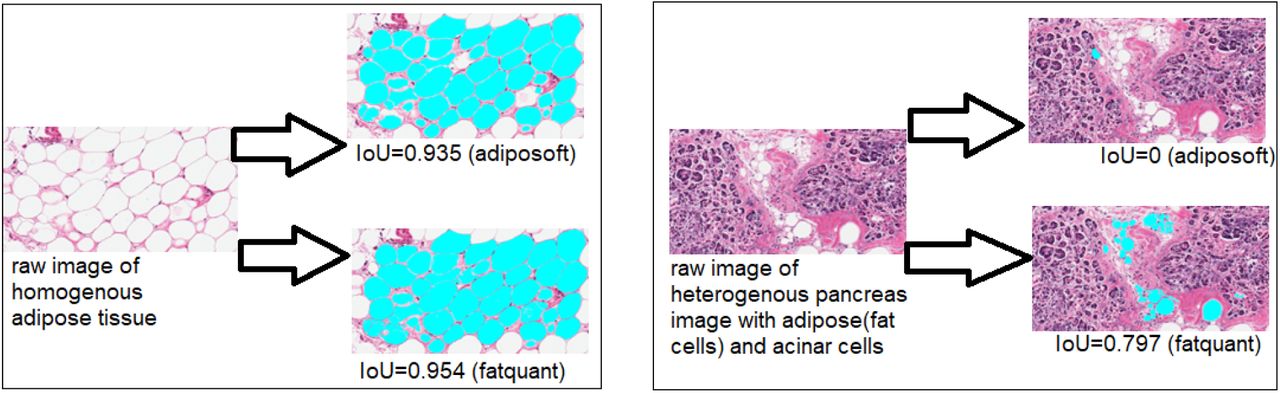
Graphical AbstractCurrently available fat quantification tools like adiposoft can analyze homogenous adipose tissue (left) with intersection over union (IoU) of 0.935 and 0.954 with adiposoft and fatquant, respectively. While in heterogenous tissue (e.g. pancreas on right) which contains adipose (fat cells), acinar cells, adiposoft fails to detect fat cells with IoU=0 while fatquant had IoU=0.797.
Competing Interest Statement
The authors have declared no competing interest.
Footnotes
we have provided additional data to support our conclusion and shared github link to access the code the data.


