

Abstract
Osteoarthritis (OA) is the most prevalent chronic joint disease which increases in frequency with age eventually impacting most people over the age of 65. OA is the leading cause of disability and impaired mobility, yet the pathogenesis of OA remains unclear. Treatments have focused mainly on pain relief and reducing joint swelling. Currently there are no effective treatments to slow the progression of the disease and to prevent irreversible loss of cartilage. Here we demonstrate that stable expression of RORβ in cultured cells results in alteration of a gene program that is supportive of chondrogenesis and is protective against development of OA. Specifically, we determined that RORβ regulates the balance of FGFRs signaling on FGFR1/FGFR3 that ERK1/2-MAPK signaling was suppressed by FGFR1(cartilage destruction) and AKT signaling was enhanced by FGFR3 (cartilage protection). These results suggest a critical role for RORβ in chondrogenesis and suggest that identification of mechanisms that control the expression of RORβ in chondrocytes could lead to the development of disease modifying therapies for the treatment of OA.
Introduction
Osteoarthritis (OA) is the most prevalent chronic degenerative joint disease where the risk of disease increases in with age and obesity 1-3. OA occurs more commonly later in life, after years of mechanical wear and tear on cartilage, a tissue that lines and cushions joints. Most therapies target pain relief and currently there are no effective treatments to slow the progression of the disease. Disease progression eventually results in irreversible loss of cartilage and when articular cartilage is significantly degraded or completely lost, joint replacement surgery is the only option 4. Although the correlation between aging and the development of OA is not completely understood, it is becoming clear that age-related changes in the musculoskeletal system, combined with mechanical injury and genetic factors all contribute to the pathogenesis of OA 5, 6. Thus, uncovering the molecular mechanism of joint degeneration requires analysis of cartilage metabolism, chondrocyte senescence, and inflammation 7. Such studies should lead to the identification of therapeutic targets for treating and preventing OA.
It has been shown that controlled fibroblast growth factor (FGF) signaling is essential for the balance of articular cartilage metabolism, as evidenced by the fact that aberrant FGF signaling contributes to progression of OA 8. FGFR1 signaling triggers upregulation of pro-inflammatory mediators, matrix metalloproteinases (MMPs), and alters anabolic activities of articular cartilage by inhibition of extracellular matrix (ECM) production and autophagy. Whereas, FGFR3 signaling is cartilage protective mainly through the inhibition of pro-inflammatory mediators and hypertrophic differentiation, as well as reduced MMP expression. For this reason, FGFR1 antagonists and FGFR3 agonists are being pursued as potential therapeutic strategies for OA 9-11.
The nuclear receptors (NRs) RORs have been shown to regulate both inflammation and metabolism. NRs are a druggable superfamily of ligand-dependent transcription factors. The NR1F subfamily contains the retinoic acid receptor-related orphan receptors (RORs), which have homology to both the retinoic acid receptors (RARs) and the retinoid X receptors (RXRs) 12. The ROR subfamily includes three major isoforms, RORα, RORβ, and RORγ. The physiological functions of RORα and RORγ have been well characterized and they have been shown to play roles in regulation of metabolism and inflammation while RORβ (NR1F2) has been significantly less well studied. RORβ is expressed in regions of the CNS that are involved in processing of sensory information and components of the mammalian clock, the suprachiasmatic nuclei, the retina, and the pineal gland. RORβ has been shown to play a critical role for the proliferation and differentiation of retinal cells in addition to the maintenance of circadian rhythms 13-15. The clock gene BMAL1 (brain and muscle Arnt-like protein 1), a target gene of the RORs, was reported that contributed to the maintenance of cartilage homeostasis 16, 17.
Here we demonstrate that stable expression of the nuclear receptor RORβ in cultured cells results in alteration of a gene program that is supportive of chondrogenesis and protective against development of OA. Specifically, RORβ balances the expression of genes implicated in cartilage homeostasis such as altering FGFR signaling towards that favorable for cartilage stability. While it remains unclear how RORβ regulates the balance of FGFR1/3 expression, the results presented here suggest RORβ is an important transcription factor involved in the control of a gene program that could prevent articular cartilage damage. Understanding the mechanism of RORβ control of this gene program could lead to the identification of novel therapeutics or drug targets for the prevention and or treatment of OA.
Materials and Methods
hRORβ/pLPCX construction and stable expressing transfection
Full length wild type human RORβ (NM_006914.3) was subcloned into the pLPCX retroviral vector (Clontech) using XhoI and NotI restriction enzymes (NEB) with forward primer 5’-CGCGCTCGAGATGCGAGCACAAATTGAAGTGATAC-’3 and reverse primer 5’-GCGGCGGCCGCTCATTTGCAGCCGGTGGCAC-’3. PCR product was obtained with Maxime PCR Premix (i-pfu) (Intron Biotechnologies). pLPCX vector was digested and then dephosphorylated before ligation using the Rapid DNA Dephos & Ligation Kit (Roche). Ampicillin resistance clones were verified with 5’ sequencing primer 5’-AGCTGGTTTAGTGAACCGTCAGATC-3’ and 3’ sequencing primer 5’-ACCTACAGGTGGGGTCTTTCATTCCC-3’. Selected clone was linearized with AseI (NEB) restriction digestion and dephosphorylated with Antarctic Phosphatase (NEB) prior usage in transfection.
MG-63 (ATCC CRL-1427™) human osteo sarcoma cells were maintained in EMEM (Eagle’s Minimum Essential Medium, ATCC) with 10% hear-inactivated fetal bovine serum (Invitrogen). Linearized hRORβ/pLPCX plasmid was transfected using lipid mediated transfection method (MG-63 transfection kit, Altogen Biosystems). 0.5 ug/ml of Puromycin was added into complete media for selection of stable expressing hRORβ/pLPCX in MG-63 cells. Highly expressing RORβ clone and mock-vector clone were used for following experiments.
mRNA sequencing analysis
Total RNA was extracted using Qiagen Kit-74106. Total RNA was quantified using the Qubit 2.0 Fluorometer (Invitrogen, Carlsbad, CA) and run on the Agilent 2100 Bioanalyzer (Agilent Technologies, Santa Clara, CA) for quality assessment. RNA samples of good quality with RNA integrity number (RIN) > 8.0 were further processed. A RNase-free working environment was maintained, and RNase-free tips, Eppendorf tubes, and plates were utilized for the subsequent steps. Messenger RNA was selectively isolated from total RNA (300 ng input) using poly-T oligos attached to magnetic beads and converted to sequence-ready libraries using the TruSeq stranded mRNA sample prep protocol (cat. #: RS-122-2101, Illumina, San Diego, CA). The final libraries were validated on the bioanalyzer DNA chips, and qPCR was quantified using primers that recognize the Illumina adaptors. The libraries were then pooled at equimolar ratios, quantified using qPCR, and loaded onto the NextSeq 500 flow cell at 1.8 pM final concentration. They were sequenced using the high-output, paired-end, 75-bp chemistry. Demultiplexed and quality filtered raw reads (fastq) generated from the NextSeq 500 were trimmed (adaptor sequences) using Flexbar 2.4 and aligned to the mouse reference database (mm9) using TopHat version 2.0.9 (Trapnell et al.). HTseq-count version 0.6.1 was used to generate gene counts and differential gene expression analysis was performed using Deseq2 (Anders and Huber). We then identified genes that were significantly downregulated or upregulated (adjusted P < 0.05) in each comparison.
Protein expression analysis
Protein was purified using RIPA lysis and extraction buffer (ThermoFisher Scientific) from MG-63 clones. Halt™ Protease and phosphatase inhibitor cocktail (100X, ThermoFisher Scientific) were added into lysis buffer. To normalize the amount of cell lysate run on SDS-PAGE, BCA (Thermo Fisher Scientific) assay was used. Anti-ROR beta antibody (rabbit monoclonal EPR15552, abcam), anti-phospho ERK1/2 (D13.14.4E, CST), anti-ERK1/2 (L34F12, CST), anti-phospho AKT (D9E, CST), anti-AKT (40D4, CST), and anti-beta Actin (8H10D10, CST) antibodies were used for primary antibody. IRDye 680RD and IRDye800CW were used for secondary antibodies. LI-COR Odyssey was used for fluorescence imaging system. Anti-aggrecan Ab (6-B-4, abcam) was used for primary antibody and FITC-anti mouse Ab was used for secondary antibody. Cell sorting was performed using LSRII (BD Bioscience).
Quantitative RT-PCR
Total RNA was extracted from RORβ OE/MG63 cells using RNeasy Plus Micro Kit (Qiagen), and the RNA was reverse transcribed using the ABI reverse transcription kit (Applied Biosystems/Thermo Fisher Scientific, Waltham MA). Quantitative PCR was performed with a 7900HT Fast Real Time PCR System (Applied Biosystems) using SYBR green (Roche). A list of primers used for these studies is shown in Table 1.
Results
Stable expression of human RORβ in MG63 cells
MG63 cells are derived from an established sarcoma cell line and is an osteoblastic model to study bone cell viability, adhesion, and proliferation. To investigate the role of RORβ in bone we transfected MG63 cells with pLPCX vector harboring the puromycin-resistance gene and human RORβ cDNA. The transfected cells were selected with puromycin and the expression of RORβ was analyzed by western blotting using human RORβ specific rabbit EPR1552 mAb and detected using the Li-Cor system (Figure 1a). Anti-beta actin (8H10D10) was used as a protein loading control. Relative mRNA expression of hRORβ was determined for clones that survived selection (Figure 1b). One clone demonstrating high expression of RORβ was selected for all subsequent studies. The expression level of RORβ in this clone was confirmed monthly. A control cell line was generated by transfection of MG63 cells with the pLPCX vector devoid of RORβ and selection with puromycin to generate the wild-type (WT) clone. The WT clone line was used as a control for all studies presented.

Selection of MG63 clone which is consistently over expressing hRORβ in MG63 cells. hRORβ was subcloned into pLPCX vector which was transfected into MG63 cells and selected by 1ug/mL of puromycin. hRORβ was consistently overexpressing in MG63 that was confirmed by protein expression level and b) relative mRNA expression level. WT: mock vector (pLPCX) transfected MG63 cells, OE: Consistently over expressing of hRORβ in MG63 cells.
Differential gene expression (DEG) analysis
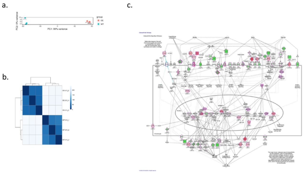
To investigate the biological function of RORβ in MG63 cells, RORβ OE-MG63 cells (cells overexpressing RORβ) and mock vector transfected MG63 cells (WT clone) were analyzed by mRNAseq. Three independent biological replicates were processed and analyzed. To investigate molecular pathways altered by RORβ expression, we analyzed log2 fold change of mRNA sequencing data using QIAGEN ingenuity pathway analysis (IPA) software with a P-value cutoff of 0.05. As shown in Figure 2c, cells overexpressing RORβ showed alteration in genes involved in signaling pathways associated with OA when compared with WT cells. As further detailed below, the expression profiling results suggest that RORβ may play a protective role in chondrocytes to prevent development of OA.

Differential gene expression sequence. a) principle component analysis (PCA) of wild type (213_1,213-2,213-3) and over expressing RORβ clone (213_4, 213-5, 213-6). Each condition is labeled with a separate color allowing for a visual method for identifying sample outliers. b) The scale is the Euclidian distance between two samples in multi-dimensional space which is also represented in different shades of blue. Each sample is most closely related to itself (with a Euclidian distance of 0) which explains the perfect relatedness (dark blue) along the diagonal. c) Suppression of osteoarthritis signaling pathway. mRNAseq data was analyzed using IPA program. The green color of genes represent significantly repressed fold changing genes on OE compared with WT and the red color of genes represent significantly increased fold changing genes on OE compared with WT in OA pathway signaling.
RORβ expression suppresses osteoarthritis inducing genes
Chondrocytes are critical to maintenance of articular cartilage where loss of chondrocytes leads to cartilage damage and this damage is often irreversible. Aging and chronic inflammation are associated with increase in several critical genes associated with the induction of OA. Matrix metalloproteinases (MMPs) including ADAM metallopeptidase with thrombospondin type 1 motif 4 (ADAMTS4) and MMP3 and the proinflammatory cytokine IL6 are well characterized pathogenic genes for OA. Interestingly, increased expression of RORβ suppressed the expression of these genes (Figure 3a) and we demonstrate that the cartilage damaging factor MMP3 was also reduced at the protein level (Figure 3b). These results suggest that RORβ may play an inhibitory role to prevent cartilage damage by suppression of chondrocyte death by blocking chondrocyte catabolic pathways.

mRNA expression level for cartilage structure genes and cartilage destruction related genes. a) Relative mRNA expression for ADAMTS4, MMP3, and IL6 which are direct/indirect cartilage destructive genes and b) the protein expression of MMP3 in OE/MG63 clone. c) Relative mRNA expression for aggrecan (ACAN) and COL2A1 which is main structure of cartilage maintain and d) the protein expression of ACAN on OE/MG63 clone. TC28α2 cells were used for positive control and MG63 cells were used for negative control. Graph with gray color is isotype control.
RORβ induces expression of articular cartilage structural genes
Articular cartilage is composed of collagens, proteoglycans, and non-collagenous proteins. Extracellular matrix proteoglycans and collagens play a critical role in the maintenance of articular cartilage structure by regulating chondrocyte proliferation and promoting cartilage repair (ref). Induction of OA is initiated by mechanical forces and inflammatory events that destabilize the normal coupling of synthesis and degradation of extracellular matrix proteins in both articular cartilage and subchondral bone. In the study presented here, we observed that the expression of the core structural genes in articular cartilage, aggrecan (ACAN) and collagen type II alpha (COL2A1), were increased by overexpressing RORβ in MG63 cells (Figure 3c). Additionally, we demonstrate that ACAN protein level was also increased when compared to the WT clone (Figure 3d). The levels of ACAN protein in RORβ OE MG63 cells was similar to that observed in TC28a2 cells, which are normal chondrocyte cells (ref). Thus, RORβ inhibits the production of MMPs and increases the expression of extracellular matrix proteins, both of which are protective to articular cartilage.
RORβ expression balances FGFR signaling
Fibroblast growth factor (FGF) signaling has a role in growth and homeostasis of joint related cells, including articular chondrocytes, synovial cells, and osteogenic cells and aberrant FGF signaling contributes to the progression of OA 18. Loss of FGFR1 in chondrocytes accelerates matrix degradation by inducing the RAF-MEK-ERK and PKCδ-p38 pathways 19 and conditional deletion of FGFR3 in mice aggravated DMM-induced cartilage degeneration 20. In addition, FGF18 attenuates cartilage degradation through FGFR3/PI3K-AKT signaling 21. As shown in Figure 4a, stable expression of RORβ reduced the ratio of FGFR1/FGFR3 towards a protective profile. This alteration of FGFR signaling impacted phosphorylation of ERK1 (decreased) and AKT (increased) (Figure 4b).

Reduction of FGFR1 expression and Erk signaling and enhanced FGFR3 expression and Akt signaling in RORβ OE cells. a) Relative mRNA expression of FGFR1 and FGFR3 along with RORβ. b) The expression of phosphorylated Erk1/2 and phosphorylated Akt in RORβ over expressing MG63 cells.
RORβ protects against IL-1β mediated inflammation and inhibits basal protease production
IL-1β is an essential mediator of acute joint inflammation induced by physical injuries and it plays a critical role in cartilage degradation. Induction of IL-1β increases the expression of catabolic matrix enzymes such as MMPs and ADAMTS, as well as the pro-inflammatory cytokine IL-6 which ultimately leads to cartilage matrix degradation 22-24. Here we demonstrate that stable expression of RORβ suppressed IL-1β induced expression of IL6, MMP3 and ADAMTS4. Additionally, expression of RORβ downregulates expression of MMP3 and ADAMTS4 as compared to WT control cells (Figure 5).

Suppression of IL1β stimulated genes in hRORβ/MG63 clone. Inflammatory cytokine IL1β was treated into WT or OE RORβ/MG63 cells for 24 hr with 2nM. The mRNA expression level of IL6, MMP3 and ADAMTS4 was significantly increased by IL1 stimulation but it was protected in RORβ OE cells. The RORβ expression did not affect by IL1β stimulation.
Discussion
Development of OA impairs the biomechanical properties of articular cartilage eventually leading to irreversible loss of cartilage resulting in debilitating joint pain and swelling. Owing to the limited regenerative capacity of articular cartilage, advanced surgical techniques have been usually required for repair of damaged cartilage 25, 26. Risk factors for the development of OA include age, obesity, physical injury, and low-grade systemic inflammation where pro-inflammatory mediators such as IL-6 and TNFa can exacerbate cartilage erosion 27, 28. While the pathogenesis of OA remains unclear, hypertrophic chondrocyte differentiation, reduced proliferation, dysregulated apoptosis, combined with the loss of collagen, proteoglycans and cartilage integrity are associated with the development of OA 3.
An important observation in patients with OA is the upregulation of FGFR1 expression which appears to accelerate matrix degradation by inducing the expression of RUNX2 /ELK1 and consequent downregulation of FGFR3 in articular cartilage. Furthermore, pharmacological inhibition of FGFR1 attenuates progression of disease in a mouse model of OA 9 and FGFR3 deficiency in myeloid cells enhances CXCRL12 dependent chemotaxis via CXCR7 (CXC-chemokine receptor 7), thereby leading to the exacerbation of joint destruction 29. In this study we demonstrate that the nuclear receptor RORβ balances FGFR1/R3 signaling towards that favorable for cartilage stability by suppressing FGFR1 expression and amplifying that of FGFR3. While it remains unclear how RORβ regulates the balance of FGFR1/3 expression, the results presented here suggest RORβ is an important transcription factor controlling a gene program that is protective against articular cartilage damage. Future studies focused on understanding the mechanism of RORβ ’s control of FGFR1/3 modulation in chondrocytes and murine models of OA could lead to the identification of novel therapeutics and drug targets for the prevention and or treatment of OA.
References


