

Abstract
Fungal plant pathogens live in the specific environment of plants. Understanding of the interaction between pathogens and their host plants might open new ways to control plant diseases. Yet, the specificities of the plant environment and its effects on fungal growth are not yet fully explored. Both pH and Eh play a key role during the interaction between the fungus and its host plants, but often studied independently or at different scales. To decipher the interrelation between plant growth and their soil environment, recent theoretical and methodological advances have been made through the joint characterization of the pH and Eh. This opens the prospect to develop similar methods for fungi. The aim of our study was to investigate whether the methods developed for soil could be transposed to fungi. We first worked on artificial media, assessing the impact of fungal growth on the media in cultures. The growth of all 16 species tested significantly altered either Eh, pH or both in agar media. Measuring Eh reveals that even the species not modifying pH can have an impact on the surrounding environment. Reciprocally, altered media were used to characterize sensitivity of fungal growth to both pH and Eh parameters. The response of the six fungi tested to the modified media was quantitative with a decrease in colony diameter. In addition, colony aspect was repeatedly and thoroughly modified. As a first step towards the same studies in conditions matching the natural environment of fungal pathogens, we tested how the measurement can be performed with fungi growing on oilseed rape plants. In infected plant stems, pH and Eh were significantly altered, in opposite directions for L. maculans and S. sclerotiorum. The observed alcalinisation or acidification correlates with canker length. Our series of experiments indicate that the procedure published for Eh and pH in soil can be extended for measurement in agar media and in infected plants. Further, the joint characterization of both parameters opens the way to a more precise understanding of the impact of fungi on their environment, and conversely, of the environment on fungal growth. The availability of methods for measurement opens the prospect to study combinations of stresses, either on agar media or in plants, and get an understanding of the involvement of pH and Eh modifications in these interactions.
Introduction
Fungal plant pathogens need to get resources from the plant and for doing so they need to live in the specific environment of plants, with an host species ranging from broad for generalists to narrow for specialist (Braunsdorf et al., 2016; Zeilinger et al., 2016; van der Does & Rep, 2017). Getting a better understanding of the interaction between pathogens and their host plants might open new ways to control plant diseases. Within species, the interaction between fungal pathogens and plant resistance has been approached through molecular dialog between partners (Lo Presti et al., 2015; Jwa & Hwang, 2017). Yet, the specificities of the plant environment and its effects on fungal growth are not yet fully explored. To decipher the interrelation between plant growth and their soil environment, recent theoretical and methodological advances have been made through the joint characterization of the pH and Eh (Husson et al., 2013; 2018). This opens the prospect to develop similar methods for fungi, because both pH and Eh are important in their metabolism.
As compared to other microorganisms, fungi are capable of growth and development over wide pH ranges. Several species, including plant pathogens, can actively modulate the pH of their environment by secreting acids or alkali (Landraud et al., 2013; Vylkova et al., 2017). The ability to control extracellular pH is an important aspect of fungal physiology that contributes to fitness within the host (Vylkova et al., 2017) via the modulation of virulence factors to best fit the host (Prusky & Yakoby, 2003). pH is revealed as one component at work during interaction between the fungus and its host plants, from field to molecular scales. At the field scale, opposite effect can be observed for wheat soilborne diseases: take-all (Gaeumannomyces graminis var. tritici) is more severe in alkaline soils (Smiley, 1974; Lebreton et al., 2014) and cephalosporium stripe (Cephalosporium gramineum) severity increase with soil acidity (Stiles & Murray, 1996). Soil pH is considered as a major structuring ambient factor of telluric microbial communities and plant microbiome (Rousk et al., 2010; Chapelle et al., 2016). In culture, pH is taken into account for the optimization of biocoltrol agents production as Trichoderma species (Singh et al., 2014). In plant tissues, pH indicators or image analysis have been proposed to monitor changes and fungal growth (Tardi-Ovadia et al., 2017). At the molecular level, pH alters gene expression (Daval et al., 2013). All these elements indicate that for fungi, pH is an important characteristic of the environment. It has however been stressed that because of their interrelation, analyses of pH gain to be associated with measures of Eh, that is oxido-reduction status (Husson et al. 2016; 2018).
So far, in plant-pathogenic fungi interactions, oxido-reduction processes are documented mostly at cellular and molecular levels (Lehman et al., 2015; Jwa & Hwang, 2017). Plants have been investigated more thoroughly. Oxido-reduction reactions and reactive oxygen species have a profound influence on almost every aspect of plant biology especially to balance information from metabolism and the environment (Dietz et al., 2016; Foyer & Noctor, 2016). The concept that reactive oxygen species (ROS) and reactive nitrogen species (RNS) are key signaling molecules that facilitate a plethora of adaptive metabolic, molecular genetic and epigenetic responses is now established (Foyer & Noctor, 2005; 2016). The balance between oxidant and antioxidant pool sizes, plays signaling roles in the regulation of gene expression and protein function in a wide variety of plant physiological processes including stress acclimation (Shigeoka & Maruta, 2014; Choudhury et al., 2017), regulation of plant development processes, organogenesis, senescence and defense against pathogens (Nosek et al., 2015; Marschall & Tudzynski, 2016; Noctor et al., 2017). As oxido-reduction processes play a major role in plant biology, developing methods to investigate if this occurs also in fungi is desirable.
Eh and pH are interacting, as oxidation leads to acidification, and the importance of measuring both parameters has been documented on soils (Husson et al., 2018). Eh and pH regulation is central in plant physiology and phenology and as the vast majority of biotic and abiotic stresses are translated into redox signals in plants (Husson, 2013). The leaf Eh and pH, were proposed in previous studies to explain the variable resistance of cereals to obligates parasites (Benada, 2017). Specifically, these studies investigated the disease gradients on plants; the change of susceptibility of organs during the ontogeny and growth; the difference in resistance in individual plant cells and the rapid change of resistance in a couple of hour (Benada, 2017). Investigating these topics further could benefit from the recent methodological developments. The aim of our study was to investigate whether the methods developed for soil (Husson et al., 2016; 2018) could be transposed to fungi. We first worked on artificial media, assessing the impact of fungal growth on the media in fungal cultures. Reciprocally, altered media were used to characterize sensitivity of fungal growth to both pH and Eh parameters. Later, we tested how the measurement can be performed with fungi growing on oilseed rape plants, as a first step towards the same studies in conditions matching the natural environment of fungal pathogens.
Materials and Methods
Culture media
To test how fungal growth modified the medium on which they grew, four independent experiments were performed with 6 fungal strains (Table 1). In the Mar2016 experiment, 13 additional strains were included (Table 1). All tests were performed on malt-agar media (Malt extract 20 g.l-1, Agar 20 g.l-1, Streptomycin 0.1 g.l-1). All culture media were autoclaved for 20mn at 120°C and cooled down before Streptomycin concentrated solution 10% w/v in water was added. Within each experiment, replicates were prepared independently and corresponding uninoculated aliquots were kept for pH and Eh initial and final measurements (Table 1). Fungal cultures were grown in sterile 6 cm diameter plastic containers (50 ml) commonly used to store and reheat food sauce.
List and origin of 19 fungal strains used in the 4 experiments (Jan2015 to Mar2016) with dates of inoculation, final measurements for media and fungal growth. “nd” stands for unknown.
To test how modified pH and Eh media altered fungal growth, within each of the 3 experiments (Jan2015, Feb2015, Jul2015) three replicates of a range of 13 malt-agar media (Malt extract 20 g.l-1, Agar 20 g.l-1, Streptomycin 0.1 g.l-1) were prepared independently. To alter Eh and pH without changing the composition of the media, we used electrolyzed water products, because when chemical compounds are added, it is difficult to disentangle the effects via pH from directs effects (Bekker et al., 2009). The range of agar media included native one (N0) and four sets of three media with part of the water (in proportions 1:3; 1:1 and 3:1) replaced by either of catholyte (pH 11;7, 11.5, 12.0 in the three experiments), basic anolyte (pH 8.1, 8.7, 9.0), neutral anolyte (pH 7.1, 7.1, 7.3) and acid anolyte (pH 3.3, 2.0, 1.5). Aliquots were prepared in 20 ml flacons for uninoculated measurements at the start and end of the experiment. Aliquots of 10 ml in 5 cm diameter Petri dishes were prepared for inoculation and fungal growth measurement. For each experiment, anolyte and catholyte were obtained from IDEEAQUACULTURE, Parc Euromédecine 2, 39 Rue Jean Giroux, 34080 Montpellier, France. Products were freshly prepared 24 to 48h before media preparation, using Envirolyte ELA900 and shipped in light-opaque containers. Eh and pH of the products were measured on aliquots before use. These products are all obtained from water, and the only compound added during the production is NaCl. We thus made a specific experiment (Sep2015, see below) to check that within the range of final NaCl concentrations in the media, fungal growth was not altered for any of the 6 fungal species (Fig. S1).
To test how modified pH and Eh media altered fungal growth of M. oryzae, within two experiments (Jan2016, Mar2016) four replicates of a range of 13 rice flour-agar media (Rice flour 10 g.l-1, Agar 15 g.l-1, Yeast extract 2 g.l-1, Penicillin 0.1 g.l-1) were prepared. The range of rice flour agar media included native one (N0), addition of Hydrogen peroxide, addition of Hydroquinone and the combination of both. This made 20 media in Jan2016 with combinations (Hydroquinone - Hydrogen peroxide) 0-0; 0- 0.05; 0-0.1; 0-0.2; 0-0.4; 0-0.8; 0-1.6; 4-0; 4-0.1; 16-0; 16-0.05; 16-0.1; 16-0.2; 16-0.4; 64-0; 64-0.1; 256-0; 256-0.05; 256-0.1; 256-0.4 and 14 media in Mar2016 with combinations (Hydroquinone – Hydrogen peroxide) 0-0; 0-1; 0-2; 0-4; 50-0; 50-2; 100-0; 100-1; 100-2; 100-4; 20-0; 200-2; 300-0; 300-4.
Fungal growth experiments
A total of 19 isolates of 16 fungal species were used, either from the INRA Le Rheu collection or kindly provided by colleagues (Table 1). For each isolate, 5 mm diameter agar plugs of fresh cultures on maltagar were prepared. Agar plug were placed in the center of Petri dishes and fungal growth was measured as two perpendicular diameters of the colony (Table 1). To assess the impact of fungal growth on the medium, plastic containers were used instead of Petri dishes in order to allow sufficient depth for the electrodes after colony growth. Agar plug was placed in the center, and final measurements were performed by sticking electrodes through the colony, on both sides of the plug. In each of the 3 replicates, there was 1 Petri dish and 1 container in Jan2015 and Feb2015; 2 Petri dishes in Sep2015; 3 Petri dishes and 3 containers in Jul2015 and 3 containers in Mar2016 experiments, respectively. All cultures and uninoculated aliquots were kept at room temperature in INRA Le Rheu, with no supplementary light. Hourly temperature (mean 21.7°C se 1.6) and humidity (mean 46.5% se 8.9) were recorded with a TEMPCO Hygro bouton 23 data logger.
M.oryzae growth experiments were performed at Africarice, Cotonou, Bénin with local strains BN1162 in Jan16 and BN1168 in Mar16. For each isolate, 5 mm diameter agar plugs of fresh cultures on rice flour-agar were prepared. Agar plug were placed in the center of Petri dishes and fungal growth was measured as two perpendicular diameters of the colony.
Oxido-reduction Potential (ORP) electrodes and voltmeter
Redox potential was measured in agar media as Described by Husson et al. (2016) using Ag/AgCl Reference electrode Radiometer analytical E21M002 and Radiometer Analytical Pt plate electrode 5×5 mm M241 Pt, with a Voltcraft VC850 multimeter (10×106 Ohm input resistance) for all fungi and a WTW 3110 (10×1012 Ohm input resistance) for M. oryzae growth experiments. The Eh value considered was the Eh (in mV) indicated on the meter after 1 minute without change in the Eh unit. After being measured according to Ag/AgCl reference electrode, all redox potentials are transformed to give Eh according to the Normal Hydrogen Electrode (ENH). Redox electrodes were calibrated at the start of the measurements and every 10-12 measurements, with Mettler Toledo Redox buffer solution 220 mV (pH 7) composed of Potassium hexacyanoferrate (III), Potassium hexacyanoferrate (II), Potassium dihydrogen phosphate and Disodium hydrogen phosphate. All measurements were conducted outdoor, in an environment identified as being free of electromagnetic interference.
pH was measured in agar media with a with a Voltcraft VC850 multimeter (10×106 Ohm input resistance) using Ag/AgCl reference electrode Radiometer analytical E21M002 and a glass pH electrode Radiometer analytical pHG301-9. pH electrodes were calibrated at the start of the measurements and every 10-12 measurements, with VWR buffer solutions at pH4, pH7 and pH10. Data were converted into pH values using linear regression of the measured values of standard solutions.
Plants experiments
Winter oilseed rape was sown in 03/10/2017 in 12 x 12 x 21 cm pots with a 1:1:1 mix of sand, peat and compost and grown outside at INRA Le Rheu (48·1°N, 1·5°W), in Brittany, France. The experiment matches the usual cultivation in this area, where winter oilseed rape is generally sown in late August – September and harvested the following July. There was three replicates of 38 plants for L. maculans experiments and 2 replicates of 38 and 44 plants for S. sclerotiorum experiments.
Natural infections with L. maculans airborne ascospores occurred on some of the plants, with typical leaf spots observed in November. Cankers became visible on some stems at flowering. Measurements were performed on infected and uninfected plants after flowering, over 4 days (02/05; 07/05; 22/05, 28/05). Plant stem was cut lengthwise, and internal canker length was recorded. Immediately after, a 2 cm portion of the stem taken at crown level was cut and quickly grinded with a mortar and pestle. The plant material was then placed in the barrel of a 2 ml syringe from which the nozzle had been cut. For the redox potential measurement with the Voltcraft VC850 multimeter, we used a 9 cm Petri dish with a filter paper moistened with 0.1M KCl solution (Fig. S2). The Ag/AgCl reference electrode was placed standing, with tip touching the moist filter paper. The Pt plate electrode was placed in the plant material in the syringe, then the syringe barrel was placed standing with tip touching the moist filter paper. After reading the redox potential, the plunger of the syringe was placed in the barrel to squeeze the liquid onto a Horiba LAQUAtwin-pH-22 meter for pH measurement.
Inoculations with S. sclerotiorum were performed at the time of petal fall. Matchsticks pieces were autoclaved in liquid 2% malt medium. Agar plugs from fresh culture were transferred (13/04 and 30/04) to the center of 9 cm diameter Petri dishes (Malt extract 20 g.l-1, Agar 20 g.l-1, Streptomycin 0.1 g.l-1) with lying matchsticks pieces, and cultured at 20°C. On 17/04 and 03/05 for each of the 2 plant replicates, a matchstick piece covered with mycelium was tightly fitted through the 3 mm diameter hole drilled at 10 cm height through the stem of 16 and 24 inoculated and 19 and 23 mock-inoculated plants. External canker length was measured 20/04 and 07/05, immediately after, a 2 cm portion of the stem taken at the inoculation point was cut and processed for Eh and pH measurement as described above.
Data analysis
Statistical analyses on artificial media experiment data were performed using XLSTAT (Addinsoft, 2016). Analysis of variance (ANOVA) was conducted on Eh, pH for comparison of agar media after fungal growth. ANOVAs were followed by a comparison of means using the Ryan-Einot-Gabriel-Welsh test Q (REGW-Q) as it provides the best compromise between the need to have a powerful test and the need to limit the familywise error rate at α, according to Howell (2009). Quadratic regressions between colony growth, pH and Eh were performed for the different 7 fungal species on altered agar media.
Statistical analyses on plant experiment data were performed using R software (R Core Team, 2013). Generalized linear modelling was used to investigate the effects of inoculation, canker on pH measured. For plant pH and Eh data, we used a Gaussian linear model on log-transformed data. Linear contrasts were calculated with as Estimated Marginal Means with emmeans package to determine specific differences among treatment combinations.
Results
Effect of fungi on pH and Eh in the agar media
Uninoculated malt-agar media were acid (pH from 4.5 to 5) and oxidized (Eh from 500 to 600) (Fig. 1). For all tested species, ANOVA analyses followed by REGW-Q comparison of means indicated that fungal growth significantly altered pH and Eh in agar media (Fig. 1; Table 2). These alterations were species-specific and in directions opposite to the slight evolution of uninoculated media between initial (empty symbols) and final (filled symbols) measurements. All Sclerotinia species acidified the media, S. minor and S. sclerotiorum did not change Eh, while S. trifolorium also reduced the media (Fig. 1A). M. oryzae, Penicillium sp. and P. brassicae alcalinised the media, with variable intensities of reduction. The main effect of other species was reduction, either mild (Bc1, Mb, Pc) intermediate (Lm, Vd) or intense (Rs, Ab, Lb, Fr). Results of independent experiments with 6 species were congruent (Fig. 1B). The Eh-pH couples obtained after the growth of the fungal species tested did not align on the theoretical correlation between Eh and pH, with a negative slope of 59mV decrease in Eh for one unit increase in pH at 25°C, according to the equation of Nernst (Fig. 1AB).
For each figure and each experiment, mean and standard error of the variable are indicated, with F and p-values of the ANOVA. Significantly different groups in the REGW-Q comparisons are added as dashed lines on the figures.

Effect of fungi on pH and Eh in plant stems
Uninfected oilseed rape stems were slightly acidic (pH from 5.5 to 6.5) like malt-agar media, but more reduced (Eh from 150 to 300) as compared to malt-agar media (Eh from 500 to 600).
Analysis of deviance showed that both inoculation status and canker length were significant predictors of changes in pH (Table 3), though in opposite directions depending on the fungal species. pH was higher following fungal infection with L. maculans (Fig. 2A; Table 3) whereas pH was lower with S. sclerotiorum (Fig. 3A; Table 3). For both fungi, canker length had a significant effect on measured pH (Fig. 2B; Fig. 3B, Table 3).


Analysis of deviance on logN transformed data (Type II tests). Pr(χ2) are p-values from a χ2 test used for significance
Analysis of deviance showed that inoculation status was a significant predictor of changes in Eh (Table 3), again in opposite directions depending on the fungal species. Eh was lower following fungal infection with L. maculans though differences between groups were not significant (Fig. 2C; Table 3) whereas Eh was higher with S. sclerotiorum (Fig. 3C; Table 3). The effect of canker length on Eh was significant for S. sclerotiorum (Fig. 3D; Table 3) but not for L. maculans (Fig. 2D; Table 3).
Effect of pH and Eh in the agar media on fungal growth
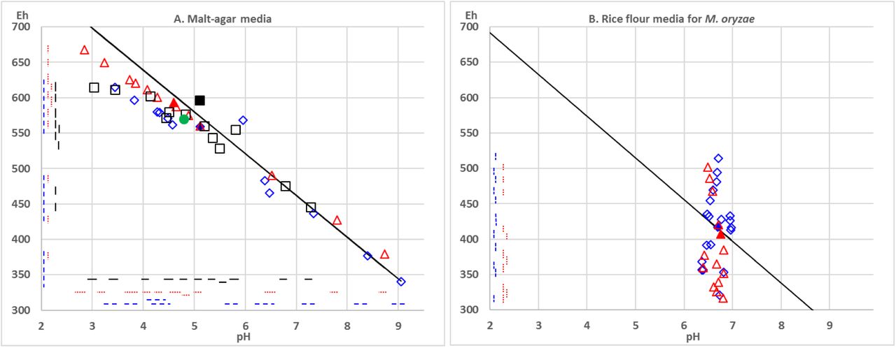
The addition of electrolyzed water products to malt-agar media extended the pH and Eh range on both sides of the native medium (Fig. 4A) and on both sides of the range from 5.5 to 6.5 observed in oilseed rape stems (Fig. 2, 3). The Eh-pH couples obtained did align on the theoretical correlation between Eh and pH, with a negative slope of −59 mV/pH unit. The native rice flour medium was pH 6.5, higher than the native malt medium at pH 4.5 to 5. It was more reduced, with Eh 400 mV compared to Eh 550-600 mV. The addition of Hydrogen peroxide, and Hydroquinone to rice flour media allowed variation in Eh (from 300 to 500 mV), but less variation in pH (6 to 7) (Fig. 4B).

For the six fungi tested, mean colony diameter varied on the range of 13 malt-agar media (Fig. 5; Table 2). Further, the colony appearance of all fungi, the pattern of sclerote production for S. minor and S. sclerotiorum, the production of yellow pigments for L. biglobosa varied in a manner congruent between replicated dishes in independent preparations (Supplementary information Fig. S3). In most cases, the fungal response to the modified media was quantitative with a decrease in colony diameter. However, in a few occasions the response was also qualitative, with the fungus either remaining on the transferred agar plug for some of the dishes, or starting to grow on some other dishes (Fig S3). The growth of A. brassicicola, L. biglobosa and V. dahliae was better at higher pH / lower Eh than at lower pH/higher Eh, with optimum pH around 7-8 and optimum Eh around 450 mV (Fig. 5). The growth of L. maculans was reduced on both sides of the range tested, with optimum pH around 6 and optimum Eh around 500 mV. The optimum were lower for S. minor with pH 5-6, and Eh below 400 mV than for S. sclerotiorum with pH around 6 and Eh around 450 mV.
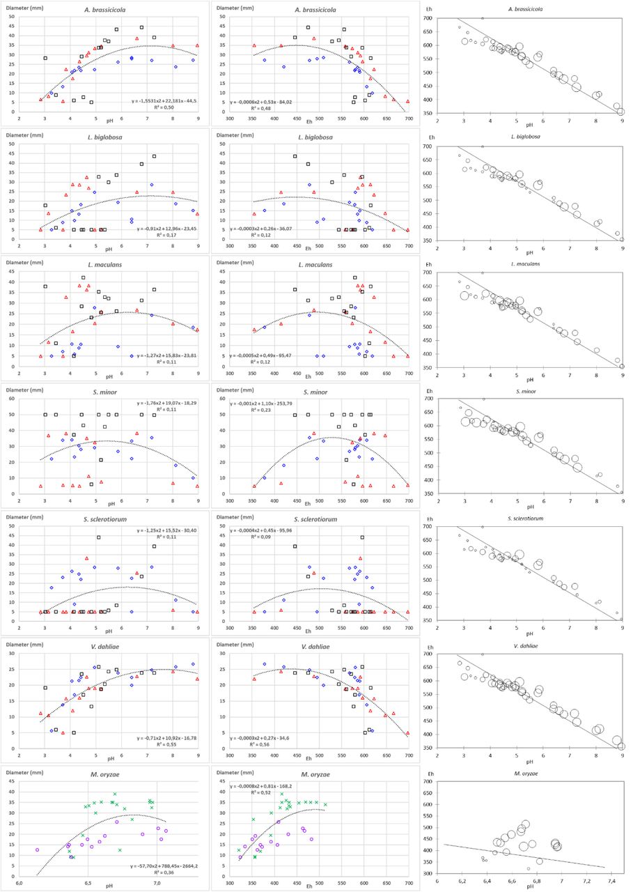
In the Sep15 experiment, the addition up to 500ppm of NaCl to the malt-agar media influenced Eh and pH (Fig. S1A). All media were more reduced (Eh from 480 to 495 mV) than the native one (Eh = 505mV). Compared to the native one (pH = 5.6), the pH was decreased (5.45 and 5.5) only for the addition of 100 and 500ppm of NaCl. The colony growth of the five fungi S. minor, L. maculans, L. biglobosa, A. brassicicola and V. dahlia was not altered by NaCl. The colony growth of S. sclerotiorum differed among media, though the only growth different from the native medium N0 was with 250 ppm of NaCl (Fig. S1B).
Discussion
The growth of all fungi tested significantly altered either Eh, pH or both in agar media (Fig. 1). This effect was consistently observed among replicates within each experiment, and among independent experiments. Changes in pH were congruent with previous literature for fungi notorious to acidify (e.g. Sclerotinia) or alkalinize (e.g. Magnaporthe, Botrytis) the media (Landraud et al., 2013; Liu et al., 2017; Vylkova et al., 2017). Changes in Eh were congruent with previous literature, with various aerobes strongly reducing the culture media, due to the consumption of oxygen (Rabotnova & Schwartz, 1962). These authors mention that as opposed to organisms able to control their internal conditions, microorganisms maintain their pH-redox homeostasis by modifying the environment where they live. In our independent experiments with six fungal species, congruent results indicate that the protocol developed for measurements in soil (Husson et al., 2016) can also be used for measurement in agar media.
Eh and pH are inversely correlated, and this process can be explained because oxidation of two molecules of water (losing 4 electrons) produces O2 and 4H+, thus leading to acidification. From the Nernst equation, it can be calculated that the theoretical slope of this correlation is a 59 mV decrease in Eh for one unit increase in pH at 25°C (Husson et al., 2018). In our experiment, we observed that without biological activity, all the agar media prepared align on this theoretical line (Fig. 4). In contrast, the final measurements in agar media after fungal growth do not match this theoretical correlation line (Fig. 1). Rabotnova and Schwartz (1962) postulate that the observed reduction of the media is due to the consumption of dissolved oxygen. Further, the available nutrients also influence the outcome, as observed with R. solani acidifying the media in presence glucose (Boswell et al., 2003). Thus, further studies on media with different compositions are needed to investigate whether the final values depend on the consumption / release of different nutrient and compounds by the fungi, or if final values are stable, thus indicating regulation.
Interestingly, the joint characterization of pH and Eh provides discrimination among species not discriminated by the pH alone. Measuring Eh reveals that even the species not modifying pH can have an impact on the surrounding environment. For plant crop species, plant pathogens on crops are controlled to avoid deleterious effects on plant yield. However, visible damage to plants does not always explain the whole of the deleterious effects. Realizing that the plant pathogens might interfere with the plant oxido-reduction status (De Gara et al., 2003; Williams et al., 2011; Kabbage et al., 2015) and thus that maintaining redox homeostasis might come at a cost for an infected plant, opens another direction of investigation to better understand pathogen damage to plants.
In infected plant stems, pH was significantly altered, in opposite directions for L. maculans (Fig. 2) and S. sclerotiorum (Fig. 3). The observed alcalinisation or acidification correlates with canker length. The direction of this change is congruent with changes observed on agar media (Fig. 1). As the measurements in plants result both from fungal growth and from the plant reaction to infection, follow-up studies are needed to disentangle the respective roles of fungal mycelium, of fungal metabolism and of plant reaction (Casdeval & Pirofski, 2003). Nevertheless, our study already confirms that direct measurement of Eh and pH is possible on infected plants. In plants, the balance between oxidant and antioxidant pool sizes, plays signaling roles in the regulation of gene expression and protein function in a wide variety of plant physiological processes including stress acclimation (Shigeoka & Maruta, 2014; Choudhury et al., 2017), regulation of plant development processes, organogenesis, senescence and defense against pathogens (Nosek et al., 2015; Marschall & Tudzynski, 2016; Noctor et al., 2017). Being able to perform direct measurements on infected plants opens the prospect of characterizing more precisely the joint effects of combinations of stresses, thus studying their interrelation.
It is known that disease severity depends on plant organ age (Kranz, 1990; Benada, 2017; Torres et al., 2017) and some pathogens are only able to infect specific of plant organs. We provide values for pH and Eh in stem, because we used stem pathogens. However, follow up studies are needed to map the pH and Eh values in the different organs of the plant at a given time, or the change in these values in a given organ over time. Also, being able to perform direct measurements on a small amount of plant material will allow detailed studies to test whether the change is only local or systemic in the plant, for example infecting one organ and measuring at another location of the same organ or in neighboring organs.
Comparing fungal growth on media with a range of pH has already been performed (Daval et al., 2013, Lebreton et al., 2014). However, when chemical compounds are added to the growth medium, it is difficult to disentangle the effects via pH from directs effects (Bekker et al., 2009). We tried to minimize this problem by using electrolyzed water products, as NaCl was the only compound added. At low concentrations, NaCl promoted growth of Botrytis cinerea (Boumaaza et al., 2015), but at high concentrations reduced growth of both B. cinerea (Boumaaza et al., 2015) and bread spoilers Penicillium roqueforti and Aspergillus niger (Samapundo et al., 2010). At concentrations up to 500 ppm, NaCl did not alter fungal growth for five species (Fig. S1). The growth of S. sclerotiorum was affected, though our highest concentration was not high enough to reduce growth. With this study, we show that from two way of altering Eh and pH, adding electrolyzed water products produce combinations that align on the theoretical slope (Fig. 4A) whereas adding Hydrogen peroxide and Hydroquinone produce combinations that do not align on the theoretical slope (Fig. 4B). Choosing one or the other allows either exploring the joint variation of Eh and pH, or focusing on the variation of Eh within a narrower pH range.
On modified agar media, we observed that most fungi could grow over a broad pH range (Fig. 5) which is congruent with previous literature. Working mostly on bacteria, Rabotnova and Schwartz (1962) observed that microorganisms that have become adapted to special living conditions, for example pathogenic bacteria, grow only in narrow pH ranges. On the other hand, saprophytes that are diffused everywhere in nature can exist within wide pH ranges. We could see no such limitation within the species tested. Among the species tested, pH optimum was the highest for V. dahliae (Fig. 5) around pH 7 to 8. This fungus is able to grow hyphae in soil, which pH is less acidic that plant environment. For A. brassicicola, optimal pH between 5.5 to 7.5 was reported (Dinh et al., 2016). This is congruent with our results (Fig. 5). In R. solani, the pH of agar media modified the ability to solubilize metal phosphates, thus affecting growth of the colony (Jacobs et al., 2002). Beside the ability to grow, the environment conditions affect fungal virulence. This was documented in B. cinerea, for which having a functional thoredoxin system, a process enabling the maintenance of redox homeostasis, was required to achieve virulence (Viefhues et al. 2014). In S. sclerotiorum, oxalic acid is required for plant infection (Wang et al., 2016) and mediates the host redox environment (Williams et al., 2011; Kabbage et al., 2015). Conversely, ageing mycelium on artificial medium loses the ability to produce oxalic acid (Wang et al., 2016). We observed both that S. sclerotiorum strongly acidifies the medium (Fig. 1) and that fungal growth is reduced at pH below 6, being stopped at pH 3 (Fig. 5). Thus, the modification of the surrounding environment by the fungus might retroact on the fungus.
As compared to pH, information on the effect of redox potential on fungal growth is scarce. In our experiment, beside data for colony diameter (Fig. 5) it was striking to see, that colony aspect was repeatedly and thoroughly modified (Fig. S3). We noticed that pigment production, mycelium color and aspect were affected. We did not attempt to quantify these changes, and further studies are needed, but it is noteworthy that pH and Eh impacted a wide range of fungal physiological processes. This is congruent with previous reports on impacts on regulation and gene expression (Daval et al., 2013; Lehman et al. 2015; Jwa & Hwang, 2017). In our experiment, the amount and pattern of sclerote production was strongly affected in S. minor, and to a lesser extent in S. sclerotiorum (Fig. S3). This is congruent with previous reports that sclerotial differentiation may be induced by reactive oxygen species. It has been shown that biogenesis of sclerotia in S. rolfsii is correlated with elevated levels of lipid peroxidation, whilst certain antioxidants (hydroxyl radical scavengers) inhibit differentiation in S. rolfsii and in other plant pathogenic sclerotium-forming fungi such as R. solani, S. minor and S. sclerotiorum (Georgiou & Petropoulou, 2001).
As a conclusion, our series of experiments indicate that the procedure published for Eh and pH in soil (Husson et al., 2016; 2018) can be extended for measurement in agar media and in infected plants. Further, the joint characterization of both parameters opens the way to a more precise understanding of the impact of fungi on their environment, and conversely, of the environment on fungal growth. This could have applications at the interface between plant pathology, microbial ecology and plant physiology. Indeed, disease severity interacts with organ age (Kranz, 1990) or senescence (Häffner et al., 2015) or nutrient status (Veresoglou et al., 2013). Contrasting disease outcomes have been described when different fungi grow on co-infected plants (Lamichhane & Venturi, 2015; Abdullah et al., 2017). The response of microbial communities or of biocontrol agents to pH have been documented (Singh et al., 2014; Chapelle et al., 2016). Furthermore, the processes of stress perception when pH is altered are being unraveled in yeast (Riback et al., 2017). However, a comprehensive understanding of these interactions or combinations of stresses is lacking (Nejat & Mantri, 2017; Pandey et al., 2017). Publications at the molecular level are congruent with an implication of oxido-reduction processes (Shigeoka & Maruta, 2014; Nosek et al., 2015; Marschall & Tudzynski, 2016; Choudhury et al., 2017; Noctor et al., 2017). These lines of evidence points to pH and Eh as key signaling factors and hub of stress in the plant-pathogen interactions. The availability of methods for measurement opens the prospect to study combinations of stresses, either on agar media or in plants, and get an understanding of the involvement of pH and Eh modifications in these interactions.
Supporting information

Effect of NaCl in the Sep15 experiment A. on the pH and Eh of the uninoculated malt-agar media; B. on the growth S. sclerotiorum colonies. Uninoculated medium (Panel A) was native (filled symbol) or with addition of NaCl up to 500 ppm (empty symbols). Dashed lines correspond to significantly different groups for pH (horizontal) and Eh (vertical) in the REGWQ analysis. In Panel B, size of circles correspond to colony diameter. Letters correspond to significantly different groups in the REGWQ analysis.
The experiment was performed on a range of 7 malt-agar media (Malt extract 20 g.l-1, Agar 20 g.l-1, Streptomycin 0.1 g.l-1) with increasing concentrations of NaCl (0, 0.1, 1, 10, 100, 250, 500ppm). Given the amount of NaCl added during water electrolysis and subsequent media preparation, final concentration was below 500ppm. Media were prepared 21/09/2015, inoculated 22/09/2015 and final assessment of colony growth was performed 25/09/2015 for A. brassicicola, S. minor, S. sclerotiorum and 08/10/2015 for L. biglobosa, L. maculans and V. dahliae.
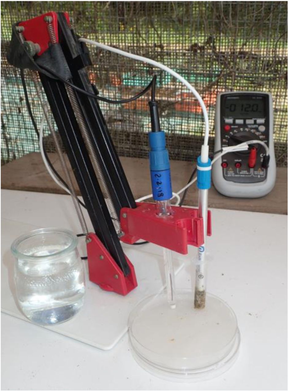
Measurement of Eh on plant stems. The plant material grinded with a mortar and pestle was placed in the barrel of a 5 ml syringe from which the nozzle had been cut. In the 9 cm Petri dish, the filter paper was moistened with 0.1M KCl solution. The Ag/AgCl reference electrode was placed standing, with tip touching the moist filter paper. The Pt plate electrode was placed in the plant material in the syringe, then the syringe barrel was placed standing with tip touching the moist filter paper.

Colony aspect at the end of the Jul15 experiment on native and altered agar media for six fungi. Each panel correspond to a single strain transferred the same day and grown in the same room, with the 13 media in columns and replicates in lines. Within the experiment, lines 1 to 3; 4 to 6; 7 to 9 correspond to the three independent replicates (i.e. is preparation in independent bottles, and inoculation with agar plugs from a different source plate). Within upper, central and lower groups, dishes were prepared from the same bottles for all fungi. Mean pH and Eh values measured on aliquots corresponding to the 13 columns are given below. Note: colony diameter had been assessed earlier than the picture for fast growing fungi. Dishes are 5 cm in diameter.
Acknowledgements
All authors declare the absence of conflicts of interest. This work benefited from the financial support of INRA – the French National Institute for Agronomical Research.
References


