

Abstract
Delamanid represents one of two novel antimicrobial classes approved to treat tuberculosis in over 40 years. Pretomanid is another promising nitroimidazole pro-drug in clinical development. Characterization of the full spectrum of mutations conferring resistance to nitroimidazoles and their related phenotypes in Mycobacterium tuberculosis will inform development of suitable genotypic and phenotypic drug susceptibility tests. Here, we used a range of pretomanid doses to select pretomanid-resistant mutants in two pathologically distinct murine TB models. The frequency of spontaneous pretomanid resistance mutations was approximately 10−5 CFU. Pretomanid demonstrated dose-dependent bactericidal activity and selective amplification of resistant mutants. Whole genome sequencing of 161 resistant isolates from 47 mice revealed 99 unique mutations, 90% of which were found in 1 of 5 genes previously associated with nitroimidazole activation and resistance. The remaining 10% harbored isolated mutations in Rv2983. Complementing an Rv2983 mutant with a wild-type copy of Rv2983 restored wild-type susceptibility to pretomanid and delamanid, confirming that loss of Rv2983 function causes nitroimidazole resistance. By quantifying F420 and its precursor Fo in Mycobacterium smegmatis overexpressing Rv2983 and an M. tuberculosis Rv2983 mutant, we provide evidence that Rv2983 is necessary for F420 biosynthesis and nitroimidazole activation, perhaps as the guanylyltransferase CofC. F420H2-deficient mutants displayed hypersusceptibility to malachite green (MG), a selective decontaminant present in solid media used to isolate and propagate mycobacteria from clinical samples. The wide diversity of mutations causing high-level pretomanid resistance and MG hypersusceptibility of most mutants poses significant challenges to clinical detection of nitroimidazole resistance using either genotypic or phenotypic methods.
Significance Nitroimidazole pro-drugs represent a promising new class of anti-tuberculosis drugs. Reliable methods to assure nitroimidazole susceptibility are critical to assure their optimal use. Yet, the spectrum of nitroimidazole resistance mutations remains incompletely characterized. Using 161 pretomanid-resistant Mycobacterium tuberculosis isolates selected in pretomanid-treated mice, we discovered a novel resistance determinant, Rv2983, required for cofactor F420 biosynthesis and characterized the remarkable diversity of mutations in this and 5 other genes involved in nitroimidazole activation. We show that F420H2–deficient nitroimidazole-resistant mutants are hypersusceptible to the selective decontaminant malachite green used in solid media to isolate mycobacteria and may evade detection on such media. These results have important implications for development and clinical use of genotypic and phenotypic methods for nitroimidazole susceptibility testing.
Introduction
Despite decades of efforts to end the global tuberculosis (TB) epidemic, Mycobacterium tuberculosis is the leading killer among infectious agents plaguing mankind (1). The emergence and spread of multidrug-resistant (MDR) and extensively drug-resistant (XDR) M. tuberculosis makes the eradication effort much more difficult because treatment requires administration of more toxic and less effective second‐ and third-line drugs for up to 2 years (1, 2). Delamanid and pretomanid are promising new bicyclic 4-nitroimidazole drugs that have shown potential in pre-clinical and clinical studies to shorten and simplify the treatment of TB, including drug-resistant forms (3-9). Delamanid received conditional approval by the European Medicines Agency (EMA) to treat MDR-TB in 2014 (10) and pretomanid is currently being evaluated in Phase 2/3 clinical trials (ClinicalTrials.gov Identifiers: NCT03338621, NCT02589782, NCT02333799, NCT03086486). Particularly notable is a novel regimen comprised of bedaquiline, pretomanid and linezolid that may represent a highly efficacious oral, short-course regimen for treatment of MDR/XDR-TB (4)(ClinicalTrials.gov Identifiers: NCT02333799, NCT03086486).
Two non-exclusive mechanisms of action have been described for these bicyclic 4-nitroimidazole drugs: inhibition of cell wall biosynthesis through inhibition of mycolic acid synthesis and respiratory poisoning through release of nitric oxide during bacterial drug metabolism (11, 12). Pretomanid and delamanid are prodrugs that require bioreductive activation of an aromatic nitro group by the 8-hydroxy-5-deazaflavin (coenzyme F420)-dependent nitroreductase Ddn in order to exert bactericidal activity (13). The reaction involves the transfer of two electrons from the reduced form of F420 (F420H2) produced by an F420-dependent glucose-6-phosphate dehydrogenase (Fgd1) (12, 14). Therefore, F420 biosynthesis and reduction are essential for the activation of delamanid, pretomanid and other nitroimidazole prodrugs. Three genes are identified as essential for F420 biosynthesis in M. tuberculosis complex (15, 16). fbiC encodes a 7,8-didemethyl-8-hydroxy-5-deazariboflavin (Fo) synthase that catalyzes the condensation of 5-amino-6-ribitylamino-2,4 (1H, 3H)-pyrimidinedione and tyrosine to form the F420 precursor Fo (17, 18). fbiA encodes a transferase that is believed to catalyze the transfer of a phospholactyl moiety to Fo to generate F420–0, while fbiB encodes a F420–0:γ-L-glutamyl ligase that catalyzes the sequential addition of a variable number of glutamate residues to F420–0 to yield coenzyme F420-5 or -6 in mycobacteria (12). In the methanogen Methanocaldococcus jannaschii, a guanylyltransferase termed CofC is believed to generate an intermediate (L-lactyl-2-diphospho-5’-guanosine,–LPPG) in the F420 biosynthesis pathway (19). A homologous enzyme, MSMEG_2392, is shown to be necessary for F420 synthesis in Mycobacterium smegmatis through transposon mutagenesis studies (20). An ortholog, Rv2983, is present in M. tuberculosis. However, the role of MSMEG-2392 and Rv2983 in F420 biosynthesis has remained unexplored.
Loss-of-function mutations in ddn, fgd1 and fbiA-C causing delamanid and pretomanid resistance are readily selected in vitro in M. tuberculosis complex (16, 18, 21-23). However, the genetic spectrum of mutations emerging during in vivo selection has not been characterized. In order to study bacterial genetic, host and pharmacological factors associated with emergence of nitroimidazole resistance in vivo, we selected pretomanid-resistant mutants using a wide range of pretomanid doses in two mouse models of TB and characterized them by whole genome sequencing (WGS). Because the lungs of TB patients feature a heterogeneous array of lesion types associated with diversified immune responses and drug penetration (24, 25), we used both C3HeB/FeJ mice, which develop caseating lung lesions in response to M. tuberculosis infection, and BALB/c mice, which do not, to investigate the impact of these caseating lesions and their associated micro-environments on mutant selection. In the present study, we found that pretomanid-resistant mutants were readily selected by monotherapy in both mouse strains. While the majority of resistant isolates harbored isolated mutations in genes previously associated with nitroimidazole resistance, all the resistant isolates lacking such mutations had mutations in Rv2983. We went on to confirm that loss-of-function mutations in Rv2983 cause high-level pretomanid and delamanid resistance through disruption of F420 biosynthesis, supporting the hypothesis that Rv2983 plays a role similar to cofC in M. tuberculosis. Furthermore, F420H2-deficient nitroimidazole-resistant M. tuberculosis mutants, including Rv2983 mutants, were found to be hypersensitive to malachite green (MG), an organic compound used as a selective decontaminant in solid media for culturing M. tuberculosis, which may have important implications for their detection in clinical samples.
Materials and Methods
Bacterial strains, media, antimicrobials and reagents
Wild type M. tuberculosis H37Rv (ATCC 27294) was mouse-passaged, frozen in aliquots and used in all the experiments. The wild type M. smegmatis strain mc2155 was obtained from the stock in the lab. Unless stated otherwise, Middlebrook 7H9 medium (Difco, BD) supplemented with 10% oleic acid-albumin-dextrose-catalase (OADC) complex (BD), 0.5% glycerol and 0.05% Tween 80 (Sigma-Aldrich) (7H9 broth) was used for cultivation. Middlebrook 7H10 agar and selective 7H11 agar (Difco, BD), prepared from powder and containing 10% OADC and 0.5% glycerol, were used for comparison of strain recovery on commercially available agar plates. Lowenstein Jensen (LJ) slants were purchased from BD. Pretomanid and delamanid were kindly provided by the Global Alliance for TB Drug Development (New York, NY).
Mouse infection models and pretomanid treatment
All animal procedures were approved by the Animal Care and Use Committee of Johns Hopkins University. Aerosol infections were performed using the Inhalation Exposure System (Glas-col Inc., Terre Haute, IN), as previously described (26). Briefly, 6-week-old female BALB/c mice (Charles River, Wilmington, MA) and C3HeB/FeJ mice (Jackson Laboratories Bar Harbor, ME) were infected with a log phase culture of M. tuberculosis that was grown in 7H9 broth to O.D.600nm= 1.0 and then diluted in the same medium prior to infection to deliver 50-100 CFU to the lungs. Pretomanid was formulated for oral administration as previously described (27). Beginning 8 weeks after aerosol infection, mice were randomly allocated into groups and treated once daily (5 days per week) for up to 8 weeks with pretomanid at doses of 10, 30, 100, 300 and 1000 mg/kg. Untreated mice were sacrificed on the day after aerosol infection and on the day of treatment initiation to determine the number of CFU implanted in the lungs and pretreatment CFU counts, respectively. Additional mice were sacrificed after 3 and 8 weeks of treatment to evaluate the treatment response. Serial 10-fold dilutions of lung homogenates were plated on 7H11 agar. Week 8 samples including those from untreated mice were also plated in parallel on 7H11 plates containing 0.25, 1 and 10 μg/ml of pretomanid to quantify the resistant CFU. Plates were incubated at 37°C for 28 days before final CFU counts were determined.
Whole genome sequencing
For each mouse lung that yielded growth on pretomanid-containing plates, individual colonies and, for a subset of mice, pools of up to 15 colonies, were randomly selected from pretomanid-containing plates and sub-cultured in 7H9 broth prior to extraction of genomic DNA using the cetyltrimethylammonium bromide (CTAB) protocol (28) and vortexing (Genegate, Inc.). 2-3 μg of genomic DNA was sheared by a nebulizer to generate DNA fragments. The DNA library was prepared using a genomic DNA sample preparation kit (Illumina, Inc.), in which adapter-ligated DNA fragments were 250-350 bp in length, and carried out on an Illumina Genome Analyzer II (Illumina, Inc). The sequencer was operated in paired-end mode to collect pairs of reads of 51-bp from opposite ends of each fragment. Image analysis and base-calling were done by using the Illumina GA Pipeline software (v0.3). The reads that were generated for each strain were aligned to the reference genome of M. tuberculosis H37Rv (29). Based on alignment to the corresponding region in the reference genome, single nucleotide polymorphism (SNP), insertion and deletion were identified on the genome of resistant strains by using a contig-building algorithm to construct a local ~200 bp sequence spanning the site of mutagenesis (30). Distribution of mutation type and mutation frequency in genes involved in nitroimidazole resistance was calculated by counting the total number of unique mutations isolated from each mouse in the same treatment group.
Complementation of an Rv2983 mutation
A 1,044-bp DNA fragment containing the open reading frame (ORF) of the wild type Rv2983 gene, including 340 bp of 5’-flanking sequence and 59 bp of 3’-flanking sequence, was PCR-amplified from M. tuberculosis H37Rv genomic DNA using primers Rv2983-1F and Rv2983-1R (Table S1). The Rv2983 PCR product was ligated into Xbal-digested E. coli-mycobacterium shuttle vector pMH94 (31) using NE builder HiFi DNA assembly kit (NE Biolabs) to generate the recombinant pMH94-Rv2983 vector. Similarly, a 388-bp DNA fragment containing the hsp60 promoter and a 645-bp DNA fragment of Rv2983 open reading frame were amplified from M. tuberculosis H37Rv genomic DNA using primer sets hsp60-F and hsp60-R and Rv2983-2F and Rv2983-2R, respectively (Table S1), and ligated into Xbal-digested E. coli-mycobacterium shuttle vector pMH94 to yield pMH94-hsp60-Rv2983. A small amount of ligation reaction was transferred into E. coli competent cells, followed by DNA sequencing of the inserts in the corresponding recombinants. The recombinants pMH94-Rv2983 and pMH94-hsp60-Rv2983 were electroporated into competent cells of Rv2983 mutant strain BA_101 (B101), harboring an A198P substitution, to enable selection of complemented candidates B101pRv2983 and B101phsp60-Rv2983 on 7H10 agar containing 25 μg/ml of kanamycin. To confirm the complementation genetically, Southern blotting was performed using a digoxigenin (DIG) DNA labeling and detection kit according to the manufacturer’s protocol (Sigma). Briefly, a 448-bp Rv2983 probe was generated by addition of DIG-dUTP (Sigma) to PCR reactions containing primer pairs Rv2983-3F and Rv2983-3R (Table S1). Acc65I-digested (NE biolabs) genomic DNA of the wild type, the B101 mutant and the B101pRv2983 and B101phsp60-Rv2983 complemented strains was separated on agarose gel and transferred onto positively-charged nylon-membrane (GE). After pre-hybridization, the membrane was hybridized with the DIG-labeled Rv2983 probe at 68°C overnight, followed by addition of anti-DIG alkaline phosphatase conjugate. After stringent washes, the membrane was incubated with the chemiluminescence substrate disodium 3-(4-methoxyspiro {1,2-dioxetane-3,2(5′-chloro)tricycloecan}-4-yl)phenyl phosphate (CSPD) and exposed on X-ray film in a dark room prior to development using a developer (AFP imaging)(32).
MIC determination
Log-phase cultures were diluted to achieve a bacterial density of approximately 105 CFU/ml in conical tubes containing 7H9 broth without Tween 80. Serial 10-fold dilutions were plated on 7H11 agar containing stepwise 2-fold increasing pretomanid concentrations ranging from 0.015 to 64 μg/ml or delamanid concentrations from 0.001 to 1.024 μg/ml. Drugs were initially dissolved in dimethylsulfoxide (DMSO) (Sigma) prior to further dilution in 7H9 broth or 7H11 agar. Cultures were incubated at 37°C for 14 days or 28 days after plating. MIC was defined as the lowest drug concentration that inhibited visible M. tuberculosis growth in conical tubes or that inhibited 99% of CFU growth on pretomanid-containing plates (33, 34). The experiments were repeated twice.
Construction of recombinants overexpressing Rv2983, with or without fbiC, in M. smegmatis
A 645-bp DNA fragment containing the Rv2983 ORF was PCR-amplified from M. tuberculosis H37Rv genomic DNA using primers Rv2983-4F and Rv2983-4R (Table S1). The amplified PCR product was ligated into the NdeI‐ and PacI-digested E. coli-mycobacterium shuttle vector pYUBDuet (35) using NE builder HiFi DNA assembly kit (NE Biolabs) and then transferred into Turbo-competent E. coli cells (NE Biolabs) prior to plating on LB agar plates containing 100 μg/ml of hygromycin B for selection of recombinants. The Rv2983 PCR product was also similarly ligated into the same NdeI‐ and PacI-digested pYUBDuet vector harboring fbiC (termed pfbiC) (35) to overexpress both Rv2983 and fbiC. After confirmation by restriction digestion and DNA sequencing, the constructs were electroporated into competent M. smegmatis cells prior to selecting recombinants on 7H10 agar plates containing 100 μg/ml of hygromycin B. PCR amplification was used to confirm the inserts on the M. smegmatis genome. pYUBDuet and pYUBDuet harboring fbiA, fbiB and fbiC (termed pfbiABC) (35) were also transferred into competent M. smegmatis cells to serve as controls.
Measurement of Fo and F420
Extraction of Fo and F420 was performed in M. smegmatis and M. tuberculosis strains according to a previous study (35), with minor modifications. Briefly, M. smegmatis strains harboring different constructs and pYUBDuet were grown in 7H9 broth in a shaker to mid-log phase (O.D.600nm = 0.7-1.0), followed by induction using 1mM isopropyl β-D-1-thiogalactopyranoside (IPTG) for 6 and 26 hours. After centrifugation for 15 min at 16000 x g, the supernatants were removed for detection of Fo, which is principally found in culture supernatant whereas F420 with 5 or 6 glutamate residues is largely retained inside cells (15, 35, 36). The cell pellets were washed with 25mM sodium phosphate buffer (pH 7.0) and re-suspended at 100 mg/mL in the same buffer, then autoclaved at 121°C for 15 min. After centrifugation at 16000 x g for 15 min at 4°C, the cell extracts were harvested for detection of F420 (35). Fluorescence of the supernatant and cell extracts was measured using an excitation wavelength of 410 nm and an emission wavelength of 465 nm. Fluorescent signals of Fo were normalized using the O.D. at 600nm. The small portion of Fo (1-7%) retained inside cells was ignored when quantifying F420 in cell extracts (37). Relative fluorescent signals were calculated in M. smegmatis harboring each of recombinants relative to pYUBDuet alone. Similarly, cell extracts and supernatant were extracted from M. tuberculosis strains grown in 7H9 broth for 6 days at initial O.D.600nm of 0.1. Relative fluorescent signals of F420 and Fo were calculated using cell extracts and supernatant relative to 25 mM phosphate buffer and 7H9 broth, respectively. M. smegmatis harboring pYUBDuet-fbiABC was used as a positive signal control for Fo and F420 due to their commercial unavailability (35). The experiment was repeated twice.
Quantification of gene expression in M. tuberculosis
6-day-old M. tuberculosis strains grown in 7H9 broth as described above were sub-cultured in fresh 7H9 at O.D.600nm = 0.05 followed by incubation at 37°C in a shaker for 2 and 4 days. Bacterial pellets were collected by centrifugation at 3500 rpm at 4°C for purification of total RNA using Trizol (Thermo Fisher Scientific) according to the manufacturer’s protocol followed by removal of DNA contamination with Turbo DNAse (Ambion). Following cDNA synthesis with random hexamers and oligo(dT)20 primer and superscript III reverse transcriptase (Invitrogen), quantitative PCR was performed to measure gene expression of M. tuberculosis using SYBR Green PCR master mix (Thermo scientific) and StepOne™ system (Applied biosystems) with primer sets listed in Table S1. The cycle threshold value (CT) measured for each gene was normalized to that of the housekeeping gene sigA (ΔCT) amplified by the primers sigA-F and sigA-R (Table S1). ΔΔCT was calculated in each of pretomanid-resistant strains relative to the wild-type H37Rv prior to calculation of the fold-change in gene expression (2^-ΔΔCT) (38). All samples were prepared in duplicate. PCR was performed from an equal amount of cDNA samples synthesized with oligo(dT)20 with primers fbiC-5-7_F and fbiC-5-7_R (Table S1) using the Q5 High-fidelity PCR kit (New England Biolabs) and C1000 Thermal cycler (Biorad). The PCR product was examined by electrophoresis on a 1% agarose gel.
Malachite green susceptibility testing
7H9 media supplemented with 10% OADC, 0.5% glycerol, 1.5% Bacto™ Agar (BD) and malachite green (MG) oxalate (Alfa Aesar) was used to prepare solid 7H9 media with differing MG concentrations. M. tuberculosis strains were grown to mid-log phase and diluted to OD600nm = 0.1 in 7H9 broth before serial 10-fold dilutions were plated in 100 or 500 μl aliquots on 7H9 agar containing MG concentrations of 0, 0.1, 0.3, 1, 3, 10, 30, 100, 300, 1000 μg/ml or 0, 3, 6, 12 μg/ml. CFU were counted after 28, 35 and 49 days of incubation. The same cultures were also plated on 7H10 and 7H11 agar plates and LJ slants. Serially diluted cultures were inoculated onto LJ slants using calibrated disposable inoculating loops (10 μl per loop, BD) as one loop per LJ slant. Plates were incubated at 37°C for 21, 28 and 35 days for CFU counts. Colony size was observed weekly until day 35, beginning 21 days after plating. The experiment was repeated two times under similar conditions.
Statistical analysis
Log10-transformed CFU counts, fold-change values of gene expression and absorbance (A410) values of fluorescent signals were used to calculate means and standard deviations for each data set. Differences between means were compared by the Student’s t test in Microsoft Excel. Differences in mutation frequencies between two mouse models were evaluated by Fisher’s exact test in GraphPad Prism 6. A p-value of < 0.05 was considered statistically significant.
Results
Spontaneous pretomanid-resistant mutants exist at a relatively high frequency in infected mice and are selectively amplified by treatment with active doses of pretomanid
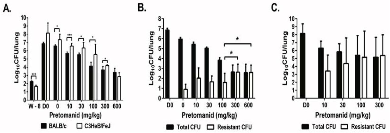
To study the dose-response of pretomanid and explore the genetic spectrum of nitroimidazole resistance selected in vivo, we established chronic M. tuberculosis infections in BALB/c and C3HeB/FeJ mice and then treated with a range of pretomanid doses for up to 8 weeks. Despite lower CFU counts on the day after infection (W-8) in C3HeB/FeJ mice (1.67 log10 CFU per lung) compared to BALB/c (2.26 log10) (p <0.001), higher CFU counts were observed in C3HeB/FeJ mice 8 weeks later on the day treatment started (D0) and after 3 weeks of treatment in almost all groups (p <0.001 - 0.05) (Fig. 1A). Three C3HeB/FeJ mice treated with 1000 mg/kg required euthanasia during the second week of treatment, prompting a dose reduction from 1000 mg/kg to 600 mg/kg in both strains. Nevertheless, a clear pretomanid dose-response relationship was observed in both mouse strains after 3 weeks of treatment (Fig. 1A). The three remaining C3HeB/FeJ mice treated with 600 mg/kg beyond the week 3 time point were euthanized after 5 weeks of treatment due to toxicity. One had no detectable CFU and two had ≤ 2.0 logi0CFU of pretomanid-resistant M. tuberculosis. After 8 weeks of treatment, total CFU counts fell in a dose-dependent manner in BALB/c mice before a plateau was reached at doses ≥ 300 mg/kg, where resistant CFU were higher and replaced the susceptible CFU (p < 0.05) (Fig. 1B). Spontaneous pretomanid-resistant CFU comprised approximately 10−5of the total CFU in the absence of drug pressure in untreated BALB/c mice and the proportion of the total CFU that was comprised of pretomanid-resistant CFU increased with dose up to the 300 mg/kg dose group. Dose-dependent bactericidal activity was also observed in C3HeB/FeJ mice (Fig. 1C). However, selective amplification of pretomanid-resistant mutants was more extensive and occurred at lower doses than in BALB/c mice (Fig. 1B and 1C). We were not able to measure the spontaneous frequency of resistant mutants in untreated C3HeB/FeJ mice because they succumbed to infection prior to week 8. Pretomanid-resistant CFU replaced susceptible CFU in C3HeB/FeJ mice receiving doses as low as 30 mg/kg and pretomanid-resistant CFU counts were roughly 10 times higher in C3HeB/FeJ mice compared to BALB/c mice (Fig. 1B and C), which indicates greater potential for selective amplification of pretomanid resistance with monotherapy in this strain. Most resistant isolates grew on plates containing 10 μg/ml of pretomanid, but some had fewer CFU on plates containing 10 μg/ml than on those containing 1 μg/ml of pretomanid.

After aerosol infection with M. tuberculosis H37Rv, BALB/c and C3HeB/FeJ mice were treated with a range of doses of pretomanid for 8 weeks and sacrificed at different time points before and after treatment for lung CFU counts. A. Mean (± S.D.) total lung CFU counts on the day after infection (W-8), on the day of treatment initiation (D0), and after 3 weeks of treatment with the indicated pretomanid dose (in mg/kg body weight). Dose-dependent bactericidal activity was observed in both strains; B. Mean (± S.D.) total and PMD-resistant lung CFU counts in BALB/c mice on day 0 and after 8 weeks of treatment with the indicated pretomanid dose. Dose-dependent bactericidal activity and selection of PMD-resistant bacteria was observed, with the resistant population overtaking the susceptible population at doses ≥ 300 mg/kg; C. Mean (± S.D.) total and PMD-resistant lung CFU counts in C3HeB/FeJ mice on day 0 and after 8 weeks of treatment with the indicated pretomanid dose. Dose-dependent bactericidal activity and selection of PMD-resistant bacteria was observed, with the resistant population overtaking the susceptible population at doses ≥ 30 mg/kg. * p< 0.05, ***p < 0.001
Whole genome sequencing of pretomanid-resistant mutants revealed diverse mutations in Rv2983 or in one of five other genes required for pretomanid activation
To characterize mutations associated with pretomanid resistance in vivo, we performed WGS on 136 individual pretomanid-resistant colonies and 25 colony pools picked from 47 individual mice harboring pretomanid-resistant CFU after 8 weeks of treatment (Table S2 and S3). Each individual isolate had an isolated mutation in Rv2983 or one of the 5 genes previously shown to be required for pretomanid activation. Overall, 99 unique mutations in these 6 genes were identified from individual and pooled isolates (Table 1 and 2). Except for mutations K9N (fgdl), R322L (fbiC) and Q120P (fbiA), which were shared by two mice each, no two mice harbored the same mutation, which emphasizes the large target size for resistance-conferring mutations. In both BALB/c and C3HeB/FeJ mice, more than half of the resistant isolates were fbiC mutants (54 and 56%, respectively) (Table 3). For the other five genes, the rank order by mutation frequency was Rv2983 (15%) > fbiA (13%) > ddn (9%) > fbiB (6%) > fgd1 (4%) in BALB/c mice and fbiA (18%) > ddn (16%) > fgd1 or Rv2983 (4%) > fbiB (2%) in C3HeB/FeJ mice. No significant differences in mutation frequencies between BALB/c and C3HeB/FeJ mice were observed, although a trend towards more Rv2983 mutations in BALB/c mice (8/54, 15% of all mutations) compared to C3HeB/FeJ mice (2/45, 4%) was detected. The mutations identified in Rv2983 included 8 point mutations resulting in the following amino acid substitutions: R25S, R25G, A68E, A132V, G147C, C152R, Q114R and A198P, as well as an insertion of C after A27 and a deletion of I129 (-ATC) (Tables 1 and 2). The overall frequency distribution of unique mutations was as follows: fbiC (55%, n = 54), fbiA (15%, n = 15), ddn (12%, n = 12), Rv2983 (10%, n=10), fgd1 (4%, n = 4), and fbiB (4%, n=4) (Fig. 2 and Table S4). There were no clear associations between pretomanid dose and the mutated gene. Mutations in fbiC comprised a higher proportion of those selected in our in vivo study compared to the proportion selected in a previous in vitro study (26%, p = 0.0001)(22). On the other hand, mutations in ddn (29%) were more frequent after in vitro selection than in our mouse models (12%) (p =0.001). In vitro mutation frequencies for fbiA, fgd1 and fbiB (19%, 7% and 2%, respectively) were similar to our findings in mice.
Mutations identified in 89 individual colonies and pooled isolates of pretomanid-resistant M. tuberculosis selected in BALB/c mice
Mutations identified in 64 individual colonies and pooled isolates of nitrioimidazole-resistant M. tuberculosis selected in C3HeB/FeJ mice
Distribution of mutation types and frequencies in genes associated with pretomanid resistance, by mouse strain
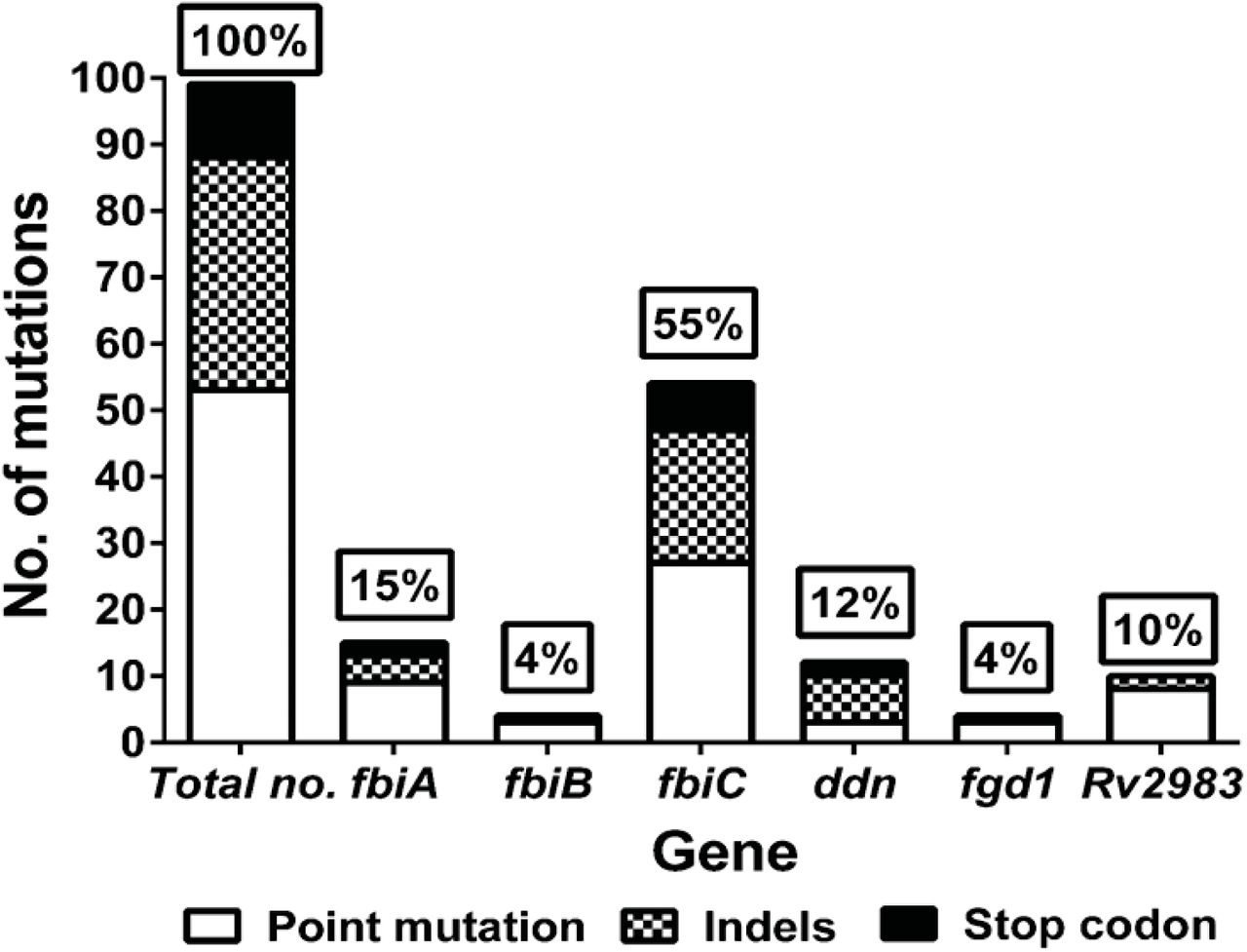
WGS was performed with 136 pretomanid-resistant colonies and 25 colony pools picked from 47 individual mice harboring pretomanid-resistant CFU after 8 weeks of treatment and identified 99 unique mutations in these 6 genes. Mutations in fbiC (55%) were the predominant cause of pretomanid resistance. For the other 5 genes, the rank order by mutation frequency was fbiA (15%), ddn (12%), Rv2983 (10%), fgd1 (4%) and fbiB (4%).
Among the 99 unique mutations, all but one (an IS6110 insertion located in 85-bp upstream of the fbiC coding sequence in isolate KA-026a (Table 2 and Table S2) were found within the coding regions of the six genes. In total, 54% (53/99) were non-synonymous point mutations (no synonymous point mutations were identified), 35% (35/99) were insertions or deletions (indels), and 11% (11/99) were substitutions resulting in a stop codon. No significant difference in the distribution of point mutation and indels was found between ours and the in vitro study by Haver, et al, in which non-synonymous point mutations and indels were 50% (75/151) and 24% (36/151), respectively. However, the frequency of stop codon substitutions in the latter study (26%, 40/151) was higher than that observed in the present study (11%, 11/99) (p = 0.004), 85% (34/40) of which were in ddn in the latter study (22). Non-synonymous point mutations predominated relative to indels and stop codon mutations overall and in each gene except for ddn (Fig. 2 and Table S4). The frequency of point mutations was similar between BALB/c and C3HeB/FeJ mice (56% versus 51%) (Table 3). A higher frequency of indels occurred in C3HeB/FeJ compared to BALB/c mice (44% versus 28%), while more stop codon mutations occurred in BALB/c, but these differences were not statistically significant. There was no clear association between dose of pretomanid and the type of mutation selected.
Comparing the 99 unique mutations identified in our study with the 151 unique mutations in 5 of the same genes selected in vitro (22), only 4 mutations were found in the same position. Only W79 stop (fbiA) and N336K (fbiC) mutations were found in both datasets while both T273 and H190 (fbiC) were mutated in the same position but with different mutations. As expected from the fact that most mutants could be isolated on plates containing 10 μg/ml of pretomanid, MICs determined against a small subset of isolates indicated high-level pretomanid resistance (Table S5 and Table 1 and 2). Taken together, these data illustrate the tremendous diversity of mutations capable of conferring high-level pretomanid resistance.
Mutations in Rv2983 cause resistance to pretomanid and delamanid
To prove that mutations in Rv2983 are sufficient for nitroimidazole resistance, merodiploid complemented strains were constructed by introducing a copy of the wild type Rv2983 gene into B101, an Rv2983 mutant (A198P), through site-specific integration (31, 32). Following confirmation of successful integration by Southern blot using a DIG-labeled Rv2983 probe (Figs. S1A and S1B), susceptibility testing by 7H9 broth dilution confirmed significantly higher nitroimidazole MICs against the Rv2983 mutant (pretomanid and delamanid MICs of 32 μg/ml and 0.064-0.128 μg/ml, respectively) and full restoration of susceptibility in the complemented strains (pretomanid and delamanid MICs of 0.25 μg/ml and 0.008 μg/ml, respectively).
Rv2983 is required for F420 biosynthesis
To demonstrate that Rv2983 is required for F420 biosynthesis, we measured the production of Fo and F420 in M. smegmatis strains overexpressing Rv2983 and in M. tuberculosis Rv2983 mutant strains compared with their corresponding control strains. Rv2983 and fbiC were successfully cloned into pYUBDuet and pfbiC (designated pRv2983 and pfbiC-Rv2983, respectively), followed by successful transformation of M. smegmatis, along with pYUBDuet and pfbiABC, which were confirmed by restriction enzyme digestion, DNA sequencing and PCR amplification (data not shown). Overexpression of Rv2983 in M. smegmatis increased F420 production but resulted in little change in Fo production compared to the control strain after 6 and 26 hours of induction with IPTG (Figs. 3A and 3B). As expected, mutation of Rv2983 in the M. tuberculosis B101 mutant markedly reduced F420 production, resulting in accumulation of Fo. Complementation fully restored the wild-type phenotype (Figs. 3C and 3D). In order to evaluate the method, we also overexpressed fbiC, which encodes the Fo synthase, with and without concomitant overexpression of Rv2983 in M. smegmatis. As expected, overexpression of fbiC increased Fo and, consequently, F420 concentrations. Relative to the control strain, F420 concentrations were similar when either fbiC or Rv2983 was over-expressed alone (Fig. 3A). Interestingly, when Rv2983 was co-overexpressed with fbiC, a dramatic increase in F420 was observed relative to over-expression of either gene alone (3.4 and 3.1-fold, respectively) after 6 hours of IPTG induction (p<0.001), with corresponding significant decreases of Fo levels after 6 and 26 hours of IPTG induction (5.8 and 3.1-fold; p<0.005 and 0.05, respectively) (Fig. 3A and 3B). These results suggest that the excess Fo produced by fbiC over-expression was efficiently converted to F420 by over-expressed Rv2983. On the other hand, although a small amount of F420 was observed in cell extracts of two Rv2983 point mutants (B101 [A198P] and KA016 [Q114R]), their F420 content was significantly lower than that of the wild type (7.3 and 7.7-fold) (p</italic> < 0.001) and complemented B101 mutant (Fig. 3C). As expected, Fo accumulated in the two Rv2983 mutant strains relative the wildtype (6.7 and 6.5-fold; p<0.05 and 0.005, respectively) (Fig. 3D), indicating that Fo was not efficiently converted to F420 in the presence of a mutated Rv2983. Two other pretomanid-resistant strains were also assessed as controls. The KA026 mutant with an IS6110 insertion 85 bp upstream of fbiC had undetectable Fo and very little F420 content, while the KA91 mutant with an IS6110 insertion at amino acid position 108 of Ddn showed a wild-type phenotype with respect to F420 and Fo concentrations (Fig.3C and D).

F420 and Fo content was measured in M. smegmatis strains harboring different recombinants relative to the control strain containing the empty vector pYUBDuet after 6 (A) and 26 (B) hours of 1mM IPTG induction; F420 (C) and Fo (D) content was measured in Rv2983 mutant strains of M. tuberculosis and control strains including B101 (ΔRv2983, A198P), KA016 (ΔRv2983, Q114R), H37Rv (wild-type), B101 complemented strain (pMH94-Rv2983), B101 complemented strain (pMH94-hsp60-Rv2983), KA026 (AfbiC, IS6110 insertion in 85-bp upstream of fbiC), and K91 (Δddn, IS6110 insertion in aa D108), after growth in 7H9 broth for 6 days. Schematic diagram (E) of proposed nitroimidazole activation pathway showing Rv2983 as putative CofC catalyzing LPPG biosynthesis. Fo, 7,8-didemethyl-8-hydroxy-5-deazariboflavin; LP, 2-phospho-L-lactate; GTP, guanosine triphosphate; LPPG, L-lactyl-2-diphospho-5’-guanosine.
To understand the effect of mutations on gene expression and its regulation, we performed RT-qPCR after sub-culturing the M. tuberculosis strains in fresh 7H9 broth. Expression of Rv2983 and fbiC increased 2.4‐ and 1.6-fold, respectively, in the Rv2983 mutant B101 relative to the wildtype H37Rv parent after 4 days of incubation in 7H9 broth. Similar increases were observed in other genes such as fbiA (2.3-fold), fbiB (2.0-fold) and fgd1 (2.2-fold) involved in F420 biosynthesis, suggesting that the reduced F420 content caused by the Rv2983 A198P mutation resulted in upregulation of the F420 biosynthesis pathway (Fig. S2A). Expression of fbiC in the KA026 mutant decreased 114-fold compared to that in the wild-type H37Rv parent after 2 days of incubation in 7H9 broth, likely resulting from interrupted fbiC transcription as a result of the insertion IS6110 at 85-bp upstream and explaining the low levels of both Fo and F420 in that mutant (Fig. S2B). A faint band representing the fbiC DNA fragment of 937-bp from the fbiC KA026 mutant relative to H37Rv further supports this conclusion (Fig. S2C).
F420-deficient pretomanid-resistant mutants are attenuated for growth in the presence of malachite green
Previous work using M. smegmatis showed that mutations in MSMEG_5126 (homolog of fbiC) and MSMEG_2392 (which shares 69% homology with Rv2983) reduce the ability to decolorize and detoxify MG, indicating that F420 is necessary for this process (20). To evaluate the role of each gene associated with nitroimidazole activation in the susceptibility to MG, log-phase cultures of 10 selected mutants were plated on 7H9 agar supplemented with a range of MG concentrations. All mutants deficient in F420 synthesis or reduction (i.e., those with mutations in fbiA-C, Rv2983 or fgd1) were more susceptible to MG, while the ddn mutant retained the same susceptibility as the wild type H37Rv parent, whose growth was almost completely inhibited at MG concentrations ≥ 30 μg/ml (Fig. 4A). The lability of F420H2 and lack of a commercial source for F420 made it unfeasible to attempt to test whether provision of F420H2 could rescue the MG-hypersusceptible phenotype of the F420H2–deficient mutants. To confirm that Rv2983 is necessary for the intrinsic resistance of M. tuberculosis to MG, we compared the growth of the B101 mutant to that of the wild type and complemented strains on MG. Again, growth of this Rv2983 mutant was inhibited by lower concentrations of MG than the wild type strain and required longer incubation times before colonies appeared on MG-containing plates (Fig. 4B-D). Plating at higher bacterial density (500 μl rather than 100 μl of cell suspension per plate) significantly increased recovery (Fig. S3A and B). Complementation of Rv2983 fully restored the wild-type growth phenotype on MG concentrations up to 6 μg/ml. Interestingly, at MG concentrations above 6 μg/ml, greater recovery was observed when Rv2983 was expressed behind the native promoter compared to the hsp60 promoter (Fig. 4B-D). Smaller colony size was observed in the Rv2983 mutant relative to the wild type and the complemented strains on plates without MG after 21 days of incubation but not after 28 days of incubation (Fig. 5). However, smaller colony size and deficient decolorization was observed for the Rv2983 mutant on plates containing MG concentrations as low as 0.25 and 1 μg/ml, even after 28 days of incubation (Fig. 5). At MG concentrations of 6-12 μg/ml, even more-concentrated aliquots of the B101 mutant culture showed markedly reduced recovery despite 42 days of incubation (Fig. 5).

A. Growth of wild-type M. tuberculosis on 7H9 agar is inhibited by malachite green (MG) in a concentration-dependent manner. F420H2–deficient, pretomanid-resistant M. tuberculosis mutants (fbiA-C, fgd1, Rv2983) are inhibited at lower MG concentrations relative to the wild type and the F420H2-sufficient, pretomanid-resistant ddn mutant. B-D. Complementation of the B101 mutant with wild-type Rv2983 restores tolerance to MG and the proportional recovery of the mutant on 6 μg/ml of MG increases as the duration of incubation increases from 28 (B), to 35 (C) to 49 (D) days of incubation. The proportional recovery of the mutant on plates containing 12 μg/ml of MG does not increase with time.

Aliquots (100 μl) of M. tuberculosis cultures (1, H37Rv wild type; 2, Rv2983 mutant B101; 3, B101 mutant complemented with Rv2983 behind native promoter; 4, B101 mutant complemented with Rv2983 behind hsp60 promoter) were spread on 7H9 agar plates containing increasing concentrations of malachite green (MG) after serial 10-fold dilutions. Colony size and MG decolorization are depicted after different incubation times. Numbers on plate labels (e.g., 0, 3, 4, 5) indicate the number of 10-fold dilutions performed before an aliquot was plated. Although the B101 mutant grows more slowly on plates without MG, growth is further slowed in the presence of ≥0.25 μg/ml of MG and the proportion of CFU recovered declines with increasing MG concentrations above 0.25 μg/ml. For plates containing 6 and 12 μg/ml of MG, plates receiving more-concentrated aliquots of the B101 mutant culture are shown to demonstrate the markedly reduced recovery of the mutant at these MG concentrations.
Because all solid media commonly used to isolate and cultivate M. tuberculosis in clinical laboratories contain MG as a selective decontaminant, the increased MG susceptibility conferred by mutations in fbiA-C, Rv2983 and fgd1 could compromise the isolation and propagation (and hence identification) of nitroimidazole-resistant mutants from clinical samples. Commercial 7H10 agar, 7H11 agar and LJ medium contain 0.25, 1 and 400 μg/ml, respectively, of MG. To assess the potential impact of these media on the isolation of an F420H2-deficient nitroimidazole-resistant Rv2983 mutant relative to an F420H2-sufficient, but still nitroimidazole-resistant, ddn mutant and the nitroimidazole-susceptible wild type and Rv2983-complemented mutant, we inoculated these media in parallel using serial dilutions of each strain. The Rv2983 mutant exhibited 10 times lower CFU counts and smaller colony size relative to other strains after 21 and 28 days of incubation on 7H10 agar plates (p <0.01) (Figs. 6A and C(a)). The result after 35 days of incubation was generally similar between the mutant and the control strains (Fig. 6A and C(b)). A similar semiquantitative growth assessment of the Rv2983 mutant on LJ media compared to other strains including a ddn mutant (K91, IS6110 ins in D108) revealed growth inhibition of the Rv2983 mutant that was ameliorated by increasing the size of the bacterial inoculum from 102 to 106 CFU/ml and increasing the incubation time from 28 to 35 days (Fig. 6D). Interestingly, no difference in growth was found on 7H11 agar (Fig. 6B), even when comparing colony size after just 21 days of incubation (Fig. 6C(c)), despite higher MG concentrations in that medium compared to 7H10. Unlike 7H10, 7H11 medium contains hydrolysate of casein, which may somehow mitigate against the MG toxicity. Taken together, these results indicate that use of 7H10 and LJ could compromise the recovery and propagation of F420H2-deficient nitroimidazole-resistant mutants from clinical specimens and that 7H11 agar may be the preferred solid media for processing specimens propagating any isolates from nitroimidazole-treated patients.

Aliquots of M. tuberculosis cultures were spread on various solid media purchased commercially after serial 10-fold dilutions. A-B. Mean CFU counts on 7H10 (A) and 7H11 (B) agar plates; C. Colonies on 7H10 agar plates after 21 (a) and 35 (b) days of incubation, and on 7H11 agar plates after 21 (c) days of incubation; D. Colonies on LJ slants inoculated with serially diluted aliquots after 28 and 35 days of incubation. 1: H37Rv wild type; 2: B101 mutant (ΔRv2983, A198P); 3: B101 mutant complemented with Rv2983 behind the native promoter; 4: B101 mutant complemented with Rv2983 behind the hsp60 promoter; 5. K91 mutant (Δddn, IS6110 ins in D108).
Discussion
As representatives of one of only two new drug classes approved for use against TB in roughly 50 years, delamanid and pretomanid are important and promising new drugs (3, 4, 6, 7, 39). The former received accelerated approval from the EMA for treatment of MDR-TB and is now used clinically, albeit sparingly, while a phase 3 trial is being completed. The latter is being evaluated in clinical trials as a component of highly promising regimens for both drug-susceptible and MDR/XDR-TB. Comprehensive knowledge of genetic mutations conferring nitroimidazole resistance in M. tuberculosis and the resultant mutant phenotypes are critical for timely and accurate diagnosis of resistance and, therefore, the safe and effective use of these drugs in the clinical setting. Previous work identified 5 genes (fbiA-C, fgd1, and ddn) involved in the activation pathway of nitroimidazole prodrugs in which mutations may confer drug resistance in M. tuberculosis complex (16, 18, 21-23, 40). In a prior study of the spectrum of nitroimidazole resistance-conferring mutations, Haver, et al found that 151 (83%) of 183 pretomanid-resistant isolates selected in vitro harbored a single mutation in one of these 5 genes (22). However, 17% of the selected strains harbored no mutations in these genes. The present study has several important new findings. First, we identified a novel nitroimidazole resistance determinant—mutations in Rv2983—that explained, in the case of our study, all of the pretomanid resistance that was not attributable to mutations in the 5 previously described genes. Second, we demonstrate for the first time that Rv2983 is required for F420 biosynthesis and likely plays a role similar to cofC in the methanogen M. jannaschii (19). Third, we show that Rv2983 and the ability to produce F420H2 are essential for full tolerance of M. tuberculosis to the selective decontaminant MG, which raises serious concerns about the ability to reliably recover most nitroimidazole-resistant mutants from clinical samples on the most widely used microbiology media.
Our study provides the first comprehensive analysis of the spectrum of nitroimidazole-resistant mutants selected in vivo and, because we used WGS, it represents the most comprehensive analysis made to-date. Despite the insignificant differences in mutation frequencies between the two mouse strains for other genes related to nitroimidazole activation, Rv2983 mutations were observed with a somewhat higher frequency in BALB/c mice, indicating a possible host-specific fitness cost that warrants further dedicated study. Like the in vitro study by Haver et al (22), we found that isolated mutations in fbiA-C, fgd1, or ddn explained the majority of the pretomanid-resistant isolates we selected. However, whereas their study left 17% of resistant isolates unexplained, we found that all of the remaining resistant isolates, representing 10% of the total number of unique mutations, harbored mutations in Rv2983, a gene not previously implicated in nitroimidazole resistance. Indeed, the proportion of resistant isolates explained by Rv2983 (10%) was similar to the proportion explained by fbiA (15%) and ddn (12%) mutations, which lagged only mutations in fbiC (55%) as the predominant cause of pretomanid resistance in our mice. Although the Rv2983 A198T mutation caused a smaller upward shift in the delamanid MIC compared to the pretomanid MIC, the delamanid MIC of 0.064-0.128 μg/ml against the mutant was still significantly higher than the recently proposed critical concentration of 0.016 μg/ml (41). Thus, the identification of Rv2983 mutations should be included in rapid molecular drug susceptibility tests and algorithms for the diagnosis of nitroimidazole resistance from genome sequence data. The 10 mutations in Rv2983 identified in this study (Table 1 and 2) represent the first step in the process of identifying specific resistance-conferring mutations to inform test development.
Rv2983 has 22% amino acid sequence identity to CofC (MJ0887) of M. jannaschii, a putative guanylyltransferase involved in the biosynthesis of coenzyme F420 from the precursor Fo. CofC is believed to catalyze the condensation of 2-phospho-L-lactate (LP) and GTP to form lactyl-2-diphospho-5’-guanosine (LPPG) and PPi (19). Subsequent condensation of LPPG with Fo catalyzed by CofD (FbiA) forms F420–0 and is followed by sequential addition of glutamate residues by CofE (FbiB) to produce F420with a poly-glutamate tail (19). Up to now, a CofC homolog has not been identified in M. tuberculosis. It is known that Fo biosynthesis is catalyzed by FbiC, but it is not clear how Fo is modified by FbiA to form F420–0. Bashiri, et al tried to understand the mechanism by overexpressing fbiC in the saprophyte M. smegmatis, which did not dramatically increase F420 biosynthesis, suggesting that an additional intermediate is required to form F420–0 from Fo (35). Using overexpression of Rv2983 in M. smegmatis and M. tuberculosis Rv2983 mutants, we provide initial evidence that Rv2983 catalyzes an important step required for synthesis of F420 from Fo in the pathogen M. tuberculosis, which adds to previous evidence that its ortholog MSMEG_2392 is involved in F420 biosynthesis in M. smegmatis (20). The validity of the method used in this study for detection of F420 and Fo was demonstrated by showing the expected results with two pretomanid-resistant strains, KA016 and KA026, harboring mutations in fbiC and ddn, respectively. Although the role of Rv2983 remains to be confirmed, our results confirm that expression of Rv2983 is necessary for efficient conversion of Fo to F420 (Fig. 3G).
Our results confirm and significantly extend prior in vitro work demonstrating the remarkable diversity of mutations capable of conferring high-level nitroimidazole resistance. Among the 99 unique mutations we identified in 47 mice, only 3 mutations (K9N in fgd1, R322L in fbiC and Q120P in fbiA) were found in more than one mouse. Furthermore, by comparing the 99 unique mutations observed in our mice with the 151 unique mutations selected in vitro (22), the same mutation occurred only twice. Thus, each of the 6 genes now implicated in nitroimidazole resistance appears to be devoid of “hot spots” for such mutations. The frequency of spontaneous mutations conferring nitroimidazole resistance in M. tuberculosis has been investigated in vitro by several studies and found to range from 1 in 105 to 7 in 107CFU (21-23, 27, 40, 42), which is consistent with our findings in the lungs of untreated BALB/c mice. The large “target size” for mutations in 6 non-essential genes drives these high frequencies of spontaneous nitroimidazole-resistant mutants in M. tuberculosis populations, which is as high or higher than that for isoniazid and other TB drugs in clinical use. Our unpublished observations suggest that similar frequencies of nitroimidazole-resistant mutants exist in sputum isolates collected from treatment-naive, drug-susceptible TB patients. Delamanid-resistant M. tuberculosis has been recovered from patients both before and after delamanid treatment (10, 43-45). To date, emergence of resistance has not been described during use of pretomanid in clinical trials, but such use has been restricted to relatively short treatment durations and/or use of highly active companion drugs. Pretomanid resistance has emerged during combination therapy in mouse models (3, 46). Thus, the relatively high frequency of spontaneous mutations conferring nitroimidazole resistance and available pre-clinical and clinical data underscore the importance of making validated drug susceptibility testing for this class widely available as clinical usage expands. The unprecedented number and diversity of resistance-conferring mutations demonstrated for nitroimidazole drugs here and by Haver et al (22), clearly challenges the development and interpretation of rapid molecular susceptibility tests, especially considering that polymorphisms in nitroimidazole resistance genes that represent phylogenetic markers but do not confer pretomanid resistance are well-described (47, 48). A similar situation exists for pncA mutations and pyrazinamide (PZA) resistance, where an efficient, yet comprehensive method based on saturating mutagenesis for distinguishing single nucleotide polymorphisms conferring resistance was recently described (49). A similar analysis of substitutions in the 6 genes related to nitroimidazole resistance would similarly advance the development of drug susceptibility testing using genome sequencing technology.
Amino acid substitutions in the PZA-activating PncA protein that confer PZA resistance are associated with diminished enzymatic activity or reduced abundance of the protein (49). Similar mechanisms could explain the overall mechanisms of the 99 mutations we identified in the six genes conferring nitroimidazole resistance. The Rv2983 A198P or Q114R substitutions may change the conformation of Rv2983 protein structure and reduce its enzymatic activity given that it has no effect on Rv2983 gene transcription. F420 biosynthesis is dramatically decreased while expression of Rv2983 and other genes increases, perhaps indicating an unknown regulatory mechanism triggered by reduced F420 biosynthesis. Mutations identified in Rv2983 in this study will provide useful information for future studies on structure-function associations of the potential M. tuberculosis CofC homolog. The KA026 mutant harboring an IS6110 insertion 85 bp upstream of fbiC exhibited significantly decreased expression of fbiC, possibly due to interruption of the promoter region, and, consequently, almost complete loss of Fo synthase activity. Hence, the upstream regulatory regions of the six genes also should be considered in future identification of resistance-conferring mutations, although their prevalence in this study was only 1%.
Our findings regarding the heightened susceptibility of F420H2-deficient mutants to MG pose a previously unappreciated challenge to the development and use of phenotypic testing methods. Indeed, we observed reduced or delayed recovery of a nitroimidazole-resistant Rv2983 mutant on commercial 7H10 and LJ media that include MG as a selective decontaminant (50). Since fbiA-C and fgd1 mutants, as well as a second Rv2983 mutant, exhibited similar hypersusceptibility to MG in 7H9 agar supplemented with MG, their recovery on 7H10 and LJ is also likely to be affected. Selective growth inhibition of nitroimidazole-resistant strains on media that are commonly used in clinical microbiology laboratories around the world raises serious concern that their recovery from clinical specimens may be impaired to such an extent that it reduces the sensitivity and accuracy of phenotypic drug susceptibility testing, especially for isolates comprised of mixed wild-type and resistant populations. This concern is further amplified by the common practice of performing susceptibility testing (including molecular testing), not on primary samples but, on isolates that have been sub-cultured one or more times on solid media. Such practices may drastically reduce the proportion of (or eradicate) F420H2-deficient mutants present in the original sample. In addition, efforts to develop MG decolorization assays for detection of drug-resistant TB are expected to be fruitless for these mutants (51-54). We did not determine the basis for the greater recovery of F420H2-deficient mutants on 7H11 vs. 7H10 media despite 4x higher total MG concentrations in the former. The principal differences between these media are the presence of pancreatic digest of casein in 7H11 and lower concentrations of magnesium sulfate countered by the addition of copper sulfate, zinc sulfate and calcium chloride in 7H10. Although this issue clearly requires further study, we presently believe that 7H10 and LJ should not be employed for phenotypic nitroimidazole susceptibility testing and that primary isolation or subculture of any isolate on such media prior to either phenotypic or genotypic susceptibility testing should be avoided whenever possible. When it cannot be avoided, larger inoculum sizes and longer incubation times may increase recovery on 7H10 and LJ. Based on our study, 7H11 agar appears to be the preferred solid medium for recovery of F420H2-deficient nitroimidazole-resistant M. tuberculosis.
A role for cofactor F420H2 in tolerance of was previously demonstrated (20). Whereas mycobacteria are normally capable of decolorizing and detoxifying MG, mutations in the saprophyte M. smegmatis orthologs of fgd1, fbiC and Rv2983 disrupt this ability and/or reduce the MIC of MG (20, 55). We now extend these observations to the pathogen M. tuberculosis and also implicate fbiA and fbiB, but not ddn, in MG tolerance. In Citrobacterspecies, an NADH-dependent triphenylmethane reductase catalyzes reduction of MG to colorless leucoMG that lacks antimicrobial activity and is sequestered in the lipid fraction of the cells (56, 57). Triphenylmethane reductase has not been identified in mycobacteria. However, the enhanced MG susceptibility of the F420H2-deficient mutants, but not ddn mutants, suggests that one or more analogous, yet unidentified, F420H2-dependent reductases is responsible for decolorizing and detoxifying MG in mycobacteria.
Prior studies indicated other fitness costs associated with F420H2 deficiency, such as hypersusceptibility to oxidative and nitrosative stresses (12, 58, 59). Nevertheless, we observed selective amplification of F420H2-deficient mutants in mice over a range of pretomanid doses that included doses producing much higher drug exposures than those produced in patients. Amplification was especially pronounced at higher drug doses, which eliminated the majority of nitroimidazole-susceptible population more effectively, and in C3HeB/FeJ mice. The lack of marked in vivo fitness defects is also supported by the fact that the proportion of all unique resistance mutations explained by each mutation did not differ much between the in vitro and in vivo conditions, except that a higher proportion of fbiC mutations predominated in mice, somewhat at the expense of ddn mutations (22). Likewise, indels and stop codon mutations comprised nearly 50% of the mutations responsible for resistance. This proportion may be biased due to the high drug doses tested in some mice because such mutations are more likely to result in complete loss-of-function and therefore more complete resistance. However, the frequency of such mutations did not appear to change in a dose-dependent manner. Interestingly, most Rv2983 mutants were selected in BALB/c rather than C3HeB/FeJ mice despite similar mutation rates in other genes between the two mouse strains. Whether this represents a real or a random difference will require further study. Clearly, clinicians must rely on effective combination drug therapy to prevent the selective amplification of nitroimidazole-resistant mutants. Heightened susceptibility to agents causing oxidative stress has been demonstrated for F420H2-deficient M. tuberculosis and M. smegmatis mutants (58). A more complete understanding of the unique vulnerabilities of such mutants should aid in the design of effective combination regimens that also optimally restrict selection of nitroimidazole-resistant mutants.
In conclusion, using BALB/c and C3HeB/FeJ mice and WGS, we characterized the pretomanid dose-response relationships for bactericidal effect and suppression of drug-resistant mutants and profiled the genetic spectrum of pretomanid resistance emerging in vivo. A novel resistance determinant, Rv2983, was identified as essential for F420 biosynthesis and activation of the novel TB pro-drugs delamanid and pretomanid. Furthermore, we provide evidence that F420H2-deficient, nitroimidazole-resistant M. tuberculosis mutants are hypersensitive to MG, raising concern that using MG-containing medium could compromise the isolation and propagation of M. tuberculosis from clinical samples and therefore hinder the clinical diagnosis of nitroimidazole resistance. These findings have important implications for both genotypic and phenotypic susceptibility testing to detect nitroimidazole resistance, which will be of increasing importance as wider use of delamanid and, if approved, pretomanid, ensues.
Funding
The authors gratefully acknowledge support in the form of funding from the Bill and Melinda Gates Foundation (OPP1037174) (ELN) and the National Institutes of Health (R01-AI111992) (ELN). G.B. is supported by a Sir Charles Hercus Fellowship through the Health Research Council of New Zealand.
Author contributions
D.R. and E.N. conceived the study and designed the experiments. S.L. and J.L. assisted with the design and conduct of the in vivo experiment. Whole genome sequencing was performed and analyzed by T.I., J.S., D.R. and E.N. In vitro experiments were performed and analyzed by D.R., J.L. and E.N. The manuscript was drafted by D.R. and E.N. with critical input from T.I., J.S., and G.B.
Competing interests
All authors declare that they have no competing interests.
Data and materials availability
All data necessary for evaluation of the conclusions are present in the paper and/or the Supplementary Materials. Pretomanid and delamanid were provided under a material transfer agreement.
Acknowledgements
The Global Alliance for TB Drug Development kindly provided pretomanid and delamanid.
References


