

Abstract
Bone Morphogenetic Proteins (BMP) are secreted regulators of cell fate in several developing tissues. In the embryonic spinal cord, they control the emergence of the neural crest, roof plate and distinct subsets of dorsal interneurons. Although a gradient of BMP activity has been proposed to determine cell type identity in vivo, whether this is sufficient for pattern formation in vitro is unclear. Here, we demonstrate that exposure to BMP4 initiates distinct spatial dynamics of BMP signalling within the self-emerging epithelia of both mouse and human pluripotent stem cells derived spinal organoids. The pattern of BMP signalling results in the stereotyped spatial arrangement of dorsal neural tube cell types and concentration, timing and duration of BMP4 exposure modulate these patterns. Moreover, differences in the duration of competence time-windows between mouse and human account for the species specific tempo of neural differentiation. Together the study describes efficient methods to generate patterned subsets of dorsal interneurons in spinal organoids and supports the conclusion that graded BMP activity orchestrates the spatial organization of the dorsal neural tube cellular diversity in mouse and human.
Introduction
The rise of methods to generate pluripotent stem cells (PSC) derived organoids, containing multiple cellular subtypes arrayed in three dimensions (3D) structures, has opened new avenues to decipher the developmental principles underlying the emergence of patterns of cellular differentiation (Huch et al., 2017; Trujillo and Muotri, 2018). These patterns are likely to be established in response to chemical and mechanical cues both externally applied to the organoids through culture conditions and internally generated by the newly created cellular diversity. However, the mechanisms by which cells in organoids interpret these cues and acquire specific cell fates remain largely to be determined.
In order to gain insight into these mechanisms, we focused on cellular subtypes generated in the dorsal part of the embryonic spinal cord (Kalcheim, 2018; Lai et al., 2016). The spinal cord itself originates from cells of the caudal lateral epiblast (or CLE) that transit through a pre-neural (PNP) state before acquiring a full neurogenic progenitor (NP) state (Gouti et al., 2015; Henrique et al., 2015). The CLE and PNP states within this developmental sequence depend on the combined activity of FGF and Wnt signalling, while the transition to the NP state is promoted by retinoic acid (RA) signalling. NP are then directed towards specific neurogenic programmes, depending on their position along the dorso-ventral (D-V) axis of the neural tube (Fig. 1A) (Kalcheim, 2018; Lai et al., 2016). On the dorsal side, NP comprise 6 pools of discrete cell types, named from dorsal to ventral dp1 to dp6 NP, as well as a group of very dorsal cells that will give rise to neural crest cells (ncc) and the roof plate (rp) (Fig. 1A). The dp4 to dp6 NP will give rise to 3 types of early born (dI4 to dI6) and two types of late born (dILA and dILB) associating interneurons (IN), while dp1 to dp3 NP generate dI1 to dI3 relay IN. These neuronal subtypes form functional circuits, in which associating IN transmit the information coming from ncc derived peripheral sensory neurones to relay sensory spinal IN, that in turn convey this information to higher centres in the brain.

(A) In vivo D-V expression patterns of progenitor (PN) and interneuron (IN) TF markers. (B) Schematic representation of differentiation conditions generating dorsal spinal cellular subtypes. (C) Immuno-detection (i-iii, viii-xiv) and ISH (iv-vii) for the indicated dorsal neural cell types. White dash lines in ii-v,vii surround ncc territory, red dash line in ii indicates cells less responsive to BMP4, the white ones in x,xiv delineate EB contour. Scale bars: 60 μm. (D) Percentage of specific neuronal cell types harbouring the indicated TF code per image field (circles: individual values, bars: mean±s.e.m.). (E) Graphs displaying the distribution and percentage of cell subtypes in day 5 organoids color-coded as in A; white circles mark cells with undetermined fate (UD). sens: peripheral sensory neurons; ncc: neural crest cells.
The commitment of NP towards these discrete dorsal fates is determined by the activity of specific combinations of transcription factors (TF), that display stereotypic temporal and spatial profiles of expression (Fig. 1A)(Kalcheim, 2018; Lai et al., 2016). The profiles of TF marking the ncc, rp, dp1 to dp3 cells are in part generated in response to gradients of diffusing Bone Morphogenetic Proteins (BMPs) secreted first from the non-neural ectoderm, the precursors of ncc and rp cells, then by the rp cells (Kalcheim, 2018; Le Dréau and Martí, 2013; Zagorski et al., 2017). Studies using classical developmental approaches led to the model in which the concentration, timing, and duration of BMP exposure are interpreted by PNP and NP cells to guide them towards a specific fate. Submitting PNP explanted from chick embryos to increasing BMP4 concentrations or exposure durations triggers progressive transcriptional switches and the generation of more and more dorsal cell types (Liem et al., 1995; Sasai et al., 2014; Tozer et al., 2013). The in vivo relevance of the levels of BMP signalling in providing positional information has been shown by applying discrete levels of BMP receptor activity to NP in chick embryos (Timmer et al., 2002; Zechner et al., 2003). Similarly, inhibiting BMP signalling using Smad6, an intracellular pathway antagonist, at discrete developmental stages reinforced the idea that duration of BMP signalling discriminates between dorsal cell types (Tozer et al., 2013). Moreover, PNP appears to harbour time regulated competence for dorsal fates specification in response to BMPs. Notably, the ability of the PNP to generate ncc in response to BMP4 is limited in time (Nitzan et al., 2016; Sasai et al., 2014).
However, recent work based on PSC-derived spinal organoids have questioned the morphogenetic potential of BMPs (Andrews et al., 2017; Gupta et al., 2018; Meinhardt et al., 2014; Ogura et al., 2018). Indeed, although exposure to BMPs triggered the dorsalization of PNP or NP, whatever the concentrations of BMPs used, many dorsal neural tube cellular subtypes were generated together in a non-organized fashion in the organoids. This raised the possibility that BMPs act as permissive rather than instructive signals during cell fate acquisition in the dorsal neural tube (Andrews et al., 2017) and that additional signals or mechanisms are responsible for the spatial organisation in vivo.
To tackle this issue, we first established robust embryoid body (EB) based protocols to drive mouse or human PSC into PNP that finally acquired a dp4 to dp6 state. We showed that in these organoids, BMP4 created concentric patterns of several dorsal neural tube cell types. These patterns stem from position specific temporal profiles of BMP transcriptional effectors Smad1/5/9 activity within the emerging organoid epithelia. The concentration, duration and timing of exposure to BMP4 modify the content and pattern of cell types produced in these organoids. Furthermore, our data revealed that across evolution the duration of time windows for which PNP and NP are competent to generate specific cell fate in response to BMP4 were modified to match with species specific temporal sequence of neural differentiation. Together, the protocols to generate specific subsets of relay spinal neurons from pluripotent stem cells will serve as useful platforms to dissect the principles underpinning their development and differentiation.
Material and methods
Cell lines maintenance and differentiation
Mouse ESC line HM1 (Selfridge et al., 1992) at passage ranging from 15 to 19 were maintained on mitotically inactive primary mouse embryo fibroblasts in EmbryoMax D-MEM supplemented with 10% ESC-qualified fetal bovine serum (Millipore), L-glutamine, nonessential amino acid, nucleosides, 0.1 mM β-mercaptoethanol (Life technologies) and 1000 U.ml-1 leukemia inhibitory factor (Millipore). Human iPSC WTSIi008-A cell and WTSIi002-A line (EBISC, European Bank for Pluripotent Stem Cells) were cultured in E8 medium on vitronectin (Life technologies) as previously described (Maury et al., 2015).
To set mouse ESC embryoid bodies differentiation, cells were trypsinized and placed twice onto gelatinised tissue culture plates to remove feeders. The differentiation was initiated by seeding 5 104 cells.ml-1 in ultra-low attachment petri dishes (Corning) and in Advanced Dulbecco’s Modified Eagle/ F12 and Neurobasal media (1:1, Life technologies) supplemented with 1x B27 devoid of Vitamin A and 1x N2 (Life technologies), 2 mM L-glutamine (Life technologies), 0.1 mM β-mercaptoethanol, penicillin and streptomycin (Life technologies). The medium was changed every day from the day 2 of differentiation. Human PSC embryoid bodies differentiation was performed as previously described (Maury et al., 2014); except to match mouse differentiation the medium did not contain LDN193189 nor SB431542. 1.75 105 cells ml-1 were seeded in ultra-low attachment 6 well plates (Corning). Medium was changed at day 2, 4, 7, 9 and 11 of differentiation. Several chemical drugs were added to the differentiation media for specific time periods to inhibit or activate key developmental important signalling pathways: 10 ng.ml-1 bFGF (FGF2, R&D), 3 μM CHIR99021 (Tocris or Axon Medchem), 10 nM Retinoic Acid (Sigma), 1 ng.ml-1 to 15 ng.ml-1 BMP4 (R&D), 10 μM DAPT (Stemgent).
Dissociation of mouse ESC derived neuroprogenitors for monolayer culture
EB at day 3 of differentiation were incubated with 0.05% Trypsin-EDTA for 3 minutes. After SVF trypsin inactivation they were dissociated by pipetting. After filtration (40 μm pore size), single cell suspension was plated at 105 cells.well-1 in a 24 well plate onto a glass coverslip coated with 20 μg.ml-1 poly-ornithine (Sigma) and 5 μg.ml-1 laminin (Life technologies). Cells were let to adhere 1h before the addition of fresh differentiation medium containing BMP4.
Expression Analyses
RT-qPCR
Total RNA was extracted from 50 to 250 EB collected at different time points using the NucleoSpin® RNA kit (Macherey-Nagel) following manufacturer’s instructions. cDNA were synthetized using SuperScript IV (Thermo Fischer Scientific), random primers and oligo dT. For real time quantitative PCR (RT-qPCR), SYBR Green I Master (Roche) and the LightCycler 480 II (Roche) were used. PCR primers were designed using Primer3 software (Table S1). Levels of expression per gene for a given time point was measured in biological duplicates or triplicates. Mouse gene expression levels were expressed relatively to TATA-box Binding Protein (TBP) mRNA levels and normalized to the expression in either GD11.5 dissected spinal cord, ESCs.
Immuno-fluorescence and in situ hybridization
EB fixation, embedding and cryo-sectioning was previously described (Maury et al., 2014), so were the immuno-labelling and in situ hybridization (ISH) protocols (Briscoe et al., 2000; Yamada et al., 1993). Details of the reagents are provided in the Table S2. Analyses were carried out using a Leica TCS SP5 confocal microscope or a Zeiss Axioplan 2 and images processed with Photoshop 7.0 software (Adobe Systems, San Jose, CA, USA) or Image J v.1.43g image analysis software (NIH).
Quantification
Number of cells immuno-labelled was calculated using Cell Profiler (Broad institute) after nuclei segmentation based on DAPI fluorescence signal and using a signal intensity threshold and was expressed as the percentage of all detected nuclei. The EB area labelled by ISH probes was estimated thanks to Image J v.1.43g image analysis software (NIH) and was graphed as the percentage of the whole EB surface. For each condition, these quantifications were performed on a minimum of 5 images per experiment and on a minimum of 2 independent experiments. Percentage of all cellular subtypes analysed were represented using a dot plot shaped as a circle, position of the cellular subtypes reflects our observations. Levels of PSmads fluorescent signal intensity per cells were evaluated by Cell Profiler and followed by background subtraction evaluated in EB not treated with BMP4. Violin plots were used to show the dispersion of these levels in 5 images per experiments and on a minimum of 3 independent experiments. The fluorescence intensity of PSmads expression along EB outer-inner axis was measured in rectangles of 16 μm wide positioned perpendicular to a EB tangent line with Image J v.1.43g. Background measurements were obtained from EB not treated by BMP4 and these were subtracted from each assayed profile. Statistical analysis and graphs were performed using Prism Graphpad software. Non-parametric t-tests were used to evaluate pair-wise comparisons between conditions. P-values are indicated as * if P ≤ 0.05, ** if P ≤ 0.01, *** if P ≤ 0.001 and **** if P ≤ 0.0001.
Results and discussion
Efficient generation of associating IN within spinal organoids
In order to generate dorsal spinal neurons in mouse embryonic stem cells (ESC)-derived organoids, we have adopted 3D embryoid body (EB) based differentiation and adapted culture conditions previously used for the targeted differentiation of PSC into ventral spinal cells (Gouti et al., 2014; Maury et al., 2015; Wichterle et al., 2002). EB were produced in neural basal medium supplemented with FGF2 for the first 3 days of culture and CHIR to activate Wnt signalling between day 2 and 3 (Fig. 1B). However, in contrast to ESC differentiated in a monolayer of cells (Gouti et al., 2014), EB cells treated with these two compounds adopted a mesodermal fate (Fig. 1SA, B, D). Addition of RA on day 2 directed cells towards a neural state (Fig. 1SA, B, D) (Gouti et al., 2015; Henrique et al., 2015). In these conditions and consistent with previous studies, NP harboured a spinal state reminiscent to that found at the cervical and brachial levels of embryos (Fig. S1C, E). Finally, in line with in vivo studies (Alvarez-Medina et al., 2008; Lee and Deneen, 2012; Valenta et al., 2011; Zechner et al., 2003), CHIR mediated Wnt signalling activation favoured a dorsal fate acquisition, so that 80% of cells exhibited a dp4 to dp6 molecular identity (Fig. 1Ci, E, S2A, Bv,ix). Accordingly, at day 7 after a 48 hour-long treatment with Notch signalling inhibitor DAPT (Fig. S3A), EB contained mainly neurons reminiscent of the dI4 to dI6 early born associating IN (Fig. 1C viii, Di, S3Bi-iv, C). Thus, the identified combination of FGF2, Wnt agonist, RA, and DAPT represents a fast and efficient means to produce mouse ESC derived organoids containing spinal associating progenitors or neurons (Andrews et al., 2017; Meinhardt et al., 2014).
Concentric patterns of dorsal neuronal cell types formed in response to BMP4 exposure
We next examined the effects of BMP4, one of the BMP ligands implicated in neural patterning (Le Dréau and Martí, 2013), by exposing day 3 NP for 24h to 5 ng.ml-1 BMP4 (Fig. 1B). Consistent with BMP4 being a dorsalising cue, it induced the expression of markers of ncc, rp, as well as relay dp1/dI1 and dp2/dI2 cells (Fig. 1Cii-vii,ix-xiv, Dii-iv, S2Bviii, Ci). Importantly, in day 4 and 5 EB, these cell-type markers displayed stereotyped concentric patterns of expression organised along the outer-inner axis of the organoid, with their relative position matching the one found along the D-V axis of the neural tube (Fig. 1Cii-vii, E). At day 7, the sensory cells produced from the outer ring of ncc detached from the EB to colonize the dish plate (Fig. 1Cx, S3vi, not shown). The rp cells clustered at the periphery of the EB and were surrounded by dI1 IN (Fig. 1Cxi, xii). Inside the EB, dI2 and few dI4-6 associating IN were intermingled (Fig. 1Cix,xiii). Altogether these data demonstrate that in addition to creating cellular diversity, BMP4 directs its spatial rearrangement so that the most dorsal cell types are found at a more peripheral position than ventral ones (Fig. 1E). This organization attenuates overtime due to ncc and post-mitotic IN delamination, which may explain why in previously generated spinal organoids in presence of BMPs and analysed solely for terminally differentiated neurons, the patterning activity of BMPs hasn’t been observed (Andrews et al., 2017; Gupta et al., 2018; Ogura et al., 2018).
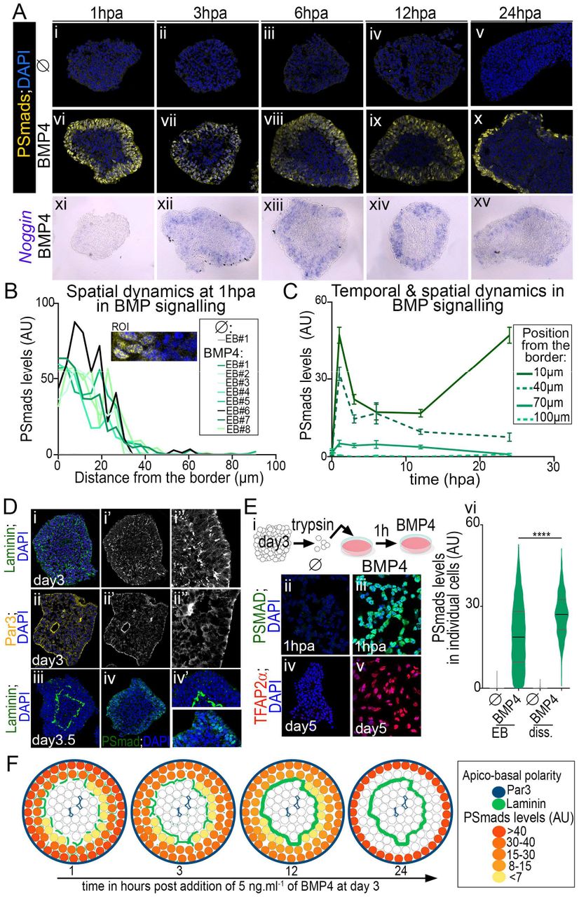
Spatial and temporal dynamics in Smad1/5/9 activity triggered by BMP4 exposure
We next sought to investigate whether BMP4 mediated EB patterning could stem from differential spatial and temporal activation of its intracellular downstream effectors. For this, we monitored the levels of the phosphorylated forms of the TFs Smad1/5/9 (PSmads) from day 3 to 4 in presence or not of BMP4 (Fig. 2A-C).

(A) Immuno-detection of the phosphorylated forms of Smad1/5/9 (PSmads) and DAPI labelling (i-x) and ISH against Noggin (xi-xv) in EB cultured without or with 5ng.ml-1 BMP4 added from day 3 and harvested 1, 3, 6, 12 or 24 hours post BMP4 addition (hpa). (B) Inward gradients of PSmads distribution measured in region of interest (ROI) defined as in inset panel and in 8 independent EB 1hpa. (C) Temporal and spatial dynamics in PSmads levels in cells positioned at discrete distances from EB borders (mean ± s.e.m.). (D) Laminin, Par3, and PSmads immuno-detection and DAPI staining in day 3 and 3.5 EB. In i’, ii’, the laminin and Par3 stainings have been isolated; i”, ii” are blown up of regions in i’, ii’. (E) (i) Schematics of experimental procedure to assess the fate of dissociated cells from day 3 EB and exposed to 5ng.ml-1 BMP4 1h post-dissociation. (ii-v) Immuno-staining of the indicated markers in dissociated cells 1hpa (ii-iii) or at day 5 (iv-v). (iv) Violin plots showing the dispersion of PSmads levels in EB cells or dissociated cells 1hpa (bars: mean±quartiles). (F) Graphs representing the temporal and spatial dynamics in PSmads levels and polarity marker distribution.
At all time-points, PSmads+ cells formed a spatially restricted 3 to 5 cells width ring at the periphery of the EB and cells deep inside the organoid were devoid of PSmads signal (Fig. 2A vi-x, B). The limited rate of BMP proteins diffusion could participate to this spatial restriction (Kicheva et al., 2007; Pomreinke et al., 2017; Zinski et al., 2017). Alternatively, the formation of an extracellular matrix separating an outer epithelium from inner epithelia within the organoid may act as a barrier hampering BMP4 diffusion (Hu et al., 2004; Ma et al., 2017; Perrimon et al., 2012; Plouhinec et al., 2013; Ramirez and Rifkin, 2009; Wang et al., 2008) (Fig. 2D, F). The emergence of epithelia was revealed by analysing the distribution profiles of Par3, a member of the apical protein complexes, and of laminin, a component of the basal lamina (Fig. 2D, not shown). PSmads+ cells delineated exactly the outer epithelium whose apical side faced the culture medium (Fig. 2D, F). The polarity of cells, which is inverted compared neural organoids grown in matrigel (Meinhardt et al., 2014), could arise from focal adhesions between the CLE and PNP cells on day 2 (not shown, (Bedzhov and Zernicka-Goetz, 2014). Furthermore, treating cells that had been dissociated from day 3 EB with BMP4 triggered a homogeneous activation of Smads and drove all cells towards a ncc state (Fig. 2E). These results further support the idea that the emerging 3D structures within organoids restrict spatially BMP4 response (Fig. 2F).
The analysis revealed also that the levels of PSmads within the outer epithelium varied in space and time. Smads activation displayed a decreasing inward gradient across this epithelium (Fig. 2B). In addition, the amplitude of this gradient decreased progressively overtime (Fig. 2C), except in the outer most layer of ncc cells, where after the initial decline, Smads activity elevated again (Fig. 2Ax,C, F, data not shown). The overall temporal adaptation of BMP signalling was supported by the progressive decrease in the levels of BMP4 target genes (Fig. 2Axv, S2Cii) and is likely to be mediated by negative feedback (Bier and De Robertis, 2015). In agreement, BMP antagonist Noggin was transcriptionally induced by BMP4 between 1 and 3 hours post BMP4 addition, when the decline in PSmads levels was observed (Fig. 2Axi-xv). Together, these data indicate that the intracellular response to BMP4 generates stereotyped temporal profiles depending on the position of cells within the organoid epithelium (Fig. 2C, F). This spatial organisation of Smads activity provide an explanation for why the use of BMP4 is not sufficient to drive cells in organoids towards a unique cell type (Andrews et al., 2017; Gupta et al., 2018) but rather creates by itself an organised cellular diversity. Furthermore, the match between this position specific-dynamics and the emergence of distinct cell types argues for a model where “positional information” results from a combination of both the levels and the duration of PSmads (Tozer et al., 2013).
Three tuneable morphogenetic parameters of BMP4 exposure discriminate between dorsal cell types and enrich organoids with specific relay IN subtypes
To test directly the morphogenetic potential of BMP4 in organoids, we sought to modulate the three main parameters known to influence cell response to morphogens (Sagner and Briscoe, 2017): the ligand concentration, the duration of exposure, as well as the time at which the ligand was added (Fig. 3, S4).

Characterization of modulating BMP4 concentration (A), BMP4 exposure duration (B) and the exposure timing (C) on the levels, the spatial distribution and temporal dynamics in PSmads. (Ai, Bi, Ci) Schematics showing the time periods of exposure to BMP4 and BMP4 concentrations used in the aside experiments. (Aii-v, Bii,iii, Cii-iv) Immuno-detection of PSmads and DAPI labelling in EB at the indicated time (hpa; hour post BMP4 addition) in the indicated conditions of exposure; insets in A and C, lower panels in B are blown ups on PSmads+ region. Scale bars: 60 μm. (Avi, Cv) Proportion of PSmads+ cells per image field. (Avii, Biv, Cvi) Violin plots showing the distribution of cells as a function of PSmads levels in organoids in the indicated BMP4 exposure condition and time of analysis.
Increasing BMP4 concentration enlarged the number of PSmads+ cells (Fig. 3Avi), the mean levels in PSmads (Fig. 3Avii), as well as the period of time for which the Smads were active (see insets in Fig. 3Aii-v). Similarly, the duration of BMP4 exposure altered the temporal dynamics of BMP4 signalling. The levels of nuclear PSmads ceased as soon as 6 hours after BMP4 removal (Fig. 3B), arguing against a long-term memory of intracellular signalling by cells exposed to BMP (Tozer et al., 2013). Importantly, the modulation of PSmads dynamics by increasing BMP4 concentration or duration extended the total amount of BMP dependent cell-types generated and promoted more dorsal cell types at the expense of ventral ones (Fig. 4A, C, D, S4A, B).

Phenotypic characterisation of EB upon the modulation of BMP4 concentration (A), of BMP4 exposure timing (B) and of both BMP4 concentration and exposure duration adding BMP4 at day 3.5 (C). (Ai, Bi) Schematics indicating when cells are exposed to BMP4 and at which concentration. (Aii-vii, Bii-vii, Cv-xii) Immuno-staining for the indicated markers in EB grown in the indicated conditions at either day 5 or day 7. (Ci-iv) ISH for the Neurog2 in day 5 EB cultured in the indicated conditions. Scale bars: 60 μm. (Cxiii, xiv) Percentage of dI2 and dI3 IN harbouring the indicated TF code per image field (circles: individual values, bars: mean ± s.e.m.). (D) Graphs displaying the distribution and percentage of cell-subtypes in day 7 organoids cultured in the indicated conditions; white circles mark cells with undetermined fate (UD).
Changing the time at which EBs were exposed to BMP4 also had marked effects on the cell fate adopted (Fig. 4B, S4C and compare Fig. 1C, D to Fig. 4D). Cells displayed a 12 hours competence time window for generating specific BMP4 dependent cell types, the most dorsal cell types requiring an earliest time of exposure than more ventral ones (Fig. 4Bii-vii). This is in agreement with the limitation in time for chick spinal NP to become ncc in response to BMP4 (Sasai et al., 2014). Importantly, the switches in cell competence were not due to alteration in the ability of cells to transduce BMP4 information intracellularly. The amplitude (Fig. 3Cvi), gradient (insets in Fig. 3Cii-iv, 3Cv) and temporal adaptation (not shown) of PSmads profiles remained invariant upon shifts in the timing of BMP4 exposure. Instead, the competence could stem from changes in the molecular state of NP during the course of their differentiation (Fig. S2Bi-vi) (Sasai et al., 2014).
Finally, in agreement with concentration, timing and duration of exposure to BMP4 as tuneable morphogenetic exposure parameters (Sasai et al., 2014; Tozer et al., 2013), we identified conditions to obtain organoids containing a dominant relay IN subpopulation at their periphery (Fig. 4C,D), including dI2 and dI3. Previous protocols had reported poor yields of these cell types (Andrews et al., 2017).
Evolutionary alterations in the length of NP competence time windows to accommodate with discrete tempo of neuronal differentiation
Dynamics of neural differentiation and tissue sizes are highly variable between species (Ebisuya and Briscoe, 2018), raising the question of whether morphogen interpretation is modulated to scale with these dynamics. To address this question, we have adapted our protocol to generate ventral spinal neurons from human iPSC, by removing the Shh agonist SAG (Maury et al., 2014) (Fig. 5Ai). As for mouse ESC, this protocol was sufficient to generate spinal organoids containing mostly dp4-dp6 NP at day 9 of differentiation and associating neurons at day 14 (Fig. 5Aii,iii). Importantly, while both human and mouse dorsal spinal organoids contained similar number of cells (not shown), the time frame of their differentiation is increased by around 2.5 fold in human compared to mouse. Hence, as for motor neuron generation, the tempo of differentiation is a major difference in dorsal differentiation between vertebrate species (Maury et al., 2015; Wichterle et al., 2002).

(A) Generation of associating progenitors and IN from human iPSC. (Ai) Schematics indicating the period of drug treatments to convert hiPSC into dorsal spinal cells in organoids. (Aii-iii) Immuno-staining for the indicated NP (ii) and IN (iii) markers in day 9 and 14 organoids, respectively. (B) Immuno-detection of PSMADs or LAMININ and DAPI labelling in day 4 organoid treated for 1 hour with BMP4. (C) Immunostaining for the indicated NP markers in day 9 EB grown in presence of BMP4 added at the indicated time points (day) and maintained up to day 9. (D) Immuno-staining for the indicated post-mitotic markers in day 14 EB grown in the indicated conditions. (E) Percentage of IN subtypes harbouring the indicated identities (circles: individual values, bars: mean±s.e.m.). Scale bars: 60 μm.
The analysis of the effects of BMP4 on iPSC derived organoids revealed that the response of human cells to BMP4 is similar to that of mouse cells. As with mouse EBs, Smads activation was restricted to the outer layer of cells, which was separated from more inner cells by a basal lamina (Fig. 5B). PSmads levels were the highest in the most peripheral cells and tended to decreased within the width of the epithelium (Fig. 5Bi,i’). Accordingly, several BMP cell types were produced in concentric manner in presence of BMP4 with the most dorsal subtypes been located at a more peripheral position than the ventral ones (Fig. 5D). Finally, as with mouse cells, both BMP4 concentration and the exposure duration influenced the proportion of BMP-dependent cell types produced (Fig. 5D, E). Human NP also displayed competence time windows for the generation of ncc or dp1-dp3 cells (Fig. 5C). Yet, these lasted substantially longer than in mouse (compare Fig. 5C to 4B) and they paralleled the species specific time frame of cell fate acquisition. This brings support to the idea that competence is a consequence of sequential transitions in the transcriptional states of differentiating NP which occur at a slower pace in human (Sasai et al., 2014).
Together our data identified crucial parameters underpinning cell fate switches triggered in organoids treated with BMP4. Firstly, the limited range of BMP4 activity in the emerging 3D structures of the organoids results in the generation of stereotyped profiles of Smads activity in a concentric manner. Changes in the concentration and exposure duration of BMP4 modulate these dynamics in time and space, within the limits imposed by the 3D structures. From these profiles, dorsal progenitor subtypes are spatially organised. This stereotypic arrangement attenuates upon cell delamination and leads to the formation of organoids with intermingled relay IN subtypes. Importantly, these results call for the elaboration of new diffusible compounds capable of activating this pathway in order to be able to reach homogeneity upon targeted differentiation. Secondly, the time of differentiation, i.e the epigenetic state of the receiving cells, appeared as the most constraining parameter to the response of cells to BMP4, with species specificities. The temporal switches in cell competence to respond to BMP4 extend the morphogenetic outputs of this ligand. It also provides robustness to its interpretation, as it no longer relies on the sole precision in diffusion gradient. This certainly explain why dynamics in competence of cells to respond to morphogens is recurrently used during pattern formation (Sagner and Briscoe, 2017). As a consequence, it will require precise temporal control of morphogen signalling by drug treatments during targeted differentiation carried out in organoids.
Contributions
Conceptualization: V.R., S.N., N.D.; Experiments: N.D., C.V., V.R., T. B.; Writing: V.R., S.N., N.D.; Supervision: V.R., S.N.; Funding acquisition: V.R., S.N.
Funding
S.N. and V.R. are INSERM researchers, while N.D. is hired by the Pasteur Institute. Work in the lab of V.R. is supported by CNRS/INSERM ATIP-AVENIR program, as well as by a Ligue Nationale Contre le Cancer grant (PREAC2016.LCC). Studies in the lab of S.N. are funded by an INSERM ATIP-AVENIR program co-sponsored with the Association Française contre les Myopathies (AFM-telethon) and a chair of excellence of the Laboratoire d’Excellence de Biologie pour la Psychiatrie (Bio-Psy) (Bio-Psy: Investissement d’avenir 11-LABX-0035).
Acknowledgement
We deeply thank C. Birchmeier, T. Müller, J.B. Brunet and A. Pierani for antibodies and the ImagoSeine core facility of Institut Jacques Monod, a member of France-BioImaging (ANR-10-INBS-04) and certified IBiSA. We are grateful to Line Manceau, Youcef Frarma, Benoit Sorre and James Briscoe for critical comments on the manuscript.
Bibliography


