

Abstract
In all metazoans, a small number of evolutionarily conserved signaling pathways are reiteratively used during development to orchestrate critical patterning and morphogenetic processes. Among these, Notch (N) signaling is essential for most aspects of tissue patterning where it mediates the communication between adjacent cells to control cell fate specification. In Drosophila, Notch signaling is required for several features of eye development, including the R3/R4 cell fate choice and R7 specification. Here we show that hypomorphic alleles of Notch – belonging to the Nfacet class – reveal a novel phenotype: while photoreceptor specification in the mutant ommatidia is largely normal, defects are observed in ommatidial rotation (OR), a planar cell polarity (PCP)-mediated morphogenetic cell motility process. We demonstrate that during OR Notch signaling is specifically required in the R4 photoreceptor to upregulate the transcription of argos (aos), an inhibitory ligand to the EGFR, to fine-tune the activity of Egfr signaling. Consistently, the loss-of-function defects of Nfacet alleles and EGFR-signaling pathway mutants are largely indistinguishable. A Notch-regulated aos enhancer confers R4 specific expression arguing that aos is directly regulated by Notch signaling in this context via Su(H)- Mam dependent transcription.
Introduction
Drosophila eye development serves as a paradigm for many developmental patterning processes and the dissection of the associated signaling pathways (Cagan and Ready, 1989a; Roignant and Treisman, 2009; Tomlinson and Ready, 1987; Wolff and Ready, 1991). The Drosophila eye consists of ~800 highly regularly arranged ommatidia, or facets, with each consisting of 8 photoreceptor (R-cell) neurons (R1-R8), arranged into a precise invariant trapezoidal pattern, and 12 accessory (cone, pigment, and bristle) cells (Tomlinson and Ready, 1987; Wolff and Ready, 1991). During larval stages, the eye develops from an imaginal disc, which is initially composed of identical pluripotent precursor cells. As a wave of cell proliferation and differentiation (referred to as morphogenetic furrow, MF) moves across the disc from posterior to anterior, it leaves regularly spaced preclusters of differentiating cells in its wake that will mature into ommatidia (Cagan and Ready, 1989a; Roignant and Treisman, 2009; Tomlinson and Ready, 1987; Wolff and Ready, 1991). At the 5-cell precluster stage, several patterning steps are apparent in addition to R-cell induction and differentiation, one being the differential specification of the two cells within the R3/R4 pair, which breaks the initial symmetry of the precluster. This differential R3/R4 specification requires the Wnt-Frizzled (Fz)/Planar Cell Polarity (PCP) pathway and its interplay with and asymmetric upregulation of Notch (N)-signaling (Blair, 1999; Cooper and Bray, 1999; Fanto and Mlodzik, 1999; Mlodzik, 1999; Strutt and Strutt, 1999). This cell fate induction step is followed by the rotation of the ommatidial precluster, referred to as ommatidial rotation, towards the dorsal-ventral (D/V) midline, the so-called equator (Mlodzik, 1999; Strutt and Strutt, 1999). As additional cells are recruited, the precluster undergoes a 90° rotation (in opposing directions in the dorsal and ventral halves of the eye) to establish the mirror-symmetric pattern most apparent in adult ommatidia along the D/V midline (Jenny, 2010) (see also Figure 1A-D).

(A) Schematic of 3rd instar eye imaginal disc. As furrow (MF) moves across the eye disc from posterior to anterior ommatidial preclusters are forming in its wake, a process that involves lateral inhibition and R8 induction. R8 subsequently induces the sequential recruitment of R2/R5 and R3/R4 precursors pairs, resulting in the 5-cell precluster. Once the symmetry of 5-cell preclusters breaks due to differential R3/R4 specification, they start to rotate towards the the dorso-ventral midline (yellow line, “equator”) until they complete a 90° rotation and are aligned perpendicular to the equator. Fmi (magenta), initially detected in junctions of both R3/R4 precursors, becomes enriched to R4 junctional surfaces as the precursors mature. DE-cadherin (green) is upregulated in R2/R5 and R8 cells. (B) Schematic and section view of the two distinct chiral forms of adult ommatidia, displaying mirror image symmetry across the equator (yellow line). (C) Wild-type third larval instar eye imaginal disc stained for Fmi (magenta) and DE-cad (green) with MF at the anterior (left). Note junctional enrichment of Fmi in R4 (white arrows in Fmi monochrome). White dashed cross-arrows indicate orientation angle of preclusters. Yellow arrow marks position of the equator near MF. (C’) Quantification of OR angles at each row plotted for wt eye discs (45<n<60 per row, 8 eye discs). (D-H) Adult eye sections with orientation schematics (arrows are as in B). Note that the equator position is not affected. (D’- H’) Histograms of ommatidial orientation angles of respective genotypes shown in D-H. Wild type (wt) (D, D’), Nfa-3 (E, E’), Nfa-sw (F, F’), mδ0.5>NRNAi (BL7078) (G, G’), and mδ0.5>mamRNAi (BL63601) (H, H’); n>300, 3 eyes per genotype.
Ommatidial rotation (OR) is a paradigm of PCP-mediated cell motility. Posterior to the MF, Wnt-Frizzled (Fz)/PCP signaling not only instructs the R3/R4 cell fate specification (Mlodzik, 1999; Strutt and Strutt, 1999), but also coordinates the direction and degree of OR. This is evident in core PCP mutants: e.g. fz, flamingo/starry night (fmi/stan), or strabismus/Van Gogh (stbm/Vang), which show defects in both R3/R4 specification (and hence ommatidial chirality) and the orientation of ommatidia (Das et al., 2002; Wolff and Rubin, 1998; Zheng et al., 1995). To date, several OR-specific regulators have been discovered based on the ommatidial misorientation phenotypes associated with their mutants (Brown and Freeman, 2003; Choi and Benzer, 1994; Chou and Chien, 2002; Fiehler and Wolff, 2007, 2008; Gaengel and Mlodzik, 2003; Mirkovic et al., 2011; Mirkovic and Mlodzik, 2006; Winter et al., 2001). For example, it is established that Fz/PCP signaling feeds into cadherin-based cell adhesion machinery through downstream effectors to precisely regulate the OR process (Mirkovic et al., 2011). EGF-Receptor (EGFR) signaling has also been shown to contribute to the process and genetic studies implicate input from EGFR signaling into cell adhesion factors (Brown and Freeman, 2003; Gaengel and Mlodzik, 2003). Genetic studies further suggest that cytoskeletal reorganization of ommatidial cells are coordinated with adhesion remodeling to drive the OR process downstream of Fz/PCP, EGFR, and potential other signaling pathways (Fiehler and Wolff, 2007; Gaengel and Mlodzik, 2003; Winter et al., 2001).
Notch (N) signaling is critical for cell fate determination in many if not all tissues in all metazoa mediating many essential cellular processes (Andersson et al., 2011; Bray, 2016). In particular in the Drosophila eye, Notch signaling is required at each step of eye development, ranging from the definition and growth of the eye field, to lateral inhibition within the MF to define correct precluster spacing, and to many aspects of cell fate induction of the individual R-cells and accessory cells including cone cell and pigment cell fate decisions (Doroquez and Rebay, 2006).
The widespread requirement in eye development means that many aspects of eye and ommatidial development are affected when Notch activity is perturbed (Cagan and Ready, 1989b; Papayannopoulos et al., 1998) causing a largely uninterpretable chaos and thus individual steps are very difficult to dissect.
Notch signaling initiates at the cell surface with the binding of a ligand (e.g. Delta) to the Notch receptor (Kopan and Ilagan, 2009; Rebay et al., 1991). Upon this interaction, the Notch receptor undergoes two sequential cleavages, releasing the Notch intracellular domain (NICD) and allowing for its translocation to the nucleus (Brou et al., 2000; De Strooper et al., 1999; Lieber et al., 2002; Mumm et al., 2000; Struhl and Greenwald, 1999). Once in the nucleus, NICD complexes with Mastermind (Mam) – a Notch pathway specific co-activator - and Suppressor of Hairless (Su[H]) - a transcription factor of the CSL family - to promote target gene expression (Bray, 2016; Struhl and Adachi, 1998; Wilson and Kovall, 2006; Wu et al., 2000). The precise signaling outcome depends on the genes that are regulated in each particular context (Bray, 2016; Bray and Gomez-Lamarca, 2018). Given the multiple requirements for Notch activity in eye and ommatidial development (Cagan and Ready, 1989b; Papayannopoulos et al., 1998), it is likely that different primary targets will be involved in implementing each distinct and individual role.
A well-established function of Notch signaling is in the context of R3/R4 cell fate specification in cooperation with Fz/PCP signaling (Cooper and Bray, 1999; Fanto and Mlodzik, 1999; Tomlinson and Struhl, 1999). At the 5-cell precluster stage, among the R3/R4 precursors, the cell that is closer to the equator ends up having higher Fz/PCP signaling activity, specifying it as an R3 cell. Fz-PCP signaling-dependent transcriptional upregulation of Delta (Dl) and neuralized (neu) in R3 subsequently induces the adjacent cell of the pair as R4 by a classical Dl-N interaction, thus activating the Notch pathway to higher levels in the R4 precursor (Cooper and Bray, 1999; del Alamo and Mlodzik, 2006; Fanto and Mlodzik, 1999; Tomlinson and Struhl, 1999; Weber et al., 2000). Since N-signaling is critical for R3/R4 asymmetry, its perturbation might also cause OR phenotypes brought about by R4 specification defects. However, strikingly, a class of hypomorphic N alleles, the so-called facet alleles Nfa-3 and Nfa-sw (Markopoulou et al., 1989), exhibits misorientation of ommatidia yet largely normal eye patterning with correct R4 specification and ommatidial chirality establishment (Figure 1E,F). This orientation-specific ommatidial phenotype is reminiscent of previously identified mutants that are linked to OR (e.g. nmo or argosrlt (Choi and Benzer, 1994; Gaengel and Mlodzik, 2003) and suggests that Notch signaling has a direct role in rotation which is independent of R4 specification per se. A potential role of Notch signaling in OR and associated morphogenesis, however, has not yet been explored.
Using a combination of phenotypic analyses, genetic interactions, cell-based and molecular studies, we define here an OR-specific role for Notch signaling after R4 fate specification, in addition to its well described function in cell fate choices in ommatidial patterning. We demonstrate that Notch signaling coordinates the morphogenetic process of OR by fine-tuning the activity of the EGFR pathway and PCP signaling. Specifically, Notch signaling in R4 leads to a direct transcriptional upregulation of argos (aos), as confirmed by Su(H) DNA occupancy and reporter expression studies. As Argos is an inhibitory ligand to EGFR and EGFR-signaling is required for OR regulation, the loss or reduction of Notch-dependent aos expression leads to an imbalance between positively and negatively acting EGFR ligands and hence to defects in the OR outcome. In addition, Notch signaling affects the levels of the PCP protein Flamingo (Fmi) in R4. This dual Notch signaling input into EGFR and PCP-pathways orchestrates a precise OR process and hence demonstrates a critical and specific role of Notch activation during OR.
Results
Notch signaling is required in R3/R4 pairs for accurate ommatidial rotation
Ommatidial rotation (OR) is a morphogenetic process that occurs during larval eye development, and it results in the final orientation of ommatidia, forming a mirror image arrangement across the dorso-ventral (D/V)-midline in the adult eyes (Figure 1A-D). OR is instructed by Fz/PCP signaling and associated pathways including Notch and EGFR-signaling that are involved in R3/R4 photoreceptor fate specification (Brown and Freeman, 2003; Cooper and Bray, 1999; Fanto and Mlodzik, 1999; Gaengel and Mlodzik, 2003; Strutt and Strutt, 2003; Tomlinson and Struhl, 1999).
At larval stages, as the morphogenetic furrow (MF) sweeps across the eye disc form posterior to anterior, it induces the formation of a new row of regularly spaced ommatidial preclusters every ~2 hours (Fig. 1A,C, and (Tomlinson and Ready, 1987; Wolff and Ready, 1991), giving rise to rows of ommatidial clusters that are ~2 hours apart from each other in developmental time and thus allowing the visualization of progressively more mature clusters in the same tissue sample (Fig. 1A,C; also (Jenny, 2010). As such, these rows reflect consecutive stages of ommatidial maturation allowing for the tracking of OR row by row as the eye disc develops (Fig. 1C-C’). The use of apical junctional markers, like E-cadherin (E-cad, enriched at the junctions of R2/R5 and R8 cell boundaries), and Flamingo (Fmi, enriched at the apical junctions of R4) allows for the tracking of OR angles of individual clusters during development (Fig. 1A,1C-C’). In wild type, ommatidial (pre)clusters initiate rotation in row 5 and largely complete the process by rows 14- 15 (Figure 1C-C’), resulting in a 90° rotation angle that aligns mature ommatidia perpendicular to the D/V midline in the adult (Figure 1B,1D-D’; note that the final angle in wild-type is an invariant 90°, Fig. 1D-D’; (Jenny, 2010).
To investigate the role of Notch signaling in OR, we first analyzed two recessive hypomorphic Notch (N) alleles: facet-strawberry (Nfa-sw) and facet-3 (Nfa-3). The facet class of a Notch alleles have been characterized and are thought to be caused by either an insertion\s of transposable element into an intronic region of Notch or deletion of non-transcribed sequences in the locus, thus not affecting the coding sequence and ultimate protein product but causing a reduction in gene expression in certain contexts (Markopoulou et al., 1989). In comparison to wild type, hemizygous Nfa-sw and Nfa-3 males showed frequent misrotations of ommatidia, including both under- and over-rotation of individual clusters. Surprisingly, besides the OR defects, these mutant eyes were normal in eye size, photoreceptor specification, chiral arrangements, and other aspects of eye development (Figure 1E-F’; also (Markopoulou et al., 1989).
Since differential R3/R4 cell specification is critical for OR (Mlodzik, 1999; Strutt and Strutt, 1999), we next asked if Notch signaling is required in this cell pair for the OR process. To separate the potential role of Notch signaling in OR from its role in the R3/R4 cell fate choice, we took advantage of the Gal4/UAS-system (Brand and Perrimon, 1993), which allows for temporal knock-down of the respective genes and employed a driver (mδ0.5-Gal4) that is active in the R3/R4 pair and is upregulated in R4, as a result of Notch-mediated R4 specification (Cooper and Bray, 1999), where it is subsequently maintained during the OR process. As it is up-regulated in response to R4 specification, gene targeting with this driver should not significantly affect cell fate specification within the R3/R4 pair. Knockdown of Notch via mδ0.5-Gal4 in R3/R4 cells phenocopied the misorientation phenotype(s) observed in the N-facet alleles, without affecting R3/R4 cell fate choices, suggesting that Notch activity is required in R3/R4 cells, and predominantly in R4, after cell fate determination to regulate OR (Figure 1G-G’, Figure S1A-A’). To ask whether this requires Notch-mediated transcriptional activation, we tested the requirement for mastermind (mam), a Notch-specific transcriptional co-activator (Wu et al., 2000). As the Notch-associated DNA binding factor, Su(H), is also required for transcriptional repression, its perturbation could yield complex effects making it a less suitable choice (Barolo et al., 2002; Furriols and Bray, 2000; Morel et al., 2001; Wu et al., 2000). Strikingly, when knocked-down in the same mδ0.5-Gal4 based assay (mδ0.5>mamRNAi), depletion of mam produced very similar phenotypes to the N knock-down, displaying OR defects with over- and under-rotated ommatidia (Figure 1H-H’, Figure S1B-B’) thus suggesting that Notch-dependent transcription is critical for accurate OR.
Notch signaling affects OR during the developmental process in eye imaginal discs
To rule out the possibility that the misorientation phenotypes observed in adults upon Notch signaling perturbation were due to a later secondary cell packing effect – Notch is required for several late steps in ommatidial patterning (Cagan and Ready, 1989b)- we followed the rotation of ommatidial clusters during development in eye discs at the time of the OR process (see for example Figure 1C for wild-type). As a standard clonal analysis cannot be used for Notch pathway components due to the multitude of steps affected by Notch signaling in the eye disc (Cagan and Ready, 1989b), we again used the mδ0.5-Gal4-mediated knockdown (KD) strategy. In contrast to wild-type eye discs, KD of Notch or mam in R4 cells led to an abnormal rotational pattern, where ommatidial preclusters displayed a significantly wider range of rotation angles as compared to wild-type (Figure 2, and Figure S2), consistent with the under and over-rotation phenotypes seen in adult eyes (Fig. 1G-H’). Taken together, these data indicate that Notch signaling controls, via its transcriptional activation function, the rotation of ommatidial clusters during early stages of the process during eye development.

(A-C) Third instar larval eye imaginal discs stained for DE-cad in wild type (A), mδ0.5>NRNAi (BL31383) (B), and mδ0.5>mamRNAi (BL28046) (C). Blue, red and green dashed cross-arrows, respectively, indicate the orientation of ommatidial preclusters for each genotype. (D) Quantification of rotation angles observed in individual preclusters in rows 5-11, plotted for control (wild type) in blue; mδ0.5>NRNAi (red); and mδ0.5>mamRNAi (green). Statistical analyses were performed for each row between control (blue) and colored genotypes. Asterisks denote significance by chi-square test (* p<0.05, ** p<0.005, *** p<0.0005). Note the generally wider spread of rotation angles in most rows in the experimental genotypes as compared to wild-type. Scale bars indicate 10 μm.
Notch signaling genetically interacts with argos in OR establishment
To get further insight into how Notch signaling might mechanistically affect OR, we tested for genetic interactions between the mδ0.5>mamRNAi genotype and known regulators of OR. Several genes have been implicated in regulating OR based on ommatidial misrotation observed in their mutants and the role of several of these has been further validated by functional and molecular studies (Brown and Freeman, 2003; Choi and Benzer, 1994; Chou and Chien, 2002; Cooper and Bray, 1999; Fiehler and Wolff, 2007, 2008; Winter et al., 2001).
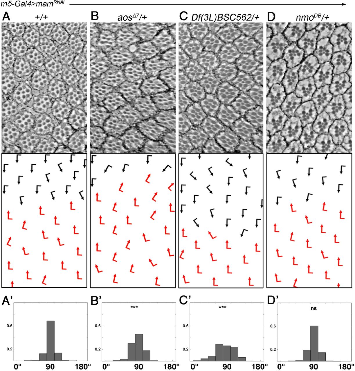
We used a mδ0.5>mamRNAi combination that caused mild OR defects at low temperatures and asked whether its phenotype can be dominantly modified by other OR-associated genes. Among the known OR regulators, we detected a specific interaction with multiple alleles of argos (aos; including a deficiency for the gene), whereas other OR-associated genes tested did not show an interaction (Figure 3, Suppl. Data, Table S1). In parallel, we also asked whether the core PCP genes could modify the mδ0.5>mamRNAi OR phenotype, as PCP factors contribute to OR (Das et al., 2002; Wolff and Rubin, 1998; Zheng et al., 1995). Whereas most core PCP genes did not dominantly affect the mδ0.5>mamRNAi phenotype, alleles of prickle (pk) did enhance the OR defects (Suppl. data, Figure S3). Neither aos nor pk heterozygosity affected OR or other aspects of eye development on its own, confirming that their interaction with the mδ0.5>mamRNAi background is not an additive feature (Figure S4). The pk−/+ effect was surprising, because pk itself does not display much of an OR phenotype in comparison to the other core PCP genes. However, pk is genetically required in R4 (Jenny et al., 2003), where mδ0.5-Gal4 is driving expression and so an R4 specific interaction could be envisioned (see Discussion).

(A-D’) Adult eye sections with ommatidial orientation schematics (arrows as in Figure 1) and orientation angle histograms of eyes of the genotypes indicated. All genotypes are mδ0.5Gal4, UAS-mamRNAi (BL28046) mδGal4>mamRNAi in the following genetic backgrounds: (A-A’) +/+ (wild-type control); (B-B’) aosΔ7/+, (C-C’) Df(3L)BSC562/+ (deletion of aos gene), and (D-D’) nmoDB/+. Asterisks denote significance by chi-square test (***p<0.0005). Note robust enhancement of the mδ0.5Gal4>mamRNAi rotation phenotype by both aos−/+ genotypes. See supplemental material for additional genotypes.
Taken together, these genetic data raised the possibility that Notch signaling modulates the OR process via the EGFR pathway, because Argos is a secreted EGFR ligand that inhibits the receptor function (Klein et al., 2004; Klein et al., 2008; Vinos and Freeman, 2000), and via R4-associated PCP signaling. As the enhancement(s) are associated with a reduction in the transcriptional output of Notch signaling, caused by the mδ0.5>mamRNAi genotype, the expression of some of these genes might be directly regulated by Notch-signaling in R4.
Notch promotes aos expression in R4
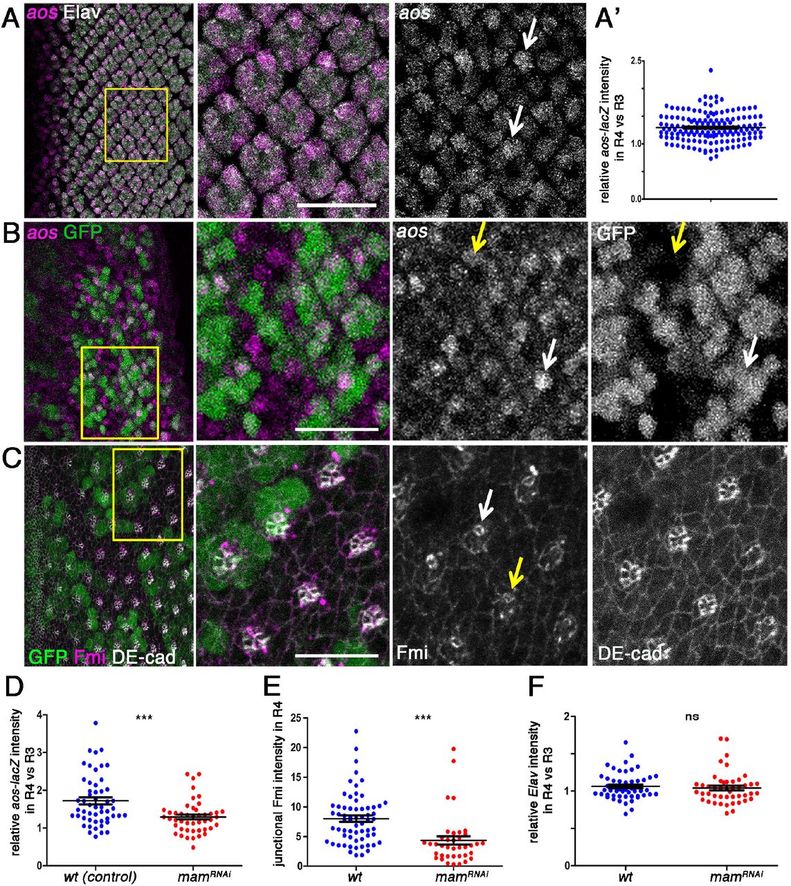
To determine how Notch signaling interacts with aos during OR, we next examined the expression pattern of aos during eye disc patterning and development. Several aos alleles have been associated with ommatidial misrotation, notably roulette (aosrlt) was identified as an OR specific mutation (Choi and Benzer, 1994), even before aos was characterized as an EGFR ligand. Aos was subsequently used as a tool to define the role of EGFR/Ras signaling in OR (Brown and Freeman, 2003; Gaengel and Mlodzik, 2003; Strutt and Strutt, 2003). Interestingly, although aos is expressed at base levels in all photoreceptors of developing ommatidia (Figure 4A, also (McNeill et al., 2008; Yan et al., 2009), it was specifically upregulated in the R4 cell during the OR process, as detected by an enhancer trap reporter for the gene (aos-lacZ; Figure 4A-A’). We thus asked whether this aos upregulation is dependent on Notch/Mam-signaling. To this end, we employed mδ0.5>mamRNAi in a mosaic clonal manner, which allows for direct comparison of wild-type and mam KD ommatidia within the same tissue. In this assay, mam KD caused a marked reduction in aos-lacZ expression levels in R4 (Figure 4B,D). This is consistent with the hypothesis that Notch signaling activation in R4 is required for aos upregulation in this cell. The levels of Elav, a nuclear neuronal (all R-cells) marker, were unchanged in the same genetic scenario (Figure 4F, also Suppl. data Figure S5), indicating that Notch signaling activity in R4 specifically affects aos transcription.

(A) Wild type third larval instar eye imaginal disc stained for Elav (gray) and aos-lacZ (magenta and monochorme panel). (A’) Quantifcation of expression level of aos-lacZ in R4 relative to R3 (see also Supplemental data for control quantification fo Elav). (B-C) Third larval instar eye imaginal discs mosaic for mδ0.5>mamRNAi (BL28046; marked by absence of GFP/green) stained for aos-lacZ (magenta in B and monochrome); Fmi (gray in C and monochrome) and DE-cad (magenta in C and monochrome). White and yellow arrows point at R4 cells in wild type and mutant tissue, respectively; note reduction of aos-lacZ and Fmi staining in mutant areas of the respective panels. (D) Quantification aos-lacZ expression in R4 relative to R3 plotted for individual clusters in wt-control (blue) and mδ0.5>mamRNAi (red). (E) Quantification of junctional Fmi intensity normalized to DE-cad staining in R4 plotted for individual clusters for wt (blue) and mδ0.5>mamRNAi (red). (F) Quantification of Elav intensity in R4 relative to R3 plotted for individual clusters with wt (blue) and mδ0.5>mamRNAi (red). Asterisks denote significance by chi-square test (*** p<0.0005). Scale bars indicate 10 μm.
Upon Notch-mediated R4 cell specification, several of the core PCP factors are enriched in R4. This is most evident with the increase of Flamingo (Fmi, also called starry night/stan) levels at the apical junctional region in R4. Fmi is an atypical cadherin that plays a central role in PCP establishment by stabilizing the core PCP complexes at junctional regions across cell membranes (Wu and Mlodzik, 2009). In particular in eye discs, before R3/R4 differentiates, Fmi is apically enriched in both precursors. As the symmetry of the precluster breaks and R4 is specified, it becomes enriched in the apical surface of R4 (Das et al., 2002) and this upregulation serves as an R4 marker (see also Figure 1A,C). In fmi mutants, ommatidia adopt a random chiral form or lose asymmetry, and additionally display misrotation defects (Das et al., 2002). As we detected a genetic interaction between mδ0.5>mamIR and pk−/+, we also examined Fmi expression as an indicator of core PCP factor levels. Upon comparing the Fmi expression pattern/junctional levels between mam KD and neighboring wild-type ommatidia in the respective mosaic eye discs (employing again mosaic clonal mδ0.5>mamRNAi-mediated KD), we observed a significant reduction in apical Fmi levels in Mam-depleted R4 cells (Figure 4B,D) suggesting a Notch-mediated upregulation of core PCP factors in R4 (see below and Discussion).
argos is a direct R4-specific transcriptional target of Notch signaling
To corroborate and refine the hypothesis that Notch-signaling directly regulates the transcription of aos, we examined whether the aos locus was occupied by with the core Notch-associated transcription factor, Su(H), in a genome-wide chromatin immunoprecipitation (ChIP) data-set from Drosophila larval central nervous system (Zacharioudaki et al., 2016). Strikingly there was significant enrichment of Su(H) within the first intron of aos overlapping with predicted conserved Su(H) binding-motifs, consistent with a direct regulation by Notch/Mam/Su(H) complexes (Fig. 5A). To ask which cells in the developing eye disc are susceptible to this regulatory input, we utilized a GFP reporter construct, encompassing the high confidence “peak” region from the Su(H) ChIP, which had previously been shown to respond to Notch in muscle progenitor cells (aos1-GFP, Fig. 5B; (Housden et al., 2014) and tested its expression in the eye discs. Strikingly, expression of aos1-GFP was detected predominantly in R4 cells, as confirmed by co-expression of the R4 marker mδ0.5-lacZ (Figure 5C). The transcriptional regulation of aos by Notch/Mam-Su(H) in R4 appears specific, as there was little or no Su(H) binding detected at the fmi or pk loci in the same ChIP data-set (Suppl. Figure S6). Taken together, these data indicate that aos is a specific transcriptional target of Notch-signaling in R4, and that the visible increase in Fmi protein in R4 is likely due to other post-transcriptional mechanisms (see Discussion).

(A) ChIP enrichment for Su(H)-occupancy at the aos locus in CNS samples (α-Su(H) enrichment relative to input, scale log2) (Zacharioudaki et al., 2016). Blue bar indicates the region of significant enrichment. Gray bars indicate the positions of Su(H)-binding motifs; bar height represents the motif-score (scale 0-5); upper graph indicates motif conservation across 12 Drosophila species. Gene regions are depicted in dark blue. Green bar indicates the genomic region used in the aos1-GFP reporter construct (Housden et al., 2014). (B-B’) Third instar eye imaginal disc stained for aos1-GFP (green), Elav (nuclei of all R-cells, gray), and mδ0.5-lacZ (magenta, R4-specific marker). B’ panels show higher magnification of boxed area in B. Yellow arrowheads highlight examples of R4 cells revealing the co-expression of aos1-GFP and the R4 marker mδ0.5-lacZ. Scale bar indicates 10 μm.
Discussion
The involvement of Notch signaling in controlling cell proliferation, cell differentiation, and patterning has been studied in a vast set of contexts ranging from neuronal development to intestinal homeostasis in flies and vertebrates (Andersson et al., 2011; Bray, 2016; Henrique and Schweisguth, 2019). Spatial and temporal control of Notch activity, along with the employment of cell/tissue specific downstream elements and crosstalk with other signaling pathways, confers the functional versatility and specific reiterative use of Notch pathway activation (Andersson et al., 2011; Bray, 2016; Doroquez and Rebay, 2006; Guruharsha et al., 2012; Henrique and Schweisguth, 2019). In the Drosophila eye alone, for instance, Notch signaling has a defined and specific function at nearly every stage of tissue development and patterning (Doroquez and Rebay, 2006). At early larval stages Notch activity in the eye is restricted to the dorsoventral equator, from which it promotes the growth of the eye disc and eye field within the disc, and the formation of the MF (Dominguez and de Celis, 1998; Kenyon et al., 2003; Reynolds-Kenneally and Mlodzik, 2005). Within and posterior to the MF, Notch signaling is first required for the spacing of the R8 precursors and thus ommatidial preclusters (Baker et al., 1990), and the subsequent specification of R3/R4 and R7 fates in a stepwise fashion (Baonza and Freeman, 2001; Cooper and Bray, 1999, 2000; Fanto and Mlodzik, 1999; Tomlinson and Struhl, 1999, 2001). As ommatidial clusters mature further, Notch signaling controls the acquisition of the cone and pigment cell fates and apoptosis of the non-committed remaining interommatidial cells to generate the precise and highly ordered pattern of a fully-developed ommatidia (Cagan and Ready, 1989b).
At each stage, Notch signaling acts in concert with multiple other pathways in a spatially and temporally restricted manner in order to achieve particular and specific readouts. For example, Fz/PCP signaling pathway triggers Delta expression in R3 to induce Notch activation and the resulting R4 cell fate in the adjacent cell of the R3/R4 pair (Cooper and Bray, 1999; Fanto and Mlodzik, 1999; Weber et al., 2000). Furthermore, at nearly every step mentioned, there is cross-talk between Notch and EGFR signaling pathways (including the R3/R4 specification steps; (Weber et al., 2008) to achieve the respective developmental outcome (Doroquez and Rebay, 2006). However, the nature of the interaction between Notch and EGFR pathways, and the downstream elements engaged, differ depending on the context. For example, in the course of R7 specification, Notch promotes the expression of the transcriptional repressor Yan which in turn needs to be post-translationally repressed by EGFR signaling to establish R7 fate (Rohrbaugh et al., 2002). On the other hand, Notch and EGFR signaling effectors combinatorially drive the expression of the Drosophila paired box gene 2 (dPax2) to promote cone cell identity (Flores et al., 2000). The interactions of Notch signaling with the EGFR and the Wnt-Fz/PCP pathways have not only been well-documented in eye patterning but also in other developmental contexts and cancer, highlighting the importance of the communication of Notch with the respective pathways during development and disease (Baker et al., 2014; Bao, 2014; Capilla et al., 2012; Haruki et al., 2005; Hasson and Paroush, 2006; Yoo et al., 2004).
Our results document a new function for Notch signaling in R4 to govern the morphogenetic process of OR, which is independent of its role in R4 cell fate acquisition. Perturbation of Notch signaling pathway components in the R3/R4 pair, and R4 in particular, after the cell fate choice is established, leads to the misregulation of the rotation process. In this context, Notch signaling regulates aos and fmi, as their expression levels in R4 are diminished upon downregulation of Notch signaling. Our data demonstrate that aos transcription is directly regulated by Notch, via Su(H)/Mam/NICD mediated transcriptional control, but the effect on fmi levels is less well defined, as there is no clear evidence to suggest direct transcriptional regulation in this case. The effect of Notch on apical Fmi levels is unlikely to be a secondary effect of aos deregulation, since Fmi has been reported to be expressed in R4 and ectopically in R3 in the absence of aos (Gaengel and Mlodzik, 2003). Yet, how Notch-signaling in R4 regulates the levels and function of the core PCP factors in R4 remains largely elusive at this time.
Based on previous reports in Drosophila, the effect of Notch signaling on aos expression is context dependent (Housden et al., 2014). Gene expression is often controlled by multiple cis-regulatory units that integrate information from various transcriptional inputs. Essentially, aos has been reported to exhibit context-dependent enhancer selection in the wing: aos contains three enhancer regions identified, that are differentially responsive to factors that act downstream of Notch or EGFR signaling pathways (Ajuria et al., 2011; Krejci et al., 2009). The presence of context-determining factors will determine whether an enhancer is primed to react to a specific signal, which may result in a gene having different responses towards the same signals depending on which enhancer is accessible (Housden et al., 2014). Notably, the aos1-GFP reporter experiment reported here, and the Su(H) ChIP data, argue that the aos1 enhancer is directly responsive to Notch signaling in R4, indicating that Notch activates aos expression in this context.
Variations in the phenotypes from mutations in rotation-specific genes are indicative of their function in OR. For example, mutations disrupting the nmo kinase result in a severe under-rotation of ommatidia arguing that it has a positive role in promoting rotation (Fiehler and Wolff, 2008; Mirkovic et al., 2011). In contrast, aosrlt mutants exhibit random rotation angles with both under- and over-rotated ommatidia (Brown and Freeman, 2003; Gaengel and Mlodzik, 2003; Strutt and Strutt, 2003). This suggests that the consequent change in EGFR signaling results in an overall misregulation of rotation, rather than promoting or inhibiting ommatidial motility per se. Consistent with the notion that Notch-signaling in R4 directly regulates aos expression, the phenotype caused by facet alleles of Notch largely mimics that of aosrlt, with an overall deregulation of the process and resulting random rotation angles. All R3/R4-specific Notch or Mam RNAi interference scenarios also mimic these phenotypes. Given that OR entails the coordination of cytoskeletal and adhesion dynamics (Chou and Chien, 2002; Fiehler and Wolff, 2007; Gaengel and Mlodzik, 2003; Mirkovic et al., 2011), our data suggest an input from Notch signaling into these molecular processes through negative regulation of EGFR-signaling and possibly also its interplay with PCP signaling.
In recent years, involvement of Notch signaling in morphogenesis has been suggested in various contexts, including Drosophila oogenesis, zebrafish sensory organ development, and human vascular barrier formation (Dobens and Raftery, 2000; Kozlovskaja-Gumbriene et al., 2017; Torres et al., 2003). These studies also suggest an input from Notch signaling into the cell adhesion and/or cytoskeletal factors, mostly through Notch-mediated transcription of genes that regulate adhesion and cytoskeletal dynamics (Pezeron et al., 2014) although a direct input from the Notch receptor into adhesion has also been revealed (Polacheck et al., 2017). Overall, the multifaceted involvement of Notch signaling in cellular (re)organization and morphogenesis is becoming increasingly evident. Future studies will be needed to provide insight into the mechanistic details of how Notch can mediate distinct morphogenetic processes. As Notch signaling has long been implicated in cancer metastasis, such studies will also hold promise for better understanding of disease and hence future therapeutic applications.
Materials and Methods
Fly strains and genetics
Flies were raised on standard medium and maintained at 25°C unless otherwise stated.
Nfa-sw and Nfa-3 were gifts from Spyros Artavanis-Tsakonas.
nmoDB/TM6b and mδ0.5-Gal4 FRT40/SM3:TM6b were from Mlodzik lab stocks.
aosΔ7/TM3, Df(3L)BSC562/TM3, pkpk-sple13/CyO, pkpk-sple6/CyO, w1118, NotchRNAi (BL31383, BL7078) and mamRNAi lines (BL28046, BL63601) were ordered from Bloomington Drosophila Stock Center.
aos-lacZ/TM6b was a kind gift from Utpal Banerjee.
aos1-GFP/TM3 was from Bray lab stocks (Housden et al., 2014).
mδ0.5>NRNAi BL31383 (mδ0.5-Gal4/+; UAS-NRNAi BL31383/+) were obtained at 25°C.
mδ0.5>mamRNAi BL28046 (mδ0.5-Gal4; UAS-mamRNAi BL28046 /+) were obtained at 18°C.
mδ0.5>NRNAi BL7078 (mδ0.5-Gal4/+; UAS-NRNAi BL7078/+) were obtained at 18°C.
mδ0.5>mamRNAi BL63601 (mδ0.5-Gal4, UAS- mamRNAi BL63601/+) were obtained at 18°C.
Control eye disc stainings were done in mδ0.5-Gal4 FRT40/+ background.
Genetic interactions were tested at 25°C between mδ-Gal4/+; UAS-mamRNAi BL28046/+ and the heterozygosity of the respective genes.
mδ0.5>mamRNAi BL28046 clones were obtained at 25°C by employing FLP/FRT mediated mitotic recombination with the following genotypes:
eyFLP/+; mδ0.5-Gal4 FRT40/ubiGFP FRT40; UAS-mamRNAi BL28046aoslacZ/+
eyFLP/+; mδ0.5-Gal4 FRT40/ubiGFP FRT40; UAS-mamRNAi BL28046/+
Immunohistochemistry and Histology
Adult eye sectioning was performed as previously described (Jenny, 2011).
Third larval instar eye discs were dissected in ice-cold PBS and fixed in PBT (PBS+0.1% Triton-X)-4% formaldehyde for 12 minutes at room temperature. For immunohistochemistry, following primary antibodies were used: rat anti-DE-cad (1:20, DSHB), mouse anti-Fmi (1:10, DSHB), rabbit anti-β-gal (1:200, ICL), rat anti-Elav (1:100, DSHB), chicken anti-GFP (1:1000, Aves Labs). Secondary antibodies were obtained from Jackson Laboratories. Eye disc images were acquired by using Leica SP5 DMI microscope.
Quantitative Analysis of Adult Eye Sections
The orientation of each ommatidium was marked based on the trapezoidal organization of the R-cells (see Figure 1B, D-H). A linear equator has been drawn along the boundary where two chiral forms meet. Clockwise and counter-clockwise angles from the equator to each ommatidia were measured for the black and red chiral forms respectively (see Figure 1B, D-H). Measurements were done by using ImageJ (National Institute of Health). The absolute values of measured angles from 3-4 independent eye sections for each genotype were pooled (300<n<550) and plotted in a polar histogram by using MATLAB. The angles were binned into 20° intervals between 0-180° and they were plotted in probability ratios from 0 to 1. For statistical analyses, the angles (α) were binned into 3 categories (α<60, 60<α<120, 120<α) for individual genotypes and chi-square test was performed.
Quantitative Analysis of Eye Discs
The orientation of each ommatidium was marked perpendicular to the plane of R2/R5 cells (See Figure 1C). A linear equator was drawn perpendicular to the MF at the dorsoventral midline. Clockwise and counter-clockwise angles from the equator to each ommatidia were measured for the dorsal and ventral halves respectively. To avoid a potential bias due to the developmental delay in rotation from equator to the poles, the measurements were limited to the first 8 ommatidia from the equator for each row. Measurements were done by using ImageJ (National Institute of Health). The absolute values of measured angles from 7-8 independent eye discs (45<n<60) were pooled and violin plotted in PRISM. For statistical analyses, the angles (α) from individual rows were binned into 5 categories (α<40, 40<α<50, 50<α<60, 60<α70 and α>70) for each genotype and chi-square test was performed.
For aos-lacZ and Elav quantifications in mδ0.5>mamRNAi BL28046 mosaic eye discs, confocal stacks were maximum projected and individual cell intensities were measured in R3/R4 pairs between rows 5-11 by using ImageJ. Intensities in GFP+ and GFP− R4 cells were normalized to their GFP− R3 neighbors within each pair. Measurements from 5 discs were pooled (45<n<60) and plotted. For statistical analyses, the normalized intensity measurements (ι) were binned into 3 categories (ι<1, 1<ι<1.5, ι>1.5) for each genotype and chi-square test was performed.
For Fmi quantifications in mδ0.5>mamRNAi BL28046 mosaic eye discs, the confocal stacks were constructed in 3D and analyzed in IMARIS. Within GFP+ and GFP− tissue, Fmi surface intensity on the apical membrane was measured for each R4 cell between rows 5-11 and normalized to the DE-cad intensity on the respective surface and plotted. Measurements from 5 discs were pooled (35<n<60) and plotted. For statistical analyses, the normalized intensity measurements (ι) were binned into 3 categories (ι<500, 500<ι<1000, ι>1000) for each genotype and chi-square test was performed.
Acknowledgements
We thank the Bloomington Drosophila Research Center, Spyros Artavanis-Tsakonas and Utpal Banerjee for fly strains and reagents. We are grateful to all Mlodzik lab members for helpful input and discussions; and Robert Krauss, Cathie Pfleger, Timothy Blenkinsop and Jennifer Zallen for helpful comments and suggestions on the manuscript. Confocal laser scanning microscopy was performed at the ISMMS-Microscopy Core Facility supported by the Tisch Cancer Institute grant P30 CA196521 from the NCI. This work was supported by a NIH/NEI grant RO1 EY13256 to M.M.
Footnotes
References


