

Abstract
The paucity of recurrent mutations has hampered efforts to understand the pathogenesis of neuroblastoma. Through analysis of RNA-sequenced neuroblastoma, we identified >900 primarily intrachromosomal fusion transcripts generated by genes in close proximity. Fusions were enriched in chromosomal regions gained or lost in neuroblastoma and included well-known neuroblastoma oncogenes. The majority of fusions contained canonical splicing sites and a subset exhibited increased sensitivity to spliceosome inhibition. As a proof-of-principle that a gene product with altered properties can be produced by these fusions, we characterized the ZNF451-BAG2 fusion which generates a truncated BAG2-protein capable of inhibiting retinoic acid-induced differentiation. Our findings elucidate a mechanism through which altered gene products, relevant for neuroblastoma pathogenesis and representing possible novel drug targets, can be generated.
Introduction
Despite intense sequencing efforts few recurrently mutated genes have been identified in neuroblastoma (1, 2), resulting in a deficiency of drug targets. Instead, high-risk neuroblastoma is characterized by large-scale chromosomal rearrangements such as chromothripsis and loss or gain of chromosomal regions (e.g. loss of 1p36 and 11q or gain of 17q and 2p) with or without MYCN amplification (1, 3). Certain other types of tumors harbor and are driven by fusion proteins generated by chromosomal translocations (e.g. BCR-ABL in chronic myelogenous leukemia) (4). An additional mode, through which fusion transcripts can be generated, is represented by cis-splicing of adjacent genes (5, 6). Besides a fusion resulting from small interstitial genomic deletions at 11q generating either a MLL-FOXR1 or a PAFAH1B2-FOXR2 fusion (7) no intra-chromosomal chimeric transcripts have been described in neuroblastoma. However, they have been shown to be present in different tumor types as well as in non-transformed tissues and be promoted by different types of cellular stress such as infections or mutations (8–12). In order to explore whether neuroblastoma tumors harbor previously undetected gene fusions, we analyzed a cohort of 172 sequenced neuroblastoma tumors. We identified an abundance of fusion transcripts, of which a significant proportion exhibited a distinct genomic distribution according to tumor risk. Identified fusions were predominantly generated by genes in close proximity and flanked by canonical splicing donors and acceptors. This pattern was distinct to fusions unique for neuroblastoma, whereas fusions we identified in normal adrenal gland or in other tumors did not exhibit such a pattern. Furthermore, a subset of identified NB specific fusions was hypersensitive to pharmacologic spliceosome inhibition in comparison to their wild type cognates. High expression levels of spliceosome factors were strongly associated with high-risk disease and spliceosome inhibition also promoted apoptosis in neuroblastoma cells. As a proof of principle, that fusions can generate novel gene products with alternative properties, we cloned and characterized the ZNF451-BAG2 fusion. The generated protein exhibited distinct protein-protein binding properties compared to wild-type BAG2 and impeded retinoic acid induced differentiation. This reveals how a fusion gene product can influence neuroblastoma response to a drug commonly used in the treatment of high-risk patients (13).
Results
Fusion transcripts are a common feature of neuroblastoma
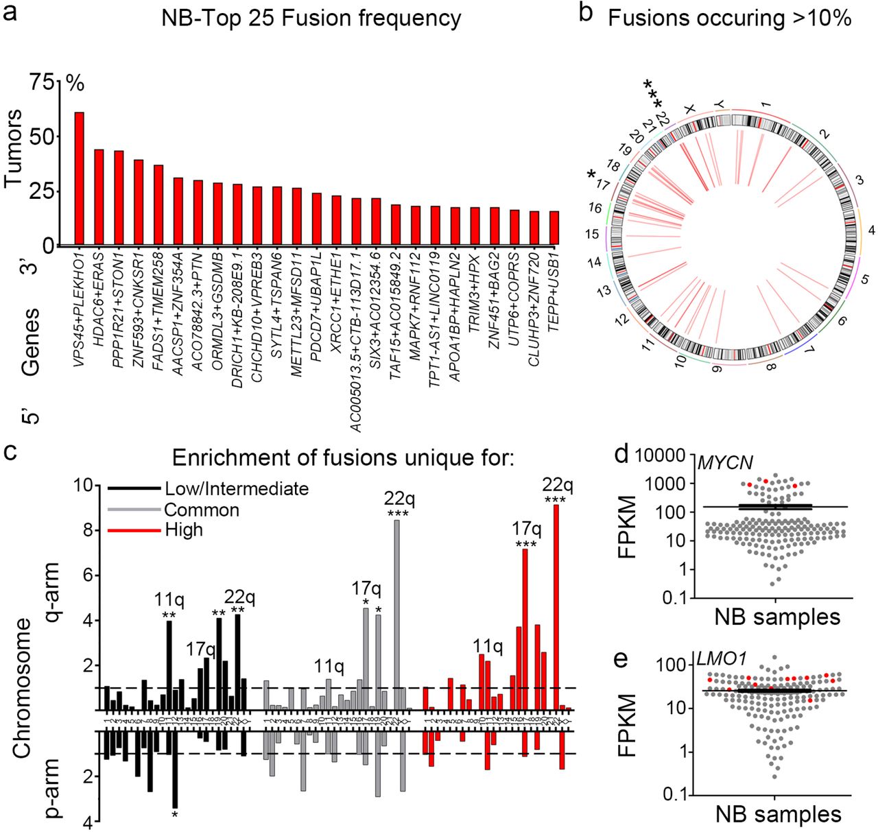
To reveal novel gene fusions in neuroblastoma we analyzed a data set (National Cancer Institute TARGET, dbGap Study Accession: phs000218.v16.p6) comprising 172 paired-end RNA sequenced neuroblastoma tumors (referred to as “NB172”), out of which 139 were diagnosed as high-risk, 19 as intermediate-risk and 14 as low-risk according to the Children’s Oncology Groups staging (COG), (Supplementary Table 1). We applied the fusion detection tool FusionCatcher (14) and identified chimeric transcripts in 163 out of 172 cases with an average of 31 distinct fusion transcripts per tumor (Supplementary Table 2). Short homologous sequences (SHS) have been suggested to serve as templates for reverse transcriptase dependent false positive chimeras/fusions (15). In order to avoid potential false positive fusions, we removed any fusion that contained genes with SHSs of five or more nucleotides, which reduced the number of identified fusions from 1073 to 924. The structural consequences of the fusions ranged from truncated proteins through bona fide fusion proteins to deletion of genes. The majority of fusions (786/924) revealed by our analysis were intra-chromosomal fusion transcripts (Supplementary Table 2-3) many of which consisted of adjacent genes. Importantly, 114 fusions occurred at a frequency of 5% or more (Top 25 in Fig. 1a, all >10% in Fig. 1b and full list in Supplementary Table 3) and all of these were intrachromosomal. There was a significant enrichment of fusion junctions at chromosomes 17 and 22 (Supplementary Fig. 1a). Furthermore, there was a significant enrichment of fusions that occurred in >10% of tumors at the same chromosomes (Fig. 1b). Gain of 17q is the most frequently occurring genomic alteration in high-risk neuroblastoma and a marker for adverse clinical outcome (3), whereas 22q alterations have been reported to be involved in the transition to metastatic and more aggressive neuroblastoma (16). We analyzed the fusion transcripts occurring exclusively in low/intermediate-risk and exclusively in high-risk tumors as well as fusion transcripts common to low/intermediate-risk and high-risk levels (Supplementary Table 4-5). Chimeric transcripts unique to low/intermediate-risk tumors exhibited significant enrichment at several chromosomal arms including 11q, a region commonly lost in high-risk neuroblastoma (Fig. 1c). In contrast, both common and high-risk unique fusion transcripts were enriched at 17q and 22q but not at 11q (Fig. 1c), with a pronounced increase in frequency of 17q fusion transcripts in high-risk tumors (Fig. 1c). Thus, with increased risk the frequency of 17q located fusion junctions also increases and was more than seven times higher than the average fusion rate per chromosomal arm. Several fusion transcripts encompassed factors involved in neuroblastoma pathogenesis, including ARID1B, CASZ1, HDAC8, LMO1, MYCN, BRCA1, TERT and PDE6G (Supplementary Table 6). Notably, tumors harboring fusion transcripts of well-known neuroblastoma oncogenes (e.g. MYCN and LMO1) also exhibited high expression levels of their wild-type cognates (Fig. 1 d-e). The high-risk susceptibility locus in LMO1 is significantly associated with MYCN-non amplified high-risk neuroblastoma but not with MYCN-amplified high-risk neuroblastoma (17), interestingly the LMO1-RIC3 fusion (Supplementary Fig. 1b) was exclusively detected in MYCN-non amplified high-risk neuroblastoma (Supplementary Table 6). The BRCA1-VAT1 fusion was also only detected in MYCN-non amplified high-risk cases; previously it has been shown that copy number amplification of BRCA1 in NB is restricted to cases lacking MYCN-amplification (18) (Supplementary Table 6).

(a) Identification of the 25 most frequent fusions by FusionCatcher in a cohort of 172 paired end RNA sequenced neuroblastoma patient samples derived from the NCI TARGET project (NB172).
(b) Circos plot of genomic distribution of top frequent fusions (> 10%) in NB172.
(c) Enrichment of fusion transcripts common or unique to low/intermediate-risk or high-risk tumors in chromosomal arms as calculated by a normalized enrichment score = (counts of fusion transcripts in each chromosomal arm / length of chromosomal arm (Mb)) / average enrichment.
(d-e) MYCN/LMO1 expression levels are high in neuroblastoma tumors (red) bearing fusion transcripts of which one fusion partner is either MYCN or LMO1, as shown by expression value FPKM (Fragments Per Kilobase per Million mapped reads).
P-values in (b-c) were calculated from Z-values, assuming standard normal distribution. *p<0.05, **p<0.01, ***p<0.001.
Validation of fusion transcripts specific for neuroblastoma
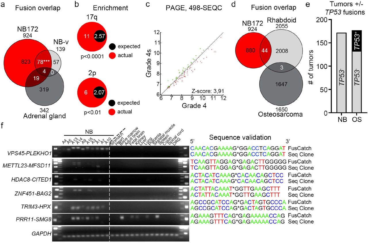
To corroborate the fusion transcripts observed in the NB172 dataset in an independent cohort, we performed paired-end RNA-sequencing in an additional cohort containing 14 neuroblastoma patient samples, together with eight neuroblastoma cell lines, NB-validation (NB-v, Supplementary table 7, Materials and Methods). We identified 139 fusions, of which 82 (~59%) were present in the NB172 dataset (Fig. 2a and Supplementary Table 8). To investigate whether the identified fusions were neuroblastoma specific, we analyzed a cohort of 161 sequenced tissue samples from human normal adrenal glands (19). Out of 342 detected fusions in the adrenal gland cohort, only 23 (~6.7%) were present in the NB172 dataset and only 4 (~1.2%) of these were present in the NB-v cohort (Fig. 2a and Supplementary Table 9). This enrichment of common fusion transcripts in the neuroblastoma cohorts vs. the adrenal gland dataset was highly significant (chi-square test with Yate’s correction, p-value<0.0001). Fusion transcript associated genes unique to and shared by the two neuroblastoma datasets were enriched at 17q and 2p, two chromosomal regions where gains are closely associated with high-risk neuroblastoma (Fig. 2b). Parametric analysis of gene set enrichment (PAGE) (20) comparing high-risk grade 4 tumors with low-risk grade 4s tumors (according to the International Neuroblastoma Staging System, INSS) in the R2 498-SEQC data base (21) showed that the NB172/NB-v common genes identified in Fig. 2a are enriched in the grade 4 high-risk tumors (Fig. 2c). To further investigate whether the identified fusions are distinct for neuroblastoma we analyzed a set of 65 sequenced rhabdoid tumors (National Cancer Institute TARGET, dbGap Study Accession: phs000470.v17.p7) wherein 2055 unique fusion transcripts were detected. However, the overlap with the fusions detected in NB172 dataset was limited to 44 transcripts (~2.1%) (Fig. 2d). In a cohort of 177 sequenced osteosarcoma tumors (National Cancer Institute TARGET, dbGap Study Accession: phs000468.v17.p7; Fig. 2d) we could detect 1650 unique fusion transcripts but there was no overlap with the NB172 cohort (Fig. 2d). In contrast to fusions detected in neuroblastoma, the majority of detected fusions in rhabdoid tumors and osteosarcoma were inter-chromosomal (Supplementary Fig. 2a). Detected fusions that occur at higher frequencies than 5% are more abundant in neuroblastoma (in 12.3% of tumors) than in rhabdoid tumor (5.7%) and osteosarcoma (0.4%) (Supplementary Fig. 2b). In osteosarcoma there was a considerable number of tumors (~24.9%, 44/177) harboring fusions predicting substantial deletion of the P53 tumor suppressor, disruption of the gene or a truncation at the C-terminal (Fig. 2e, Supplementary Table 10). Notably, TP53 is one of the most frequently altered genes in osteosarcoma (22). As a comparison we could not detect any fusions containing TP53 in the neuroblastoma tumors (0/172) (Fig. 2e).

(a) Venn diagram of identified fusions in three datasets, NB172 (NCI TARGET), Validation-NB (NB-v, 14 neuroblastoma patients plus 8 neuroblastoma cell lines) and adrenal gland (161 samples from normal human adrenal gland).
(b) Enrichment of fusion transcript genes unique to the NB172 and the NB-v cohorts on chromosomal arms 2p and 17q.
(c) Parametric analysis of gene set enrichment (PAGE) of the 498-SEQC neuroblastoma dataset show an enrichment of NB172 and NB-v unique common fusions in high-risk grade 4 tumors compared to low-risk 4S tumors.
(d) Venn diagram of identified fusions in three datasets, NB172 (NCI TARGET), Rhabdoid (65 Rhabdoid tumor patient samples) and Osteosarcoma (177 Osteosarcoma patient samples).
(e) Comparison of the number of neuroblastoma (NB) and osteosarcoma (OS) tumors with at least one fusion transcript containing TP53.
(f) Validation of fusion transcripts by RT-PCR and sequencing in Validation-NB neuroblastoma patients, indicated normal tissues were used as controls; DRG, human dorsal root ganglion.
P-values in (a and b) were calculated by chi-square test with Yate’s correction.
For further validation, we designed primers spanning the fusion junctions of selected chimeric transcripts VPS45-PLEKHO1, METTL23-MFSD11, HDAC8-CITED1, ZNF451-BAG2, TRIM3-HPX and PRR11-SMG8 (Supplementary Table 11). We proceeded to perform RT-PCR in 10 neuroblastoma tumor samples and in a panel consisting of cDNA from 14 untransformed human tissues. Our expression analysis revealed that all selected candidates, except PRR11-SMG8, were expressed in a neuroblastoma specific manner (Fig. 2f). For validation, PCR-products including four additional fusions, TAF15-AC015849.2, FADS1-TMEM258, CHCHD10-VPREB3 and LMO1-RIC3, were excised, inserted into the pCR-Blunt II-TOPO vector and subsequently sequenced. All the sequenced PCR-products exhibited an identical sequence of nucleotides to that of the fusions identified by FusionCatcher (Fig. 2f and Supplementary Table 11-12). A previous report identified the VPS45-PLEKHO1 fusion in non-transformed tissue (12), but it was not detected in our panel of non-transformed tissues.
Transcriptional profile of ZNF451-BAG2 positive neuroblastoma predicts poor clinical outcome and high tumor risk
One of the 25 most frequently occurring identified fusion transcripts encompassed the BCL2 associated athanogene (BAG2) which encodes a co-chaperone, BAG2, involved in targeting misfolded proteins for degradation through an ubiquitin independent pathway (23). BAG2 levels have previously been shown to increase upon neuronal differentiation in neuroblastoma cells (24). In addition, BAG2 clears phosphorylated TAU from neuronal microtubule (23), potentially promoting stabilization of axons, an important feature of neuronal differentiation. We thus selected ZNF451-BAG2 to investigate if the presence of a fusion transcript actually would generate a protein with altered functional properties.
To elucidate whether expression of the ZNF451-BAG2 fusion correlates with altered expression levels of transcripts predicting clinical outcome we analyzed nine RNA-sequenced neuroblastoma tumors of the NB-v cohort that had been validated by RT-PCR (Fig. 2f). Tumors harboring the ZNF451-BAG2 fusion (3/9 tumors, A13, A14 and A15) had significantly elevated expression of 32 genes and 34 genes with lower expression (Fig. 3a). To correlate these differentially expressed genes with clinical outcome we utilized a cohort of 498 sequenced neuroblastoma tumors (498-SEQC) available in the R2 database (21). The majority of genes with elevated expression was also enriched for in high-risk tumors whereas the opposite was the case for genes with lower expression (Fig. 3b). Consequently, several genes with elevated expression in ZNF451-BAG2 neuroblastoma were strong indicators of shorter overall survival e.g. ENOSF1 (exemplified in Fig. 3c). In contrast, genes that are strong predictors of longer overall survival showed decreased expression in ZNF451-BAG2 expressing neuroblastoma cases (exemplified in Fig. 3d). In addition, k-means analysis revealed that a subset of ZNF451-BAG2 associated transcripts clustered the 498-SEQC cohort into two groups (Fig. 3e). Group 1, with predominantly low expression levels, consists mainly of low-risk tumors, with low stages according to INSS and lack of MYCN amplification whereas the opposite is evident for group 2 wherein expression levels of ZNF451-BAG2 associated transcripts are high (Fig. 3e). Consequently, patients with group 2 tumors have a significantly shorter overall survival (Fig. 3f). Gene set enrichment analysis (25) of transcriptional differences between ZNF451-BAG2 expressing NB and those lacking ZNF451-BAG2 expression showed a significant enrichment of cell cycle associated gene sets and a depletion of apoptosis related gene set (Fig. 3g-h).

(a) Number of differentially expressed genes (DEGs) in RT-PCR validated neuroblastoma samples with or without ZNF451-BAG2 fusion as shown in Fig. 2e.
(b) Distribution of DEGs from (a) in 498 sequenced neuroblastoma (498-SEQC) according to high-risk vs low-risk disease. Red dots designate genes up in (a) and green dots designates genes down in (a). The two most significant genes in high-risk (ENOSF1) vs low-risk (RAMP3) disease are indicated.
(c) Overall survival probability according to ENOSF1 expression.
(d) Overall survival probability according to RAMP3 expression.
(e) K-means analysis of the 498-SEQC dataset, utilizing the DEGs in tumors harboring the ZNF451-BAG2 fusion (a), generates two clusters with significant differences in Risk, INSS and MYCN amplification.
(f) Overall survival probability of the two clusters identified in (e).
(g-h) Gene set enrichment analysis (GSEA) showing enrichment of genes in the cell cycle associated gene set “HALLMARK_G2M_CHECKPOINT” (g) and depletion of genes in the “HALLMARK_APOPTOSIS” gene set in ΔBAG2 containing neuroblastoma.
The ZNF451-BAG2 fusion generates a truncated BAG2 protein, present in a subset of neuroblastoma tumors
The ZNF451-BAG2 fusion spans the 3’ UTR or exon 14 of ZNF451 and the second exon of BAG2, potentially generating a truncated BAG2 transcript lacking the first exon (Fig. 4a). Its first exon encodes part of a coiled-coil domain that is absent in the ZNF451-BAG2 fusion (Fig. 4a). Full length BAG2 encodes a 23.8 kDa protein, whereas the ZNF451-BAG2 fusion transcript encodes a smaller 19.6 kDa protein (ΔBAG2) (Fig. 4a). The ZNF451-BAG2 chimera was present in 31 of the 172 sequenced tumors (18%). Alignment of wild-type BAG2 protein (BAG2) across different species showed that in ΔBAG2 the highly conserved N-terminal coiled-coil domain was truncated (Supplementary Fig. 3a), implying functional relevance of the truncated region for BAG2. To understand if tumors wherein ZNF451-BAG2 was identified (Fig. 2f) also had detectable levels of ΔBAG2 protein, we performed immunoblotting with an antibody targeting BAG2. All probed (n=13) tumors contained BAG2 protein at varying levels however only five tumors also co-expressed detectable levels of ΔBAG2, while no detectable levels of ΔBAG2 were observed in tissue from six human normal adrenal glands (Fig. 4b and Supplementary Fig. 3b).

(a) Schematic representation of ZNF451-BAG2 fusion, the resulting truncated BAG2 is referred to as ΔBAG2.
(b) Endogenous tumor ΔBAG2 protein expression is associated with high levels of phosphorylated TAU (p-TAU) in neuroblastoma tumors as detected by immunoblotting.
(c) SKNFI cells were transfected with p3XFLAG-CMV14-empty/BAG2/ΔBAG2/BAG2+ΔBAG2 for 48 hours. Cells were harvested and proteins were extracted; whole-cell lysates were used to detect BAG2, FLAG, p-TAU on Ser404, total TAU and ACTIN.
(d) FLAG-tagged proteins were immunoprecipitated from whole-cell lysates as prepared in (c) using Anti-FLAG M2 magnetic beads and eluted. Immunoprecipitated proteins were western blotting to detect HSC70, BAG2.
(e-l) Constitutive lentiviral overexpression of ΔBAG2, but not wildtype BAG2 (backbone pLVX-EF1α-IRES-mCherry) inhibited RA-induced differentiation (6 days of treatment) in SK-N-FI cells.
(m-r) Doxycycline inducible lentiviral overexpression of ΔBAG2 (backbone pLVX-Tet-one-puro-IRES-mCherry) inhibited RA-induced differentiation (4 days of treatment) in SK-N-BE(1) cells. Protein levels of BAG2 and ΔBAG2 were analyzed by Western blotting (e and m). Immunostaining was performed using antibody against neuronal marker β3-tubulin (TUJ1).
Data in l and r is represented as mean of transduced cells with TUJ1+ neurites/total number of transduced cells +/− SEM, each data-point represents this ratio in a single 10× microscopic field (n=10-20). ***p<0.001, one-way ANOVA with Tukey’s multiple comparison test.
ΔBAG2 impairs clearance of phosphorylated TAU and binding to HSC70
BAG2 has been shown to be important for clearance of phosphorylated forms of the TAU protein (pTAU) and thus been implicated as a stabilizer of microtubules (23). To test if this capacity was attenuated by the presence of ΔBAG2 we probed the levels of pTAU in a panel of neuroblastoma tumors and normal adrenal glands. This showed a clear association between the presence of pTAU and endogenous ΔBAG2 (Fig. 4b and Supplementary Fig. 3b). To validate that this was caused by ΔBAG2 protein expression, we cloned and validated the ΔBAG2 transcript where after we expressed it and BAG2 alone or in combination in SK-N-FI neuroblastoma cells. Upon BAG2 overexpression, the levels of pTAU were significantly reduced whereas total TAU was present in amounts similar to those in control-transduced cells (Fig. 4c). ΔBAG2 overexpressing cells retained pTAU levels and more importantly, upon co-expression BAG2 failed to clear pTAU (Fig. 4Cc), implying that ΔBAG2 can act as a negative regulator of BAG2 function. BAG2 has been shown to bind the heat shock cognate 70 (HSC70) (26), a chaperone protein important for pTAU ubiquitination (27) and axon outgrowth (28). Overexpression of BAG2 and ΔBAG2 in SK-N-FI neuroblastoma cells followed by BAG2 immunoprecipitation via FLAG revealed that BAG2 but not ΔBAG2 binds a 70 kDa protein (Supplementary Fig. 3c). Since BAG2 has been reported to bind HSC70 (26), we performed additional immunoprecipitation followed by immunoblotting with a HSC70 specific antibody. This revealed that BAG2 binds HSC70, whereas ΔBAG2 does not (Fig. 4d).
ΔBAG2 impedes differentiation of neuroblastoma in response to retinoic acid
To investigate whether ΔBAG2 impinges on the capacity of neuroblastoma cells to differentiate, we treated CTRL (empty vector), BAG2 or ΔBAG2 expressing SK-N-FI cells (Fig. 4E) with retinoic acid (RA), a compound used in adjuvant therapy of high-risk neuroblastoma patients (13). After six days of RA treatment, cells transduced with either CTRL (Fig. 4f-g, l) or BAG2 (Fig. 4h-i, l) acquired neuronal morphology with long neurites. In contrast, ΔBAG2 transduced cells exhibited a weaker response to RA, with significantly less neurite formation (Fig. 4j-l). To validate this effect we transduced SK-N-BE(1) neuroblastoma cells with a doxycycline-inducible version of the ΔBAG2 fusion genes (Fig. 4m). Upon doxycycline induction, ΔBAG2 expressing cells exhibited a reduced capacity to respond to RA (Fig. 4n-r).
Alternative splicing affects the generation of neuroblastoma specific fusions
Previously it has been suggested that cis-splicing between adjacent genes can generate fusion transcripts in prostate cancer (5, 6). To elucidate whether there was a correlation between distance and frequency we plotted the distance between 5’ and 3’ of the fusion junction in the intrachromosomal fusion transcript versus the fusion frequency in the NB172 and NB-v data sets (Fig. 5a). Fusions occurring at high frequency in both data sets were enriched between 1 to 100kb, whereas transcripts separated by more than 100kb occurred at lower frequencies and almost exclusively in the larger NB172 data set (Fig. 5a). The spatial proximity of identified transcripts suggests that these fusions are the result of cis-splicing (5, 8). Inspection of nucleotide sequences located at 5’ and 3’ of the fusion junctions for canonical splicing donors (GT) and acceptors (AG) revealed that 81.9% carried GT and AG at the 5’ and 3’ fusion sites (GT*AG) (Fig. 5b). Notably, 87% of intra-chromosomal fusions had GT*AG at the fusion junction whereas only 45.9% of inter-chromosomal fusions contained canonical splice sites at the junctions. This pattern was unique to neuroblastoma as fusions detected in normal adrenal gland, osteosarcoma and rhabdoid tumor exhibited no enrichment of splice sites (Fig. 5b). Aberrant RNA splicing has been suggested as a driving event for several cancers and mutations in genes coding for components of the spliceosome have been identified in several tumors (29). Furthermore in breast cancer, it has been shown that an intact spliceosome is required to tolerate oncogenic MYC hyperactivation (30). We compared expression levels of genes of the KEGG, “Spliceosome” gene category between neuroblastoma and normal adrenal gland. Out of 134 genes 46 had significantly higher expression levels in the neuroblastoma, whereas only three had lower levels of expression (Fig. 5c). To further elucidate whether there were clinically relevant differences in expression of spliceosome genes between low- and high-risk neuroblastoma we performed k-means analysis of the 498-SEQC neuroblastoma cohort. Together with previous observations (31) our analysis elucidates how the differential expression of spliceosome factors clearly identifies tumors of different clinical outcome, with high expression levels of splicing factors predicting high-risk tumors with bleak clinical outcome and substantially shorter overall survival (Fig. 6d-e). Furthermore, previous genome sequencing studies of neuroblastoma patients revealed mutations in several spliceosome factors in primary tumors and de novo mutations in spliceosome factors occurred in relapsed tumors (1, 2, 32, 33) (summarized in Supplementary Table 13). To investigate whether inhibition of spliceosome activity would selectively impede the generation of fusion transcripts but not their wild type cognates, we treated neuroblastoma cells (LAN-1 and SK-N-BE(1)) with the spliceosome inhibitor pladienolide B (34).

(a) Plot of the frequency versus the distance from 5’ to 3’ in the intrachromosomal fusion transcript identified in the NB discovery cohort (NB172); each dot represents a unique fusion transcript; fusion transcripts re-identified in the NB validation cohort (NB-v) were marked as red.
(b) Distribution of unique intrachromosomal vs interchromosomal fusion junctions flanked by GT_AG or other nucleotide motifs in neuroblastoma, rhabdoid tumor, osteosarcoma and normal adrenal gland.
(c) Differential expression of genes in the KEGG Spliceosome pathway between neuroblastoma dataset (NBL172) versus human normal adrenal gland dataset.
(d) K-means analysis of the 498-SEQC dataset, utilizing the genes in the KEGG spliceosome pathway, generates two clusters with significant differences in Risk, INSS and MYCN-amplification.
(e) Overall survival probability of the two clusters identified in (d).
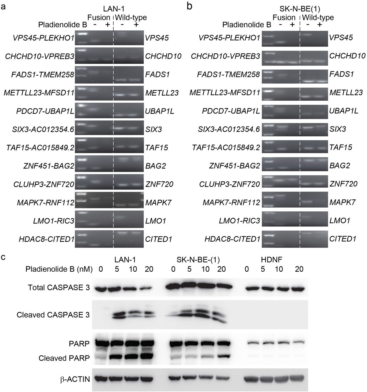
(a-b) Splicing-dependent generation of several high frequent fusion transcripts in LAN-1 (a) and SK-N-BE(1) (b) neuroblastoma cells. SK-N-BE(1) and LAN1 cells were treated with splicing inhibitor Pladienolide B at 100 nM (SK-N-BE(1)) and 5-50 nM (LAN-1) for 6 hours; RNA was isolated from harvested cells and reversely transcribed to cDNA; RT-PCR were performed with primers spanning the fusion junctions (fusion transcript) or exon-exon boundary (wild-type cognate).
(c) Induction of apoptosis in LAN-1 and SK-N-BE(1) cells, but not in human normal diploid fibroblasts (HDNF) upon pladienolide B treatment (0-20 nM) for 48 hours, as detected by western blotting of full-length CASPASE-3, cleaved CASPASE-3, full-length PARP and cleaved PARP; β-ACTIN was used as a loading control.
Upon treatment, there was a loss of expression for a majority of selected high frequency fusion transcripts whereas expression of most wild type genes constituting the fusions were unaffected at these concentrations (Fig 6a-b and Supplementary Fig. S4). To elucidate if selective loss of fusion transcripts upon spliceosome inhibition was associated with increased apoptosis we treated the neuroblastoma cell lines LAN-1 and SK-N-BE(1) with increasing concentrations of pladienolide B. As controls we utilized non-transformed human diploid fibroblasts (HNDF). Already at 5nM the neuroblastoma cell lines exhibited increased levels of cleaved caspase-3 and cleaved PARP, whereas control cells showed no signs of cell death. At 20nM cell death in both neuroblastoma cell lines was further increased but control cells were still non-responsive (Fig. 6c).
Discussion
Our study shows that a high frequency of neuroblastoma specific fusion transcripts could constitute an overlooked process through which altered transcripts are generated. It has been shown that upon cellular stress (e.g. viral infection, replicative or osmotic stress and mutational events) transcriptional termination can be blocked, increasing the probability of generating “downstream of genes”-transcripts (11). Thus, a proportion of fusions could represent passengers that occur as a response to cellular stress combined with neuroblastoma associated events such as gain or loss of chromosomal regions (e.g. 17q and 2p). In contrast to such passengers, certain fusions could be early events actually preceding and promoting other transformative events. Pharmacologic inhibition of splicing selectively repressed expression of several top frequent fusion transcripts but not of their wild-type cognates and there was a high frequency of splicing donor/acceptor sites in neuroblastoma specific fusions but not in fusions detected in normal adrenal gland, osteosarcoma or rhabdoid tumors. This pattern implies that a substantial proportion of the detected fusions are of the same cis-splicing type as previously reported in prostate cancer (5). Our analysis reveals that high expression levels of splicing associated factors is a distinguishing feature of high-risk neuroblastoma, representing a strong predictor of tumor grade. Regardless of the mechanisms underlying the generation of these fusions, they are not necessarily dependent on amino acid changing mutations but can still provide a source of modified gene products with the potential to promote neuroblastoma but also reveal novel drug targets. Given the low frequency of recurrent mutations in neuroblastoma, such a pool of altered gene products could indeed be relevant for tumor pathophysiology. A background of expressed fusion transcripts potentially augments the effect of oncogenic drivers. Interestingly, our analysis shows that when established drivers of neuroblastoma (e.g. MYCN and LMO1) are part of the fusion transcripts the expression levels of wild type cognates are elevated. In addition, a panel of neuroblastoma specific fusions occurring at high frequency could serve as biomarkers for diagnosis and the presence of risk specific fusions could sub-divide neuroblastoma patients for precision therapy. Hence, fusions that are passenger events rather than oncogenic drivers can still be of clinical relevance. One concern with previously reported fusions is the relatively few cases that have been independently validated. In 2015, only 3% of fusions identified by deep sequencing could be reproducibly detected (4). Arguably, the “non-genomic” characteristic of this type of intrachromosomal fusions potentially augments the detection of false positives. The risk that a proportion of fusion transcripts constitute false positives as the result of spurious transcription or of sequencing errors is reduced by our crosswise analysis with other tumors and healthy tissues. There is a clear enrichment of common fusions unique for the neuroblastoma data sets that do not appear in any of the other tumors nor in normal adrenal glands. It should however be noted that the normal adrenal gland is not perfect as control tissue due to the cellular heterogeneity of the organ. Nevertheless, neuroblastoma specific fusions are enriched for genes located at chromosomal regions (2p or 17q) which are commonly gained in high-risk neuroblastoma. The selective loss of several fusion transcripts upon spliceosome inhibition is an additional strong indication that fusion transcripts indeed are present. Our validation of ten fusions through PCR and subsequent sequencing further underscores that these fusions can generate alternative gene products. Interestingly, tumor specific distribution of fusions is reflected in the osteosarcoma tumors where the TP53 tumor suppressor is a fusion partner in an disproportional amount of the detected fusions, mirroring the importance of inactivating mutations in TP53 for this disease (22). It has previously been reported that the presence of short homology sequences (SHS) can generate false RNA-chimeras due to template switching during the reverse transcriptase reaction (35). Such a mechanism would presumably generate random fusions between transcripts containing matching SHSs with no preference for any particular chromosomal location nor any preference for intrachromosomal vs interchromosomal fusion transcripts. The non-random enrichment of fusions at chromosomal locations that mirror the disease as well as the enrichment of intrachromosomal fusions between closely located genes suggests that reverse transcriptase induced template switching is not the cause of these fusions. Furthermore, it is plausible that fusions detected due to random template switching should correlate with high expression levels, which we do not detect. Our study shows that an altered protein with novel properties can be generated as a consequence of an intrachromosomal, splicing dependent fusion and that this altered protein influences the response to a drug (RA) commonly used to treat high-risk neuroblastoma patients. However, to fully evaluate the importance of identified fusions further functional experiments are required. A previous study indeed shows how the SLC45A3-ELK4 read-through fusion transcript is elevated in prostate cancer tissue, is androgen-regulated and can be detected in an non-invasive assay from biopsies of men at risk of having prostate cancer (9). Even though it is possible that a portion of individual fusions are passenger events rather than oncogenic drivers a continued effort is justified to understand the regulation of fusions and downstream consequences of fusions in neuroblastoma as well as in other tumors.
Materials and Methods
Human tissue Samples
Neuroblastoma primary tumors came either from the Swedish NB Registry (ethical permission (DN03-736) granted by Karolinska Institutets Forskningsetikommitté Nord, (clinical information described in Li et. al. (36), or from the Irish NB cohort (described in Supplementary table 7), with ethical approval of the Medical and Research Ethics Committee of Our Lady’s Children’s Hospital, Crumlin, Dublin, Ireland. Informed consent from families of subjects was obtained for samples. Six histologically confirmed normal human adrenal glands were included as controls (covered by existing ethical approvals; Dnr 01-136 and Dnr 01-353 KI forskningsetikkommitté Nord). Human total RNA from different normal tissues from Clontech (Human Total RNA Master Panel II,Cat#. 636643).
Paired-end RNA-Seq
RNA was isolated using the PerfectPure RNA Cultured Cell Kit (cell lines) and PerfectPure RNA Tissue Kit (patient samples) from 5 PRIME. RNA-seq libraries were prepared using TruSeq RNA Library Preparation Kit v2 (Illumina); paired-end RNA sequencing (125 bp) were performed in SciLifeLab (Stockholm).
Data Analysis
FusionCatcher (14) was applied to detect the fusion transcripts in paired-end RNA-seq data. Reads were mapped to hg19 and differential expression analysis was performed as described in(37). For enrichment analysis in Figure 1c, only fusions occurring above ~3% in each risk group were included (1 case in Low/Intermediate-risk, 4 cases in High-risk and 5 cases for the common fusion transcripts shared by Low/Intermediate-risk and high-risk groups).
Cloning and expression of wildtype BAG2 and ZNF451-BAG2
cDNA was synthesized from total RNA by the iScript cDNA Synthesis Kit (Bio-Rad). Coding regions of wildtype BAG2 and ZNF451-BAG2 were amplified from SK-N-AS cells for subcloning into p3XFLAG-CMV14 (Sigma) and pLVX-EF1α-IRES-mCherry (Clontech) using primer pair 1 and 2 respectively (Supplementary Table 11), inserted to pCR-Blunt II-TOPO vector via zero Blunt TOPO PCR Cloning Kit (ThermoFisher Scientific) and sequenced. BAG2 and ZNF451-BAG2 were subcloned into p3XFLAG-CMV14 vector using EcoRI and BamHI and pLVX-EF1α-IRES-mCherry lentiviral vector using EcoRI and BamHI. To generate pLVX-Tetone-puro-IRES-mCherry-empty/BAG2/ZNF451-BAG2 vectors, pLVX-Tetone-puro-empty (Clontech) construct was linearized with AgeI, blunted and digested with EcoRI; pLVX-EF1α-IRES-mCherry-empty/BAG2/ZNF451-BAG2 vectors were linearized with MluI, blunted and digested with EcoRI to release IRES-mCherry-empty/BAG2/ZNF451-BAG2; then linearized and blunted pLVX-Tetone-puro-empty construct was ligated with fragment of IRES-mCherry-empty/BAG2/ZNF451-BAG2 separately.
Lentiviruses expressing pLVX-EF1α-IRES-mCherry-empty/BAG2/ZNF451-BAG2 and pLVX-Tetone-puro-IRES-mCherry-empty/BAG2/ZNF451-BAG2 were produced in 293FT cells using lipofectamine 2000 based protocol.
Cell culture
All neuroblastoma cell lines (SH-SY5Y, SK-N-SH, SK-N-FI, SK-N-BE(1), SK-N-AS, LAN-1, CHP-212 and IMR-5) were maintained in RPMI1640 medium supplemented with 10% FBS, 1% penicillin/streptomycin and 1% L-glutamine and grown in 5%CO2 at 37°C.
Differentiation assay and immunofluorescence staining
For short-term RA induced differentiation assay, SK-N-FI cells were seeded in 6 well plates; after 24 hours, cells were infected by lentiviruses expressing pLVX-EF1α-IRES-mCherry-empty/BAG2/ZNF451-BAG2 for 48 hours; then cells were trypsinized and 20,000 cells were reseeded into 6 well plates with coverslips; 24h later, cells were treated with 1 μM retinoic acid (RA) or DMSO as control for 6 days. For doxycycline-inducible system, 20,000 SK-N-BE(1) cells with stable overexpression of pLVX-Tetone-puro-IRES-mCherry-ZNF451-BAG2 were seeded in 6 well plates with coverslips, and after 24h, cells were pre-treated with 1 μM doxycycline or DMSO as a control for one day; then cells received one of the following 4 different treatment for 4 days: DMSO, 1 μM RA, 1 μM doxycycline or 1 μM RA+1 μM doxycycline.
Cells on coverslips were fixed in RPMI1640 medium containing 2% PFA for 5 minutes at room temperature (RT), washed once with PBS at RT, fixed with 4% PFA for another 15 minutes at RT and washed twice with cold PBS; then cells were permeabilized with PBS containing 0.25% Triton X-100 for 15 minutes at RT and blocked with 3% BSA for 1h at RT. Immunofluorescent stainings were performed with the following primary antibody Tuj1 (Covance, 1:1000) overnight at +4 °C.
Quantification
Cells were stained as in Fig. 4g-l and o-r. Images were taken by confocal microscopy (10×). For quantification, images were coded and a researcher who had not participated in staining and image acquisition manually counted the ratio of transduced cells extending TUJ1+ neurites/total number of transduced cells/microscopic field. For DOX− SK-N-BE(1) the ratio of cells extending TUJ1+ neurites/total number of cells/microscopic field were counted. Microscopic fields containing less than three transduced cells were discarded. Grubb’s test was performed for outlier detection (alpha p<0.05). For statistical analysis in Fig. 4l and r one-way ANOVA followed by a Tukey’s multiple comparison test was performed.
Western blotting
Immunoblotting was performed using standard protocols. Following primary antibodies were used: BAG2(sc-390262, 1:200), phospho-ser396-Tau(sc-101815, 1:1000), HSC70(sc-7298, 1:200) from Santa Cruz; phosphor-ser404-Tau(44-758G, 1:200) from ThermoFisher Scientific; total Tau(A0024, 1:1000) from Dako; beta-actin(AC-15, 1:3000), FLAG(F1804, 1:1000) from Sigma; PARP (9542s), Caspase-3 (14220s) and cleaved caspase-3 (9664s) from Cell Signaling.
Co-immunoprecipitation
SK-N-FI cells were seeded in 15 cm dishes 24 hours before transfection. Transfection of p3×FLAG-CMV14-empty/BAG2/ZNF451-BAG2 constructs was performed using lipofectamine 2000. 48 hours post-transfection, cells were harvested and proteins were extracted using lysis buffer containing 10 mM TRIS-HCl (pH7.4), 150 mM NaCl, 0.5% NP40, complete protease and phosphatase inhibitors (Roche). FLAG-tagged fusion proteins were immunoprecipitated using Anti-FLAG M2 magnetic beads (Sigma) according to manufacturer’s instructions. Bound proteins were examined by silver staining or Western blotting using standard protocols.
Funding
The Johan Holmberg lab is supported by the Swedish Children Cancer Foundation, the Swedish Cancer foundation, Knut and Alice Wallenberg Foundation, Swedish Research Council (VR), The Strategic Research Programme in Cancer (StratCan, SFO) and The Swedish Brain Foundation.
Author contributions
Y.S. and J.H. designed the study. Y.S. performed the majority of the experiments with help from V.R., E.M., S.L., P.B., J.Y., and I.W. Y.S. generated the libraries for RNA-sequencing. Y.S. and J.H performed the analysis with help from O.B.R. C.C.J, A.S., C.L., P.K. and M.J.S supplied the clinical material. Y.S. and J.H. wrote the manuscript with input from all authors.
Data and materials availability
Data needed to evaluate the conclusions in the paper are present in the paper and/or the Supplementary Materials. Raw and processed NGS data will be deposited in the GEO database.
Declaration of interests
The authors declare no competing interests.
Acknowledgements
We thank members of the Holmberg and Schlisio groups for valuable comments.
References


