

Abstract
The delta glutamate receptors, GluD1R and GluD2R, are mysterious members of the ionotropic glutamate receptor family in that they are not gated by glutamate1,2. One theory is that they are scaffolding proteins or synaptic organizers strictly, rather than ion conducting channels. Although mutant forms and wild type channels have been reported to conduct3,4,5, conduction, gating, and biophysical properties of native GluD1R remain unexplored. Here we show that the inward current induced by activation of α1-adrenergic receptors (α1-ARs) in the dorsal raphe nucleus (DR) is mediated by GluD1R. Native GluD1R channels are functional ion channels that are constitutively active under basal conditions and α1-ARs increase the tonic current. This inward current is responsible for the α1-AR-dependent induction of persistent pacemaker-type firing of neurons in the DR. Given the extensive distribution of these receptors, the ionotropic nature of GluDR is proposed to be widespread in the nervous system.
The dorsal raphe nucleus (DR) is the largest serotonergic nucleus in the brain and the predominant source of central serotonin (5-HT). Activation of Gαq/11 protein-coupled α1-adrenergic receptors (α1-ARs) in the DR produces a slow and long-lasting membrane depolarization6. In vivo, tonic noradrenergic input that activates α1-ARs is required for 5-HT neurons to fire action potentials7,8. Despite having a crucial role in regulating 5-HT neuron excitability, the ion channels responsible for the depolarization remain unknown.
Activation of α1-AR produces an EPSC
Electrophysiological recordings were made from DR neurons in acute brain slices from wild type mice at 35° C in the presence of NMDAR, AMPAR, KainateR, GABA-AR, and 5-HT1AR antagonists. With cell-attached recordings, a train of 5 electrical stimuli (60 Hz) produced firing in previously quiescent neurons, which was blocked by application of the α1-AR antagonist, prazosin (100 nM, Fig. 1a). The excitation produced 20±5 action potentials (APs) that lasted 9.0±3.0 s, with a latency of 650.6±0.1 ms from onset of stimulation to the first AP (Fig. 1b-e). In whole-cell recording using a potassium-based internal solution, the same train of electrical stimuli produced prolonged AP firing (Fig. 1f). In voltage-clamp mode (Vhold −65 mV), the same stimulation produced a slow and long-lasting (27.4±2.3 s, n=10) excitatory postsynaptic current (EPSC, Fig. 1f) that was eliminated by application of prazosin (Fig. 1g). Prazosin had no effect on basal whole-cell current (−3.8±3.4 pA, p=0.232, n=10, not shown) indicating a lack of persistent inward current due to noradrenaline (NA) tone. Application of tetrodotoxin (1 μM) reversibly abolished the α1-AR-EPSC, demonstrating a dependence on presynaptic action potentials (Fig. 1h). Disruption of the vesicular monoamine transporter with reserpine (1 μM) or removal of external Ca2+ also eliminated the α1-AR-EPSC, indicating NA release is vesicular (Fig. 1i,j). To test whether α1-AR-EPSCs were dependent on G protein-signaling, recordings were made with an internal solution containing GDP-βS-Li3 (1.8 mM) instead of GTP. Disruption of G protein signaling with intracellular dialysis of GDP-βS-Li3 eliminated the α1-AR-EPSC within 5-20 min post-break-in (p=0.004, n=9), whereas dialysis with LiCl alone had no effect on the α1-AR-EPSC amplitude (p=0.625, n=4, Fig. S1a). These findings demonstrate a cell-autonomous requirement of G protein signaling in the generation of the α1-AR-EPSC.

a, With GDP-βS-Li-containing internal solution, the amplitude of the α1-AR-EPSC ran down within ~5-10 minutes of break-in to whole-cell mode; shown in a plot compared with control internal solution containing LiCl (left) and in grouped data (right, p=0.004, n=9, 1st: first EPSC; post: post-dialysis). Line and error bars represent mean±SEM, * denotes statistical significance.
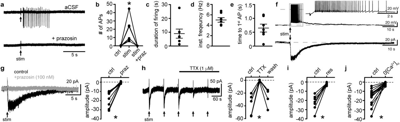
a, Representative traces of cell-attached recording where stimulation of the brain slice (5 stims at 60 Hz) produced AP firing that was abolished by application of the α1-adrenergic receptor antagonist, prazosin (100 nM). b, Plot of number of APs showing the stimulation-induced increase in frequency (p=0.004, n=6). c, Plot of duration of AP firing. d, Plot of mean instantaneous frequency of AP firing over the first 10s of firing. e, Plot of the latency from stimulation onset to the first AP. f, Example whole-cell recordings in the same cell, where electrical stimulation of the slice produced prolonged AP firing in current-clamp (upper trace) and a slow EPSC in voltage-clamp mode (lower trace). g, Application of prazosin eliminated the slow EPSC shown in a representative trace (left, baseline adjusted) and in grouped data (right, p=0.002, n=10). h, Application of tetrodotoxin (TTX, 1 μM) reversibly eliminated the α1-AR-EPSC shown in a representative trace (left, α1-AR-EPSC evoked every 90s (arrows)) and in grouped data (right, p=0.009, n=4). i, Plot of the inhibition of α1-AR-EPSC amplitude by application of reserpine (res, 1 μM, p=0.016, n=7). j, Plot of the inhibition of α1-AR-EPSC amplitude by removal of external Ca2+ (0[Ca2+]o, p=0.0001, n=14). Line and error bars represent mean±SEM, * denotes statistical significance.
Biophysical properties of the channel
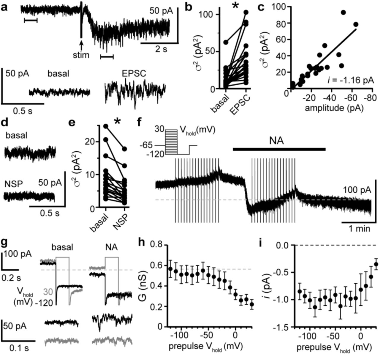
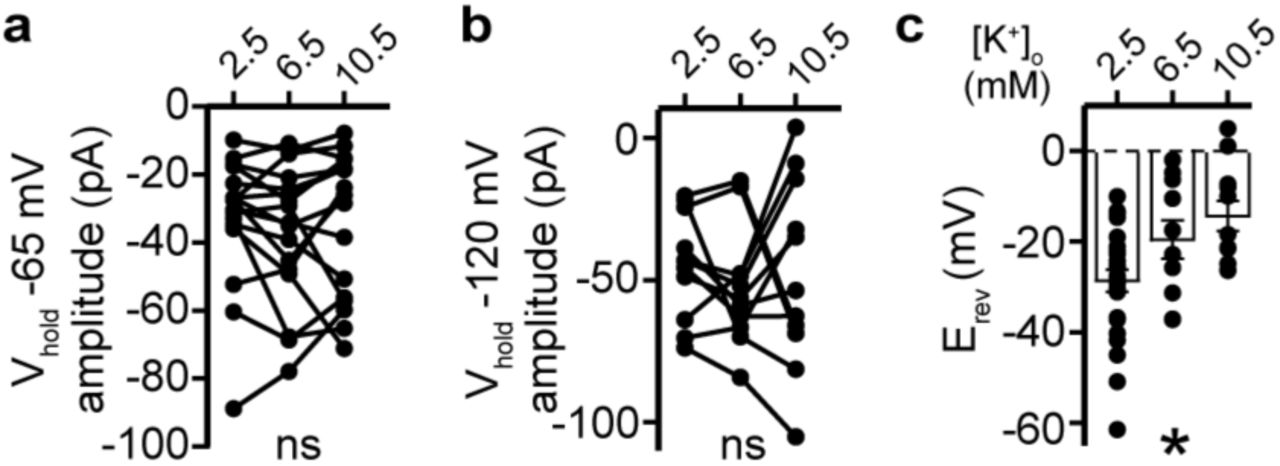
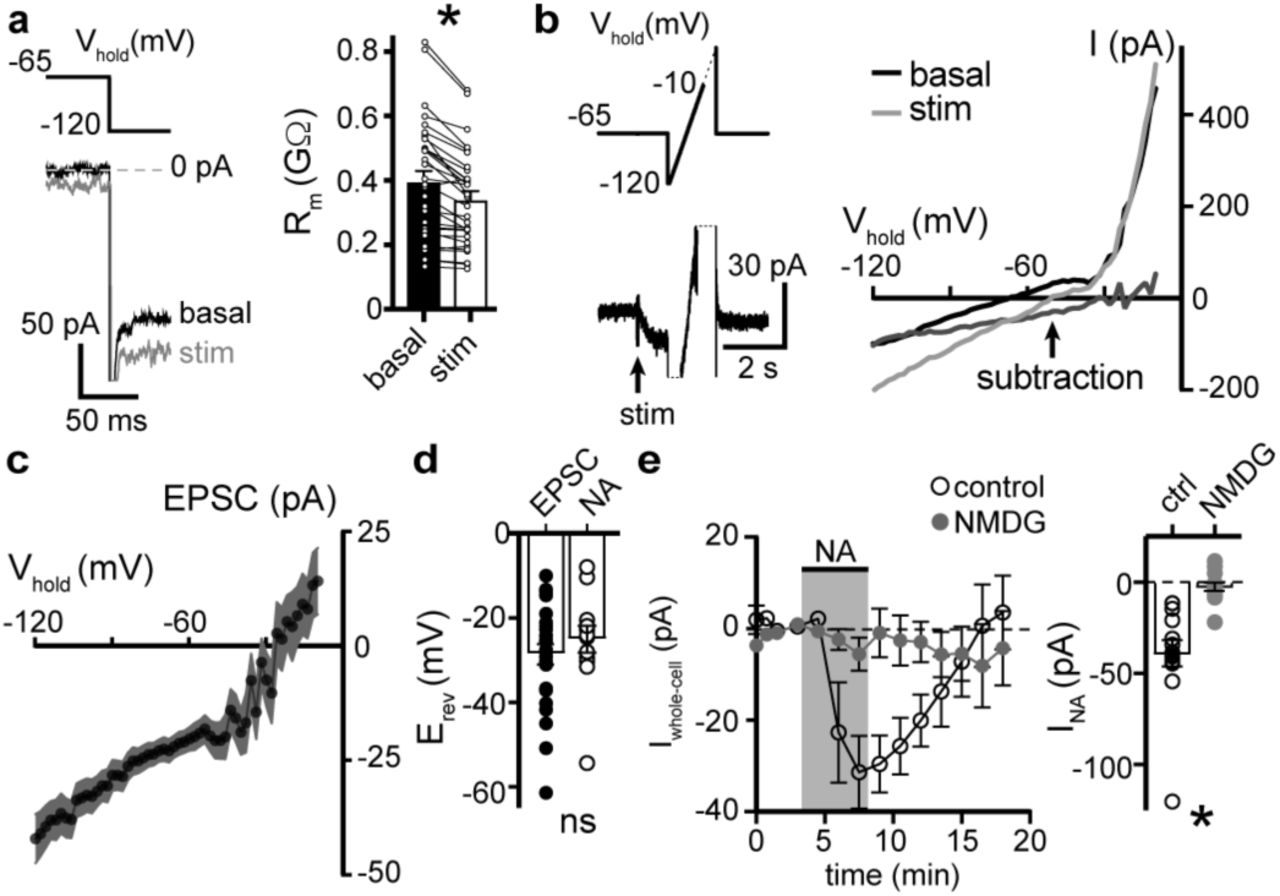
Under our recording conditions, membrane resistance (Rm) decreased during the α1-AR-EPSC, indicative of ion channels opening (Fig. 2a). Membrane noise variance (σ2) increased during the EPSC compared to membrane noise under basal conditions (Fig. S2a,b). The α1-AR-EPSC σ2-amplitude relationship was well-fit by linear regression, yielding a measurement of −1.16 pA unitary current (Fig. S2c). Voltage ramps from −120 to −10 mV (1 mV/10 ms) before and during the α1-AR-EPSC (Fig. 2b) showed that the current reversed polarity at −28.6±2.4 mV (Fig. 2b-d). Exogenous application of NA (30 μM) produced an inward current (INA) with a similar reversal potential (Erev, - 25.1±2.9 mV, Fig. 2d). Replacing 126 mM [Na+]o with N-methyl D-glucamine (NMDG) completely abolished inward INA, suggesting Na+ as the prominent charge carrier (Fig. 2e). Increasing [K+]o from 2.5 to 6.5 or 10.5 mM, expected to shift EK from −107 to −81 and −69 mV, respectively, had no effect on the α1-AR-EPSC amplitude at Vhold −65 mV nor −120 mV, but produced a depolarizing shift in Erev of the α1-AR-EPSC (Fig. S3a-c), suggesting the channel is also permeable to K+. Removal of external MgCl2 had no significant effect on Erev, nor on the amplitude of INA (Fig. S4a,b). Removal of external CaCl2 also had no significant effect on Erev, but augmented inward INA, (Fig. S4a,b). Taken together, the data suggest that α1-AR-dependent current, whether produced by vesicular NA release or exogenous NA is carried through a mixed cation channel, with inward current carried predominantly by Na+ entry.

a, Representative trace of membrane noise during the α1-AR-EPSC, brackets denote segments shown below on an expanded scale. b, Membrane noise (variance, σ2) increased during the α1-AR-EPSC (p<0.0001, n=22). c, Plot of α1-AR-EPSC variance versus mean amplitude, linear fit represents mean unitary current (i, r2=0.713). d, Representative trace of membrane noise under basal conditions and after NASPM application (NSP, 100 μM). e, Membrane noise (variance, σ2) decreased following NASPM (NSP, p<0.0001, n=19). f, INA was isolated using a two-pulse voltage protocol by subtracting current measured during voltage steps (shown in left inset) at the peak of inward current from NA application from current measured in control conditions just prior to NA (30 μM) application. g, Representative traces of current at Vhold −120 mV following 150 ms conditioning pre-pulses to −120 mV (black) or to 30 mV (grey) in control conditions (basal) and after NA. Traces below shown membrane noise of current at Vhold −120 mV on an expanded scale. h, Conductance (GNA) was measured at peak INA from Vhold −120 mV, calculated using a Erev of −25.1 mV. Plot of GNA versus Vhold of the conditioning pre-pulses, demonstrating decreased GNA with depolarizing pre-pulses (linear trend of −0.022 nS/mV, p<0.0001). i, Plot of unitary current (i) measured at Vhold −120 mV versus Vhold of the conditioning pre-pulses demonstrating a decrease in i with depolarizing pre-pulses (linear trend of 0.026 pA/mV, p=0.002). Line and error bars represent mean±SEM, * denotes statistical significance.

a, Plot of α1-AR-EPSC amplitudes measured at Vhold −65 mV, in 2.5, 6.5, and 10.5 mM [K+]o (p=0.162, n=17). c, Plot of α1-AR-EPSC amplitudes measured at Vhold −120 mV, in 2.5, 6.5, and 10.5 mM [K+]o (p=0.692, n=11). d, Plot of α1-AR-EPSC Erevs with varying concentration of external K+ ([K+]o), demonstrating a depolarizing shift in Erev as external K+ was increased (p=0.010, n=26, 10,&11). Line and error bars represent mean±SEM, * denotes statistical significance, ns denotes not significant.

a, Plot of reversal potentials (Erev) of INA, demonstrating no significant difference between control conditions (con), or after removal of external Ca2+ (0[Ca2+]o) or Mg2+ (0[Mg2+]o). b, Plot of the amplitude of INA (Vhold −65 mV) demonstrating an augmented INA amplitude in 0[Ca2+]o (p=0.017, n=14), but not in 0[Mg2+]o, (p>0.9999, n=10) as compared with control conditions (n=14). Line and error bars/shaded area represent mean±SEM, * denotes statistical significance, ns denotes not significant.

a, Membrane resistance (Rm, ΔV −65 to −120 mV) decreased during the α1-AR-EPSC indicating an opening of ion channels, as shown in an example trace (left) and in grouped data (right, p<0.0001, n=31). b, Representative trace of current-voltage relationships of the α1-AR-EPSC (subtraction), determined by subtracting current measured during slow voltage ramps (1 mV/10 ms, analyzed from −120 to −10 mV) at the peak of the α1-AR-EPSC (stim) from current measured in control conditions just prior to stimulation (basal). Current generated during ramps were truncated for clarity. c, Current-voltage relationship of the α1-AR-EPSC from grouped data. d, Plot of reversal potentials (Erev) of the α1-AR-EPSC and INA (p>0.999, n=26&14). e, Replacing 126 mM NaCl with NMDG eliminated inward INA, shown in a time-course plot (left) and grouped data (right, Vhold −65 mV, p<0.0001, n=14&13). Line and error bars/shaded area represent mean±SEM, * denotes statistical significance, ns denotes not significant.
Measurements of Erev assume voltage-independence of the channel and the signaling mechanism between α1-AR and the channel. To test for voltage-dependence, we employed a two-pulse voltage-step protocol. Current was measured at Vhold −120 mV following a conditioning pre-pulse (−120 to 30 mV, 150 ms) before and after application of NA (Fig. S2f,g). INA was isolated by subtracting the current under basal conditions from the current during NA. Conductance (GNA) was calculated, using an Erev of −25.1 mV. Conditioning depolarizing pre-pulses incrementally reduced GNA and the increase in membrane noise induced by NA measured at Vhold −120 mV (Fig. S2h-i), demonstrating voltage-dependence of inward INA, such that prior depolarization reduced conductance.
α1-ARs modulate tonic GluD1R current
Activation of Gαq/11 protein-coupled receptors (GqPCRs), namely metabotropic glutamate mGluRs, muscarinic acetylcholine M1 (mAChRs), or α1-ARs produces slow, noisy inward currents throughout the central and peripheral nervous system. Multiple mechanisms have been reported to underlie the inward current including: inhibition of K+ current (including leak, Ca2+-activated, and Kv7/M-current)9–12, modulation of TTX-sensitive persistent Na+ current13, and activation of transient potential receptor canonical (TRPC)14,15, Na+-leak (NALCN)16, or GluDR channels4,5. GluD1R-encoding (Grid1) mRNA is expressed widely throughout the brain, with notably high levels in the DR17,18. To assess involvement of GluD1R, we applied a channel-selective toxin. Akin to other Ca2+-permeable ionotropic glutamate receptors, GluDR channels are blocked by the Joro spider toxin, 1-Naphthyl acetyl spermine (NASPM)5,19. Application of NASPM (100 μM, 6 min) blocked the α1-AR-EPSC (96.0±12.5% reduction), which recovered to baseline after >30 min of wash (Fig. 3a,b,d,f). NASPM alone produced an apparent outward current (INSP) of 20.5±3.7 pA with an Erev of −31.4±4.8 mV (Fig. 3a,c,d; Fig. S5) and a reduction in membrane noise (Fig. S2d,e). Upon washout, INSP reversed with a similar time course of recovery of the α1-AR-EPSC (Fig. 3d). INSP was associated with an increase in Rm (Fig. 3e) indicating a closure of channels. Replacing 126 mM [Na+]o with NMDG eliminated INSP (Fig. S5a). Thus, INSP was due to block of tonic Na+-dependent inward current. INSP was not dependent on prior electrical stimulation of the brain slice, as the magnitude of INSP was similar between stimulated and unstimulated brain slices (Fig. S5b,c). Given that NASPM is an open-channel blocker20, we tested whether electrically evoking an α1-AR-EPSC during application of NASPM was required for block. After obtaining a steady α1-AR-EPSC baseline, NASPM was applied for 6 min without stimulating the brain slice. The α1-AR-EPSC was blocked when stimulation was reapplied (Fig. 3f), indicating that the channels underlying the α1-AR-EPSC were blocked. Thus, the α1-AR-EPSC is carried by channels that are at least transiently open at rest and may be the same channels that underlie the NASPM-sensitive tonic inward current.

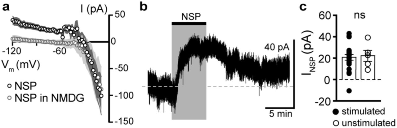
a, Current-voltage relationship of the apparent outward current produced by NASPM (NSP, 100 μM). Replacing 126 mM NaCl with NMDG eliminated the apparent outward current, suggesting a block of a tonic inward Na+ current. b, Representative trace of whole-cell voltage-clamp recording of the apparent outward current induced by application of NASPM in an unstimulated brain slice. Dashed line is at 0 pA (Vhold −65 mV). c, Plot of amplitude of NASPM-induced apparent outward current in stimulated and unstimulated brain slices demonstrating no effect of prior electrical stimulation (p=0.850, n=21&5). Line and error bars/shaded area represent mean±SEM, ns indicates not significant.

a, Example whole-cell voltage-clamp recording of the basal whole-cell current and the α1-AR-EPSC evoked every 90s (arrows) prior to, during, and after application of NASPM (NSP, 100 μM). b, NASPM completely eliminated the α1-AR-EPSC shown in representative traces (left, baseline adjusted) and in grouped data (right, p=0.002, n=10). c, Application of NASPM produced an apparent outward current (p<0.0001, n=21). d, Time course of the inhibition of the α1-AR-EPSC amplitude (bottom) and apparent outward current (top) by application of NASPM. e, Membrane resistance (Rm, ΔV −65 to −75 mV) increased during application of NASPM, indicating the apparent outward current was due to ion channels closing (p=0.004, n=10). f, Time course of the inhibition of the α1-AR-EPSC amplitude by application of NASPM, demonstrating identical block of the α1-AR-EPSC whether or not α1-AR-EPSCs were evoked during NASPM application. Line and error bars represent mean±SEM, * denotes statistical significance.
GluDRs bind the amino acids D-serine and glycine which inhibit constitutively open mutant GluDR current21,22. Application of D-serine (10 mM, 13.5 min) reduced the α1-AR-EPSC amplitude by 49.7±9.6% (Fig. S6a,c). Application of glycine (10 mM, 4.5 min) reduced the α1-AR-EPSC amplitude by 70.9±11.0% (Fig. S6b,c). Lastly, application of the glutamate receptor antagonist kynurenic acid (1 mM, 10.5 min) reduced the α1-AR-EPSC amplitude by 65.6±8.3% (Fig. S6c).

a, Application of D-serine (10 mM) reversibly reduced the α1-AR-EPSC, shown in a representative trace (left) and in grouped data (right, p=0.001, n=7). b, Application of glycine (10 mM) reversibly reduced the α1-AR-EPSC, shown in representative traces (left, baseline adjusted) and in grouped data (right, p=0.015, n=7). c, Summarized data of percent remaining of the α1-AR-EPSC amplitude after NASPM (NSP, 100 μM), D-serine (D-S, 10 mM), glycine (glyc, 10 mM), or kynurenic acid (KA, 1 mM). Line and error bars represent mean±SEM, * denotes statistical significance, ns denotes not significant.
Next, a viral genetic strategy was used to functionally delete GluD1R by targeting the encoding gene, GRID1, via CRISPR/Cas9 (Fig. S7a,b). In brief, a cocktail of AAV1 viruses encoding Cas9, and mouse Grid1 guide RNA with enhanced green fluorescent protein (eGFP) reporter (GRID1) was microinjected into the DR of wild type mice. A separate cohort received microinjections of a cocktail of AAV1 viruses encoding Cas9 and eGFP reporter (control). Brain slices were prepared after >4 weeks and the DR was microdissected and frozen on dry ice to assess the mutation of GRID1. Restriction enzyme site-digested PCR confirmed in vivo mutation of GRID1 at the expected site (Fig. S7c). In a separate cohort, brain slices were prepared and whole-cell voltage-clamp recordings were made from infected and uninfected neurons visualized in brain slices by expression of eGFP. In eGFP+ neurons from control mice, electrical stimulation produced a decrease in Rm and an α1-AR-EPSC, and application of NA caused inward INA (Fig. 4). However, in eGFP+ neurons from GRID1 mice, electrical stimulation did not change Rm (Fig. 4a) and no α1-AR-EPSC was detected above baseline noise (Fig. 4b). In addition, inward INA was substantially smaller in eGFP+ neurons from GRID1 mice, as compared to eGFP+ neurons from control mice (Fig. 4c). In the same slices from GRID1 mice, eGFP-neurons still had an α1-AR-EPSC and inward INA (Fig. 4a,c). Lastly, application of NASPM produced an apparent outward current in eGFP+ neurons from control mice, but not from GRID1 mice (Fig. 4d). Taken together, these results demonstrate that conduction through GluD1R is necessary for the α1-AR-EPSC and the NASPM-sensitive tonic inward current.

a, A graphic representation of the third exon of mGrid1, marked with the location of the gRNA (390F) and the overlapping BglI restriction site. Filled arrows represent the location of primers used for PCR amplification while the hollow arrow indicates the primer used for sequencing (Table S1). b, A nucleotide representation of gRNA target site with the translated peptide sequence underneath. The BglI recognition site (GCCNNNN^NGGC) is contained by the grey box and overlaps with the predicted Cas9 cleavage site (|). c, Fragment analysis on BglI-digested Grid1 PCR products amplified from the DR of mice injected with (AAV-Cas9 and AAV-empty) or (AAV-Cas9 and AAV-GRID1). “% PCR undigested” refers to the amount of the undigested PCR product relative to the total PCR product as determined by AATI fragment analysis.

a, Membrane resistance (Rm, ΔV −65 to −120 mV) decreased after stimulation in infected neurons from mice injected with AAV-Cas9 and AAV-empty (control, p=0.0002, n=13), but not in infected neurons from mice injected with AAV-Cas9 and AAV-GRID1 (GRID1, p=0.562, n=16). b, Representative traces (left) and grouped data (right, p<0.0001, n=16&16) demonstrating the presence of an α1-AR-EPSC in infected neurons from control mice, but not from GRID1 mice. Neighboring uninfected neurons from GRID1 mice (uninf.) had an α1-AR-EPSC that was indistinguishable from infected neurons from control mice (p>0.999, n=7). c, Targeting GluD1R reduced the inward current to NA (INA, 30 μM) as compared to infected neurons from control mice, as shown in representative traces (left) and grouped data (right, p=0.009, n=16&16). Capacitive transients and ramp-induced currents have been truncated for clarity. d, Targeting GluD1R reduced the tonic inward current revealed by application of NASPM (100 μM, INSP) as compared to infected neurons in control mice (p=0.009, n=5&11). Line and error bars represent mean±SEM, * denotes statistical significance, ns denotes not significant.
Relevance of GluD1R to the dorsal raphe
In vivo, 5-HT neurons in the DR require NA release and subsequent activation of α1-ARs to maintain AP firing8. Activation of α1-ARs in the DR by exogenous agonist was reported to depolarize neurons through net reduction of K+ conductance and by activation of an unidentified non-K+ conductance23. More recently, Brown et al., reported that α1-ARs activation in the DR induces Na+-dependent inward current with an Erev of −23 mV24, similar to our findings. Our study identifies GluD1R as the channel that carries this mixed cation current, indicating that modulation of GluD1R is a key constituent in driving persistent AP firing of the 5-HT neurons. In principle, inward GluD1R current may bring the membrane potential to threshold, but recruitment of other voltage-gated ion channels is expected to underlie the persistent pacemaker-like activity.
Ionotropic nature of GluD1R
GluDR have been characterized as synaptic organizers, regulating LTD, endocytosis and trafficking of AMPAR, formation of excitatory and inhibitory synapses, and spine density3,25–27, without the requirement of ion conduction. Similarly, NMDAR signal through non-ionotropic or “metabotropic” mechanisms where ion conduction is not required, to regulate LTD, AMPAR endocytosis, and spine morphology28. The ionotropic nature of GluDR does not conflict with its known role as a scaffold protein, but rather expands the similarities between NMDAR and GluDR.
The largest obstacle towards understanding the ionotropic nature of GluDR is the lack of known agonist and inability to gate the intact channel. The majority of studies have been performed on constitutively open mutant or chimeric channels29,30. Two prior studies have demonstrated that in heterologous systems and brain slices, activation of metabotropic glutamate receptors (mGluR) produces an inward current carried by GluD1R5 or GluD2R4, concluding mGluR activation triggers rather than modulates gating of GluDR. The congruous explanation of our results is that, in DR neurons, GluD1R are functional and open under basal conditions, carrying subthreshold, tonic Na+ current. Activation of α1-ARs, by exogenous agonist or synaptic NA release, excites DR neurons by increasing GluD1R current.
In general, the kinetics of iGluR synaptic currents are controlled by the lifetime of the receptor-agonist complex and the rates of desensitization and deactivation. In brain slices, NMDAR carry tonic inward current31 attributed to slow desensitization and ambient levels of glutamate and glycine. What remains to be understood are the conditions that permit GluD1R opening, and why their activation has been largely elusive in heterologous expression systems. Reminiscent of times before the discovery of glycine as a necessary co-agonist at NMDAR32,33, it may be that an endogenous agonist needed for gating is present in brain slices. Alternatively, it is possible GluD1R are gated by an intracellular factor or require expression of accessory or interacting proteins34. Tonic activation of α1-ARs cannot explain the tonic inward current as α1-AR antagonism did not change basal whole-cell current.
The mechanism by which α1-ARs increase GluD1R current also remains to be described and may be distinct from tonic activation. It is well-established that GqPCRs, especially mGluR and mAChR, bidirectionally change NMDAR and AMPAR currents, producing the two major forms of synaptic plasticity, long-term potentiation (LTP) and long-term depression (LTD), in part through a variety of postsynaptic mechanisms35. To our knowledge, the duration of the α1-AR-EPSC (~27 s) is exceptional for any synaptic current and more closely resembles the duration of short-term synaptic plasticity; for instance, endocannabinoid-mediated short-term depression36. GqPCRs can activate phospholipase C which hydrolyzes the integral membrane lipid, phosphatidylinositol 4,5-bisphosphate (PIP2). PIP2 stabilizes Kv7 channels such that PIP2 hydrolysis following mAChR activation accounts for inhibition of M-current37. By the same signaling cascade, GqPCRs stimulate the production of endocannabinoids that can act directly on membrane ion channels38. Thus, one possibility is that α1-ARs modulate GluD1R through membrane lipid signaling. Largely, it remains to be seen whether the intracellular signaling cascades known to affect NMDAR and AMPAR currents generalize to GluDR.
Using heterologous systems or constitutively open mutants, GluDR current reverses polarity around 0 mV39, akin to AMPAR and NMDAR, while our results show an Erev of ~ −30 mV. The difference may be due space-clamp error in brain slices, since the magnitude of subtracted current is small relative to total current at depolarized potentials. Activation of α1-ARs in the DR also inhibits A-type K+ current40. Thus, Erev measurements may include inhibition of a voltage-gated K+ conductance that is not contributing directly to the α1-AR-EPSC but is activated during voltage ramps at Vhold >-65 mV. Tail current analysis revealed voltage-dependence of INA, such that prior depolarization reduced conductance. These data may reflect block of GluD1R by endogenous intracellular polyamines, as established for calcium-permeable AMPAR and KainateR41. Another important consideration is that our measurements may be subject to voltage-dependence of the signaling pathway between α1-ARs and GluD1Rs. There are reports of inward currents generated by GqPCR activation with Erevs between −40 and −23 mV13,24,42, measured under different recording conditions, suggesting a commonality that is unlikely to be due to space-clamp error alone. Taken together, measurements here should be considered an approximate estimate of GluD1R, and more precisely as the current-voltage relationship of the α1-AR-GluD1R signaling complex.
Summary
Native GluD1R are functional ion channels that carry ionic current. GluD1R underlie the α1-AR-mediated depolarization of DR neurons that drives AP firing in vivo. Many of the biophysical properties of the GluD1R channel are shared with other members of the ionotropic glutamate receptor family. This study lays the foundation to investigate the ionotropic role of GluD1R in excitatory synaptic transmission and neuronal excitability, expanding upon the wealth of knowledge of pharmacology and regulatory elements established for NMDAR and AMPAR signaling.
METHODS
Animals
All studies were conducted in accordance with the National Institutes of Health Guide for the Care and Use of Laboratory animals with the approval of the National Institute on Drug Abuse Animal Care and Use Committee. Wild type C57BL/6J (>3 months old) mice of either sex were used. Mice were group-housed on a 12:12 h reverse light cycle.
Brain slice preparation and electrophysiological recordings
Mice were deeply anesthetized with isoflurane and killed by decapitation. Brains were removed quickly and placed in warmed modified Krebs’ buffer containing (in mM): 126 NaCl, 2.5 KCl, 1.2 MgCl2, 1.2 CaCl2, 1.2 NaH2PO4, 21.5 NaHCO3, and 11 D-glucose with 5 μM MK-801 to reduce excitotoxicity and increase viability, bubbled with 95/5% O2/CO2. In the same solution, coronal DR slices (220 μm) were obtained using a vibrating microtome (Leica 1220S) and incubated at 32 °C >30 minutes prior to recording.
Electrophysiological recordings were made at 35 °C with a Multiclamp 700B amplifier (Molecular Devices), Digidata 1440A A/D converter (Molecular Devices), and Clampex 10.4 software (Molecular Devices) with borosilicate glass electrodes (King Precision Glass) wrapped with Parafilm to reduce pipette capacitance. Pipette resistances were 1.8-2.8 MΩ when filled with an internal solution containing, (in mM) 104.56 K-methylsulfate, 5.30 NaCl, 4.06 MgCl2, 4.06 CaCl2, 7.07 HEPES (K), 3.25 BAPTA(K4), 0.26 GTP (sodium salt), 4.87 ATP (sodium salt), 4.59 creatine phosphate (sodium salt), pH 7.32 with KOH, mOsm ~285, for whole-cell patch-clamp recordings. Series resistance was monitored throughout the experiment. Cell-attached recordings were made from quiescent neurons in slice, using pipettes filled with modified Krebs’ buffer. Reported voltages are corrected for a liquid junction potential of −8 mV between the internal solution and external solution. All drugs were applied by bath application. All experiments were conducted with NMDAR, AMPAR, KainateR, GABA-AR, and 5-HT1AR antagonists in the external solution. In addition, a α2-adrenergic receptor antagonist was added for experiments where NA was applied and a glycine receptor antagonist was added when glycine was applied. Unitary current was calculated from fluctuation analysis, as previously described43, assuming the macroscopic current arises from independent, identical channels with a low probability of opening, according probability theory; i = σ2/[I(1 - p)] where i is unitary current, σ2 is the variance, I is mean current amplitude, and p is probability of opening.
Vector construction
gRNA identification
CRISPR SpCas9 gRNA target sites were identified in the mouse Grid1 gene (NC_000080.6) using CRISPOR44. The seed sequence (GAACCCTAGCCCTGACGGCG) was chosen based on its relatively high specificity scores and the observation that it contains a Bgl I restriction enzyme site (GCCNNNN^NGGC) that overlaps with the Cas9 cleavage site.
mGrid1 genotyping
C57BL/6J mouse genomic DNA was isolated from tail biopsies or brain pieces containing microdissected DR by digestion in DNA lysis buffer (50 mM KCl, 50 mM Tris-HCl (pH 8.0), 2.5 mM EDTA, 0.45% NP-40, 0.45% Tween-20, 0.5 ug/mL proteinase K) for 3 h at 55 °C, and 1 h at 65 °C. Lysates were then used as templates to amplify a 654 basepair fragment including the 390F gRNA target site using Q5 HotStart Master mix (New England Biolabs). A portion of the finished PCR reaction was treated with Bgl I restriction enzyme (New England Biolabs) for 60 minutes and processed on an AATI fragment analyzer.
Construction and packaging of AAV vectors
The AAV vector plasmid encoding SpCas945 (pX551) expressed from the MeCP2 promoter was a gift from Feng Zhang (Addgene plasmid # 60957, AAV-Cas9). The AAV packaging plasmid encoding a nuclear envelope-embedded eGFP reporter (Addgene 131682) was constructed by amplifying the KASH domain from (Addgene 60231, a gift of Feng Zhang) and fusing it (in-frame) to the end of coding region for eGFP in (Addgene 60058, pOTTC407) using ligation-independent cloning (AAV-empty, Fig. S8). gRNA was cloned into a mU6 expression cassette and then moved into an AAV backbone expressing a nuclear envelope-embedded (KASH-tagged) eGFP reporter (Addgene 131683) by PCR amplification and ligation-independent cloning (AAV-GRID1). Insert-containing clones were verified by sequencing and restriction fragment analysis prior to virus production. All AAV vectors were produced using triple transfection method as previously described46. All vectors were produced using serotype 1 capsid proteins and titered by droplet digital PCR.

A graphic representation of AAV vectors used in conjunction with AAV-Cas9 to encode eGFP reporter only (top, AAV-empty) or mouse Grid1 guide RNA with eGFP reporter (bottom, AAV-GRID1).
Stereotaxic intracranial microinjections
Mice were anesthetized with a cocktail of ketamine/xylazine, immobilized in a stereotaxic frame (David Kopf Instruments), and received one midline injection of a 1:1 (v/v) cocktail of viruses AAV-Cas9 and AAV-empty or AAV-GRID1 for total volume 400 nL delivered over 4 minutes. The coordinates for injection were AP −4.4; ML 1.19, 20° angle; DV −3.62 mm, with respect to bregma. Prior to surgery, mice were injected subcutaneously with warm saline (0.5 mL) to replace fluid lost during surgery and given carprofen (5 mg/kg) post-surgery for pain relief. Mice recovered for >4 weeks to allow expression. Infected neurons were identified in the slice by visualization of eGFP.
Materials
MK-801, noradrenaline, NASPM, NBQX, WAY-100635, and picrotoxin were purchased from Tocris Bioscience. All other chemicals were obtained from Sigma-Aldrich.
Data analysis and visualization
Data were analyzed using Clampfit 10.7. Data are presented as representative traces, or in scatter plots where each point is an individual cell, and bar graphs with means ± SEM. Unless otherwise noted, n = number of cells. Erevs were determined by linear regression for each cell. Recordings in which current did not cross 0 pA were omitted from analysis. To minimize space-clamp errors, analysis of current during voltage ramps was limited to −10 mV where the currents were typically less than 500 pA. Ramp currents were averaged in 2 mV bins (20 ms). Data sets with n>30 were tested for normality with a Shapiro-Wilk test. When possible (within-group comparisons), significant differences were determined for two group comparisons by paired t-tests, Wilcoxon matched-pairs signed rank test, and in more than two group comparisons by nonparametric repeated-measures ANOVA (Friedman test). Significant mean differences in between-group comparisons were determined for two group comparisons by Mann Whitney tests, and in more than two group comparisons by Kruskal-Wallis tests. ANOVAs were followed, when p<0.05 by Dunn’s multiple comparisons post hoc test. Linear trends were analyzed using a mixed model ANOVA. A difference of p<0.05 was considered significant. Exact values are reported unless p<0.0001 or >0.999. Statistical analysis was performed using GraphPad Prism 8 (GraphPad Software, Inc.).
AUTHOR CONTRIBUTIONS
S.C.G. designed the electrophysiology experiments. S.C.G. and H.S.H. performed the experiments. S.C.G. and K.M. analyzed the data and prepared the figures. S.C.G. and K.M. wrote the manuscript with the help of all co-authors.
COMPETING INTEREST DECLARATION
The authors declare no competing interests.
Primer sequences used in PCR amplification (amp) and sequencing (seq).
ACKNOWLEDGMENTS
This work was supported by the Intramural Research Program at the National Institute on Drug Abuse (S.C.G., H.S.H, K.M.) and the National Institutes of Health Center on Compulsive Behaviors (S.C.G.). The selection and functional validation of the Grid1 gRNA and the construction and packaging of AAV viral vectors, were performed by the Genetic Engineering and Viral Vector Core of the National Institute on Drug Abuse. The opinions expressed in this article are the authors’ own and do not reflect the views of the NIH/DHHS. We thank Drs. John T. Williams and Bruce P. Bean for comments on this manuscript.
Footnotes
↵* lead contact


