Review

Oxidative Stress Responses in the Human Fungal Pathogen, Candida albicans

1
Departamento de Biologia Celular e Genética, Instituto de Biologia Roberto Alcantara Gomes, Universidade do Estado do Rio de Janeiro (UERJ), Rio de Janeiro 20550-013, Brazil
2
Institute for Cell and Molecular Biosciences, Faculty of Medical Sciences, Newcastle University, Newcastle upon Tyne NE2 4HH, UK
*
Author to whom correspondence should be addressed.
These authors contributed equally to this work.
Biomolecules 2015, 5(1), 142-165; https://doi.org/10.3390/biom5010142
Submission received: 12 January 2015 / Revised: 11 February 2015 / Accepted: 12 February 2015 / Published: 25 February 2015
(This article belongs to the Special Issue Oxidative Stress and Oxygen Radicals)

Abstract

:
Candida albicans is a major fungal pathogen of humans, causing approximately 400,000 life-threatening systemic infections world-wide each year in severely immunocompromised patients. An important fungicidal mechanism employed by innate immune cells involves the generation of toxic reactive oxygen species (ROS), such as superoxide and hydrogen peroxide. Consequently, there is much interest in the strategies employed by C. albicans to evade the oxidative killing by macrophages and neutrophils. Our understanding of how C. albicans senses and responds to ROS has significantly increased in recent years. Key findings include the observations that hydrogen peroxide triggers the filamentation of this polymorphic fungus and that a superoxide dismutase enzyme with a novel mode of action is expressed at the cell surface of C. albicans. Furthermore, recent studies have indicated that combinations of the chemical stresses generated by phagocytes can actively prevent C. albicans oxidative stress responses through a mechanism termed the stress pathway interference. In this review, we present an up-date of our current understanding of the role and regulation of oxidative stress responses in this important human fungal pathogen.

1. Candida albicans Is a Major Fungal Pathogen of Humans

The polymorphic fungus, Candida albicans, is a constituent of the normal human microbiome. This fungus, together with other Candida family members, is present on the skin and in the oral cavity and gastrointestinal and urogenital tracts of most healthy individuals [1,2]. In the healthy host, C. albicans normally exists as a benign commensal organism. However, as an opportunistic pathogen, this fungus can also cause superficial infections, such as oral or vaginal candidiasis, or life-threatening systemic infections [2]. Perturbation of the microbiome through antibiotic usage or mild to severe defects in immune defences, such as in patients with HIV, can result in superficial oral and vaginal infections (thrush), termed oral (OC) and vulvovaginal (VVC) candidiasis, respectively. OC occurs in about 90% of HIV-infected persons as an AIDS-defining illness [3]. Defective immunity in premature infants and the elderly can also result in OC [4,5]. Significantly, 75% of women of childbearing age suffer from VVC, 45% of whom go on to have a least one recurrent infection [6]. Superficial candidiasis can also manifest as chronic infections of the skin and nails, resulting in mucocutaneous candidiasis (CMC) [7]. Although superficial infections are remarkably commonplace, they are non-life threatening and can be easily treated.
In contrast, systemic candidiasis is associated with unacceptably high crude and attributable mortality rates of 42 and 27%, respectively, despite the availability of antifungal drugs, such as the polyenes, azoles and echinocandins. These mortality rates exceed those attributed to sepsis caused by the most aggressive bacterial and viral pathogens [8] and are attributed to difficulties in diagnosing fungal systemic infections and the consequential delays in treatment [9]. Patients who are severely immunocompromised, such as those on immunosuppressive treatments for cancer or transplant surgery, are at risk of systemic candidiasis [10]. In such patients, the innate defence mechanisms, which are vital to prevent invasive disease, are significantly compromised [11]. Consequently, the fungus can survive in the bloodstream and subsequently colonise a number of internal organs [2]. Other risk factors include invasive clinical procedures or trauma, which disrupt the protective anatomical barrier of the mucosa, and the use of venous catheters, which can allow access of the fungus to the bloodstream [10]. Indeed, overall, Candida spp. are the fourth most common nosocomial (hospital acquired) systemic infection in the United States [8]. Clearly, C. albicans poses a significant medical problem, and thus, it is important that we understand what makes this fungus such a successful pathogen.

2. Reactive Oxygen Species Are a Core Component of the Immune Cell Armoury

In healthy hosts, the first line of defence against C. albicans is through phagocytosis by innate immune cells, including macrophages and neutrophils. A major antimicrobial defence mechanism mounted by these phagocytes is the production of reactive oxygen species (ROS) through a process known as the respiratory burst. Following stimulation by cytokines, phagocytic cells activate the assembly of the NADPH oxidase complex, which results in the generation of superoxide (O2•). Given the potency of the ROS produced by NADPH oxidase [12], activation of this multi-subunit enzyme is tightly regulated. The NADPH oxidase complex consists of the Nox2 (gp91phox) catalytic subunit, the p22phox transmembrane protein and three cytosolic subunits, p47phox, p67phox and p40ph°x. Nox2 and p22phox make up the membrane-associated cytochrome b558 heterodimer. Activation of Nox2 is dependent on the interaction with the cytosolic components, in particular p67phox, which translocate to the membrane following phagocytosis [13]. This interaction is dependent on the binding of the small GTPase Rac to p67phox, which induces a conformation change in this subunit, thus promoting its interaction with Nox2 [14]. Activation of Nox2 drives the production of superoxide via the NADPH-driven reduction of molecular oxygen. This is generated at an extremely high rate of 5 to 10 nmol per s within the neutrophil phagosome [15], and it has been estimated that approximately 4 mol L−1 of O2• is produced per bacterium engulfed in the phagocytic vacuole [16]. The superoxide is then dismutated to hydrogen peroxide (H2O2) by superoxide dismutase or to hydroxyl anions (OH) and hydroxyl radicals (OH) via the Haber-Weiss reaction. The importance of the NADPH oxidase-mediated respiratory burst as an antimicrobial mechanism is manifested in patients with chronic granulomatous disease (CGD). CGD is a human genetic disorder characterized by a deficiency in the NAPDH oxidase complex and is associated with recurrent and life-threatening bacterial and fungal infections [17]. Significantly, patients with CGD have an increased susceptibility to Candida infections [18]. Interestingly, in addition to the fungicidal roles of ROS, recent work has revealed that the ROS produced by NADPH oxidase also functions to recruit phagocytes to C. albicans infection foci. This NADPH oxidase-regulated recruitment of phagocytes is important for efficient phagocytosis, containment of the fungus within the phagocyte and survival of the host [19].
Other toxic chemicals are subsequently derived from the ROS in the phagosome [20]. For example, H2O2 can react with chloride ions (Cl) to form hypochlorous acid (HOCl) in a reaction catalysed by myeloperoxidase (MPO). In addition, the nitric oxide radical generated by the action of the inducible nitric oxide synthase (iNOS) interacts with superoxide to produce the highly toxic peroxynitrite (ONOO) [21]. Recently, work has also revealed that the combination of reactive oxygen species together with the cationic stress generated during phagocyte maturation underlies the potency of phagocytes in C. albicans killing [22]. Thus, phagocytic cells synthesize an array of toxic chemicals that work in combination to promote fungal killing. It is also noteworthy that, in addition to ROS production within the phagosome, phagocytes secrete ROS into the external milieu [23]. Consistent with this, C. albicans cells have been shown to mount an oxidative stress response prior to phagocytosis [24]. Furthermore, C. albicans will also come in contact with ROS produced by H2O2-producing bacteria in the mouth and gut. Several commensal bacteria, for example Enterococcus faecalis [25] and Lactobacillus species [26], secrete ROS into their surroundings, and this may have an inhibitory effect on the growth of C. albicans in host niches, other than the phagosomal environment. Consistent with this, using a Caenorhabditis elegans model of polymicrobial infection, E. faecalis was shown to reduce the virulence of C. albicans [27].
The ROS generated within the phagosome creates a toxic environment that induces oxidative stress in C. albicans. Indeed, exogenous ROS can induce programmed cell death in this fungal pathogen [28]. ROS interact with proteins, lipids and nucleic acids [29], causing irreversible damage to the pathogen. DNA damage caused by ROS can result in chemical base changes, structural alterations, single- and double-strand breaks and cross-linkage. Lipid peroxidation occurs by a free radical chain reaction, which culminates in peroxidation events at many fatty acid side chains, leading to the damage of the cell membrane. ROS reactions with proteins can lead to the formation of protein-protein cross-links, oxidation of the peptide backbone and reversible or irreversible oxidation of amino acid side chains. Although this can be deleterious to protein function, as discussed below, several oxidative stress-sensing proteins are activated by the reversible oxidation of cysteine residues.

3. Response of Candida albicans to ROS

3.1. Transcriptional Responses to ROS

A well-characterized response of eukaryotic microbes to ROS is the rapid induction of mRNAs that encode oxidative stress detoxification and repair proteins. Interestingly, C. albicans is considerably more resistant to oxidative stress than the benign model yeasts, Schizosaccharomyces pombe and Saccharomyces cerevisiae [30,31]. However, the basis for this resistance does not appear to be due to differences in transcriptional responses to oxidative stress, as all three fungi appear to induce a similar set of core antioxidant genes following exposure to H2O2 [32,33]. These include catalase (CAT1), glutathione peroxidase (GPX) and superoxide dismutase (SOD) antioxidant-encoding genes, in addition to genes encoding components of the glutathione/glutaredoxin (GSH1, TTR1) and thioredoxin (TSA1, TRX1, TRR1) systems, which play critical roles in repairing oxidatively-damaged proteins, protein folding and sulphur metabolism. Such oxidative stress-responsive genes are also induced in C. albicans following exposure to macrophages or neutrophils [34,35,36,37,38], illustrating that this pathogen induces the respiratory burst in these phagocytes. The analyses of GFP-reporter fusions, under the control of oxidative stress-responsive promoters, have also revealed that C. albicans is exposed to significant levels of ROS prior to phagocytosis [24]. In contrast, however, oxidative stress responses do not appear to be induced once C. albicans cells have established systemic kidney infections [34,39,40]. Thus, inducible oxidative stress responses appear vital for C. albicans to survive phagocytosis by innate immune cells, but are seemingly less important for the fungus to develop systemic infections. Indeed, whilst a number of genes encoding key antioxidants (such as CAT1, TRX1, GRX2, SOD1, SOD5) are important for virulence in systemic models of infection [41,42,43,44,45], others (including TSA1, GPXs) are dispensable [46,47].

3.2. Transcriptional Responses to ROS Are Inhibited in the Presence of Cationic Stress

In healthy individuals, C. albicans cannot evade the oxidative-killing mechanisms mounted by innate immune cells. Such cells prevent infection by employing a battery of toxic chemicals in addition to ROS. For example, phagocytes expose C. albicans to cationic fluxes (K+) and acidification, as well as to superoxide anions [16,21,48]. However, as C. albicans is resistant to each of these individual stresses in vitro [30,31], a key question, therefore, is: what accounts for the potency of innate immune defences? Although host microenvironments are complex and dynamic, our understanding of C. albicans stress responses is based on studies of individual stresses. Significantly, however, recent work has revealed that C. albicans is exquisitely sensitive to combinations of oxidative and cationic stresses [49], which are encountered following phagocytosis. Cationic stress can be imposed in vitro by exposure of the fungus to either NaCl or KCl and in the phagocyte is caused by increased flux of K+ into the phagosome [16]. Strikingly, exposure to cationic stress results in the inhibition of C. albicans oxidative stress responses. This phenomenon has been termed “stress pathway interference” [22]. The combinatorial stress-mediated synergistic killing of C. albicans contrasts starkly with the stress cross-protection described in model yeasts, whereby exposure to one stress protects against subsequent exposure to a different stresses [50]. The existence of stress pathway interference was revealed through gene expression analysis in C. albicans. Transcript profiling showed that H2O2-induced gene expression is severely attenuated, and intracellular ROS levels increase dramatically, following combinatorial oxidative and cationic stress. For example, key antioxidant genes, such as CAT1 encoding catalase and TRR1 encoding thioredoxin reductase, which are significantly induced following H2O2 stress, fail to be induced following exposure of cells to H2O2 in the presence of cationic stress [22]. This cationic stress-mediated inhibition of oxidative stress responses appears to be of physiological relevance, as the high fungicidal activity of human neutrophils is dependent on the combinatorial effects of the oxidative burst and cationic fluxes [22]. However, as discussed above, oxidative stress-responsive genes are induced following co-culture of C. albicans with phagocytes [34,35], so how is this reconciled with the combinatorial stress-mediated inactivation of such genes? It has been suggested [22] that the activation of C. albicans antioxidant genes during interaction with phagocytes may be due to exposure to extracellular ROS prior to engulfment [24]. Furthermore, as exposure of cells to combinatorial oxidative and cationic stresses prevents the normal activation of oxidative stress-responsive genes, this may explain why C. albicans oxidative stress genes are not expressed in certain host niches, such as during systemic infections of the kidney, despite the presence of neutrophil infiltrates [34].

3.3. Extracellular Antioxidant Enzymes as a Pathogen-Specific Adaptation Mechanism

As C. albicans appears to mount standard transcription responses to oxidative stress, the high level of resistance of this pathogen to ROS could, instead, be related to the evolutionary expansion of the SOD family and the fact that this pathogen expresses SODs and other antioxidant enzymes on the cell surface. C. albicans contains six SOD enzymes distributed between different cellular compartments. Sod1–3 are intracellular enzymes, while Sod4–6 are glycosylphosphatidylinositol (GPI)-anchored cell wall-associated enzymes. The Cu-/Zn-containing Sod1 is induced following phagocytosis and is required for C. albicans to resist macrophage-mediated killing [43]. The extracellular Sods also have vital roles in the detoxification of superoxide radicals generated by phagocytes; co-culture of macrophages with C. albicans cells lacking Sod4 and Sod5 leads to massive extracellular ROS accumulation in vitro [23]. Consequently, inactivation of Sod4 and Sod5 results in C. albicans cells that are exquisitely more susceptible to phagocyte-mediated killing [23,35]. Interestingly, the expression of Sod4 and Sod5 is dependent on the morphology of C. albicans, as Sod4 is expressed in yeast cells, whereas Sod5 is a hyphal-induced gene [44,51]. Sod5 is also induced following phagocytosis by neutrophils independently of hyphae formation [35]. Recently, structural analysis of Sod5 revealed that it represents a novel class of superoxide dismutases that only depends on Cu for activity. Furthermore, it is secreted in its apo-form and can readily capture extracellular copper without the aid of a Cu chaperone, which rapidly induces activity [52]. It is suggested that this novel mode of activation is uniquely adapted to the host environment, as macrophages release copper in an attempt to kill invading microbes through copper toxicity [53]. In addition to specific Sods, two key peroxidase enzymes have also been found at the cell surface of C. albicans; the thiol-specific peroxidase Tsa1 [47,54] and the peroxide detoxifying enzyme catalase [54]. Tsa1 and Cat1 were identified as major plasminogen-binding proteins in isolated cell wall protein preparations [54], and the cell wall localization of Tsa1 has also been illustrated using fluorescence microscopy [47]. These extracellular mechanisms for protection against ROS likely reflect an adaptation of this pathogenic fungus to prevent the intracellular accumulation of toxic levels of ROS.

3.4. Morphogenesis as an Oxidative Stress Response

Following phagocytosis, C. albicans can evade oxidative-killing by macrophages and neutrophils by switching from budding to filamentous cells, which can pierce the phagosomal membrane [37]. Not only does this allow the pathogen to escape, but this also results in the C. albicans-mediated killing of the phagocyte [55]. It has recently been demonstrated that the ability of C. albicans to mount robust oxidative stress responses is vital for this polymorphic pathogen to filament in the phagosome [46,56]. C. albicans mutants that are sensitive to ROS in vitro fail to filament once phagocytosed and, thus, are trapped within the macrophage and unable to evade phagocyte-mediated killing. Consistent with the requirement of fungal oxidative stress defences to allow filamentation and macrophage escape, the phagocyte NADPH oxidase is important in inhibiting filamentation in vivo [57]. The ROS produced by the NADPH oxidase also function to recruit phagocytes, thereby increasing phagocytosis and inhibiting filamentation [19]. Thus, the outcome of the battle between C. albicans and innate immune cells appears dependent on the NADPH oxidase-regulated functions of the phagocyte and the robustness of the fungal oxidative stress responses.
The mechanisms underlying C. albicans filamentation following phagocytosis remain to be fully explored. A recent study reported that the ROS-induced induction of arginine biosynthesis genes is important for hyphal formation following phagocytosis of C. albicans [58]. Moreover, exposure of C. albicans to the ROS H2O2 triggers the filamentation of this polymorphic fungus in vitro [42,59]. A close examination of the morphology of these cells revealed that H2O2-induced filaments are hyperpolarized buds, which are morphologically distinct from hyphae and pseudohyphae filamentous forms [42]. The hyperpolarised bud is a relatively recently characterized filamentous form of C. albicans and is normally associated with either mutations or chemicals that perturb cell cycle progression [60,61]. The observation that H2O2 stimulates hyperpolarised bud formation provided the first example of a physiologically relevant condition that induces this filamentous form of growth in C. albicans. Does exposure of ROS following phagocytosis trigger the formation of hyperpolarised buds allowing this pathogen to pierce the phagocyte membrane and escape? Evidence so far indicates that ROS-stimulated hyperpolarized bud formation may not contribute to C. albicans filamentation within the macrophage. For example, ROS-sensitive C. albicans mutants that cannot filament within the macrophage [56] can form H2O2-induced hyperpolarized buds in vitro [42]. Nonetheless, ROS-stimulated filamentation of C. albicans may be beneficial for survival in other host niches, such as the gut, where C. albicans co-exists with ROS-producing bacteria.

4. Signalling Pathways that Mediate C. albicans Responses to ROS

To date, three signalling pathways have been demonstrated to be directly activated in response to ROS in C. albicans. These include the Cap1 transcription factor, the Hog1 stress-activated protein kinase and the Rad53 DNA damage checkpoint kinase. Here, we discuss the role and regulation of these pathways in oxidative stress responses in C. albicans. Other signalling pathways not known to be activated by ROS, but which contribute to oxidative stress tolerance in C. albicans are also briefly summarized.

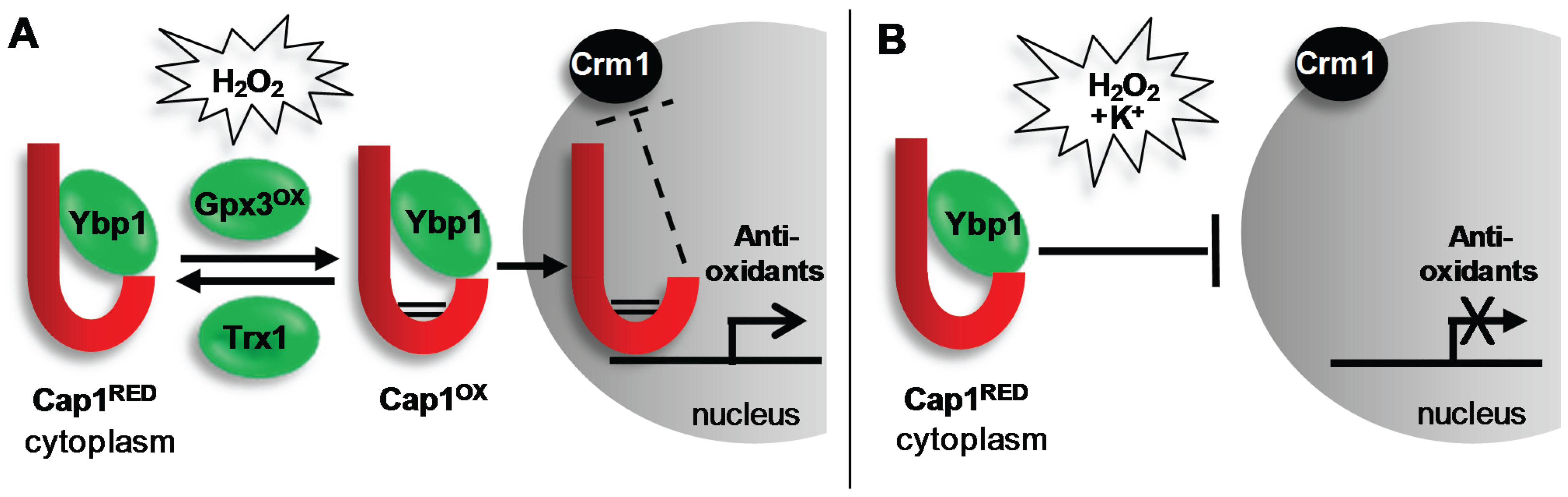
4.1. The Cap1 ROS-Responsive Transcription Factor

In C. albicans, the Cap1 transcription factor is the major regulator of the oxidative stress-induced transcriptome and proteome, both in vitro [62,63] and ex vivo, following exposure to neutrophils [35]. Cap1 is a bZip transcription factor of the AP-1 family and is closely related to the S. cerevisiae Yap1 and S. pombe Pap1 proteins, which have well-characterized roles in oxidative stress and multi-drug resistance [64,65]. Similarly, C. albicans cap1Δ cells are sensitive to several reactive oxygen species and drugs [33,66,67]. Chromatin immunoprecipitation (CHiP) analysis to determine direct targets of Cap1 identified many key antioxidant genes, including CTA1 and TRX1, and those involved in the response to drugs, such as MDR1 [68]. Cap1 plays a role in recruiting the Ada2 component of the SAGA/ADA histone acetylase co-activator complex to the promoters of oxidative stress and drug-responsive target genes [69,70]. Cells lacking Ada2 are highly sensitive to ROS, and the oxidative stress-induced transcription of key Cap1 target genes is significantly impaired; therefore, Cap1 recruitment of the SAGA complex appears to be a vital component of the oxidative stress response in C. albicans.

4.1.1. Regulation of Cap1

Similar to that reported for S. cerevisiae Yap1, C. albicans Cap1 rapidly accumulates in the nucleus in response to H2O2 [56,67]. Under non-stressed conditions, Yap1 shuttles between the cytoplasm and the nucleus due to the interaction of a nuclear export sequence (NES), located at the C-terminus of these transcription factors, with the Crm1 nuclear export factor [71]. However, following exposure to H2O2, Yap1 is activated by oxidation of specific cysteine residues, resulting in disulphide bond formation between two cysteine-rich domains (n-CRD and c-CRD). This triggers a conformational change within Yap1 that masks the NES, thereby preventing its interaction with Crm1. The inability to be recognized by Crm1 leads to the nuclear accumulation of Yap1, the nuclear-dependent phosphorylation of this transcription factor and the induction of Yap1-dependent genes [72]. Conversely, activation of Yap1 is counteracted by the thioredoxins Trx1 and Trx2, which function to reduce oxidised Yap1 [72]. This basic mechanism of regulation is conserved in C. albicans (Figure 1). Mutation of the c-CRD affects Cap1 regulation [67], and Cap1 is rapidly oxidised following exposure to H2O2 [42]. In addition, following the nuclear accumulation of Cap1, this transcription factor becomes phosphorylated, and the induction of Cap1-dependent genes is observed. Furthermore, as seen in S. cerevisiae, thioredoxin functions to reverse the H2O2-induced oxidation and activation of Cap1 [42].
Fungal AP-1-like transcription factors are not directly oxidised by H2O2, but instead, specific peroxidase enzymes sense and transduce the H2O2 signal to these transcription factors (Figure 1). Similar to that observed in S. cerevisiae [72], Cap1 oxidation requires Gpx3, a glutathione peroxidase (Gpx)-like enzyme [56]. Studies with Yap1 showed that this transcription factor undergoes multiple oxidation events, with Gpx3 initiating Yap1 oxidation [73,74,75]. Similarly, multiple oxidized forms of Cap1 are also observed [56]. Gpx3-mediated oxidation of Yap1 and Cap1 also requires a second protein, Ybp1, which binds to and forms a complex with the AP-1-like factors [56,76]. A recent study has provided insight into an additional function of Ybp1 in both C. albicans and S. cerevisiae, as Cap1 and Yap1 are highly unstable in ybp1Δ cells [56]. Ubiquitin-mediated degradation of oxidised AP-1-like factors has recently been shown to be an important regulatory mechanism [77,78]; therefore, Ybp1 binding to the reduced cytoplasmic pools of Yap1 or Cap1 possibly functions to prevent this proteasome-mediated degradation [56].
Figure 1. H2O2-induced activation of Cap1 is inhibited in the presence of cations. (A) Exposure of C. albicans to H2O2 promotes the Gpx3/Ybp1-mediated oxidation and activation the Cap1 transcription factor (Cap1ox). Cap1ox can no longer interact with the Crm1 nuclear export factor resulting in its nuclear accumulation, and the subsequent Cap1-dependent induction of genes with antioxidant functions necessary for cell survival. Following cellular adaptation, Cap1ox is returned to the inactive reduced form (Cap1RED) by thioredoxin (Trx1); (B) Remarkably, when C. albicans cells are exposed to H2O2 in the presence of cations, Cap1 fails to accumulate in the nucleus and therefore antioxidant gene expression is not induced leading to cell death. This is important as, following phagocytosis, C. albicans is exposed simultaneously to ROS and cationic fluxes. See text for details.
Biomolecules 05 00142 g001
In S. cerevisiae, Yap1 functions alongside the Skn7 response regulator transcription factor, to regulate antioxidant gene expression [79]. An orthologue of Skn7 has been identified in C. albicans [80]. The overall domain architecture is conserved and comprised of a DNA-binding domain, a coiled-coil domain and a receiver domain (analogous to those in response regulator proteins of two-component signal transduction pathways). It is not known whether Skn7 acts alongside Cap1 in C. albicans. However, C. albicans cells lacking Skn7 display increased sensitivity to ROS, including H2O2, consistent with this transcription factor regulating oxidative stress-induced gene expression [80].
Interestingly, Cap1 fails to be activated following exposure to combinatorial oxidative and cationic stress (Figure 1), which underlies the lack of antioxidant gene expression following this combinatorial stress treatment (Section 3.2). In contrast with that seen following oxidative stress, following combinatorial cationic plus oxidative stress treatments, Cap1 fails to accumulate in the nucleus [22]. Consequently, Cap1 is not phosphorylated, and Cap1-dependent oxidative stress genes are not induced [22]. The impact of stress pathway interference upon Cap1 signalling underlies the potency of combinatorial cationic plus oxidative stress, as ectopic expression of the Cap1-dependent catalase gene, CAT1, rescues the hypersensitivity to the combinatorial stress [22]. However, the mechanism underlying combinatorial stress-mediated inactivation of Cap1 is not known. Cations inhibit catalase function, which results in high levels of intracellular ROS [22]. Whether high levels of ROS result in Cap1 inactivation or whether cations inhibit Cap1 activation in other ways remains to be determined.

4.1.2. Role of Cap1 in Virulence

Loss of Cap1 or its regulators Gpx3 and Ybp1 attenuates virulence in some, but not all infection models. For example, cells lacking Cap1, Gpx3 or Ybp1 are unable to kill macrophages, due to the inability of these mutant strains to filament following phagocytosis [56]. Consequently, cells lacking Cap1 or its regulators are sensitive to macrophage- and neutrophil-mediated killing [35,56,81]. Cap1, Gpx3 and Ybp1 are also vital for C. albicans virulence in a Galleria mellonella model of infection [56], and Cap1 is important for virulence in a Caenorhabditis elegans infection model in nematode hosts that have a functional NADPH oxidase [81]. In contrast, Cap1, Gpx3 and Ybp1 are dispensable for C. albicans virulence in murine systemic infection models [56,81]. Similar findings were reported for Skn7 [80]. The observation that Cap1 is dispensable for virulence in murine systemic models of infection was unexpected, as certain genes that are induced by Cap1 in response to H2O2, such as CTA1 and TRX1, are important for C. albicans survival in such models [42,45]. This indicates that Cap1-independent basal levels of such genes may be important for virulence in such models and that Cap1-mediated gene expression is not vital for the establishment of systemic infections.

4.2. The Hog1 SAPK

Stress-activated MAPKs are conserved signalling molecules that promote the ability of cells to adapt to environmental change [82]. They are components of a three-tiered core signalling module that comprises the SAPK itself, a MAP kinase kinase (MAPKK) and a MAPKK kinase (MAPKKK). Activation of the MAPKKK results in the phosphorylation and activation of the MAPKK, which, in turn, culminates in the phosphorylation of the SAPK on conserved threonine and tyrosine residues located within the TGY motif in the phosphorylation lip of the catalytic domain. This induces the activation and nuclear accumulation of the kinase [83] and the proline-directed phosphorylation of Ser/Thr residues on diverse substrates, including transcription factors, kinases, cell cycle regulators and membrane proteins, thus eliciting appropriate cellular responses. In C. albicans, Hog1 is robustly phosphorylated and rapidly accumulates in the nucleus following exposure of cells to H2O2 [33]. In addition, cells lacking Hog1 display increased sensitivity to a range of ROS, indicating that Hog1 activation is a critical component of the oxidative stress response in C. albicans [84,85]. Interestingly, Hog1 is only activated following exposure of C. albicans cells to relatively high levels of H2O2 compared to the analogous Sty1 SAPK in the model yeast, S. pombe. This may reflect an adaption of this pathogenic fungus to restrict Hog1 activation to ROS-rich environments during infection [85]. Despite the increased H2O2 sensitivity exhibited by hog1Δ cells and significant phosphorylation of Hog1 in response to H2O2, transcript profiling experiments revealed that Hog1 is largely dispensable for H2O2-induced gene expression [33]. Although a small subset of H2O2-responsive genes were identified that showed Hog1-dependent induction, subsequent analysis failed to identify any genes coding for proteins with known antioxidant functions [33]. This is in contrast with S. pombe, where Sty1 is required for the activation of the core stress genes in response to H2O2, including genes encoding important antioxidants, such as catalase and glutathione peroxidase [86]. What, therefore, is the role of Hog1 in the C. albicans oxidative stress response if it is not required for the induction of antioxidant gene expression? One possibility is that Hog1 contributes to the oxidative stress response at a post-transcriptional level in C. albicans. Indeed, the S. pombe Sty1 SAPK has been shown to interact with translation factors [87]. However, Hog1 does not play a major role in regulating the oxidative stress-induced proteome, although proteomic experiments did indicate that Hog1 might be required to ensure the prolonged expression of some proteins during recovery from H2O2 stress [88]. Loss of Hog1 has been shown to affect respiratory function [89], although it is unclear whether this underlies the sensitivity of hog1Δ cells to ROS. One downstream target of Hog1 regulated by H2O2 stress is the Mkc1 cell integrity MAPK. Mkc1 is rapidly phosphorylated in response to H2O2 stress in a Hog1-dependent mechanism, although Mkc1 is not required for cell survival in response to H2O2 stress [90]. In addition, the Sko1 transcription factor is a target of the Hog1 SAPK in C. albicans, as this becomes phosphorylated following stress in a Hog1-dependent manner [91]. However, consistent with Hog1 not playing a major role in regulating oxidative stress-induced gene expression, the H2O2-induced transcriptome is not dependent on Sko1 [92]. Thus, in C. albicans, Hog1 regulation of the oxidative stress response must involve targets in addition to Mkc1 and Sko1 (Figure 2).
Figure 2. H2O2-induced activation of the Hog1 SAPK. In response to H2O2, Hog1 becomes rapidly phosphorylated and accumulates in the nucleus, and C. albicans cells lacking Hog1 are sensitive to oxidative stress. Proteins required for H2O2-induced activation of Hog1 are shown in green. These include the response regulator Ssk1 (but no other two-component protein), the redox sensitive antioxidants Tsa1 and Trx1, and the mitochondria biogenesis factor Fzo1. Following H2O2-induced activation, Hog1 phosphorylates the Mkc1 MAPK. However, cells lacking Mkc1 are not sensitive to oxidative stress, suggesting that an, as yet, unknown Hog1 substrate(s), mediates oxidative stress resistance.
Biomolecules 05 00142 g002

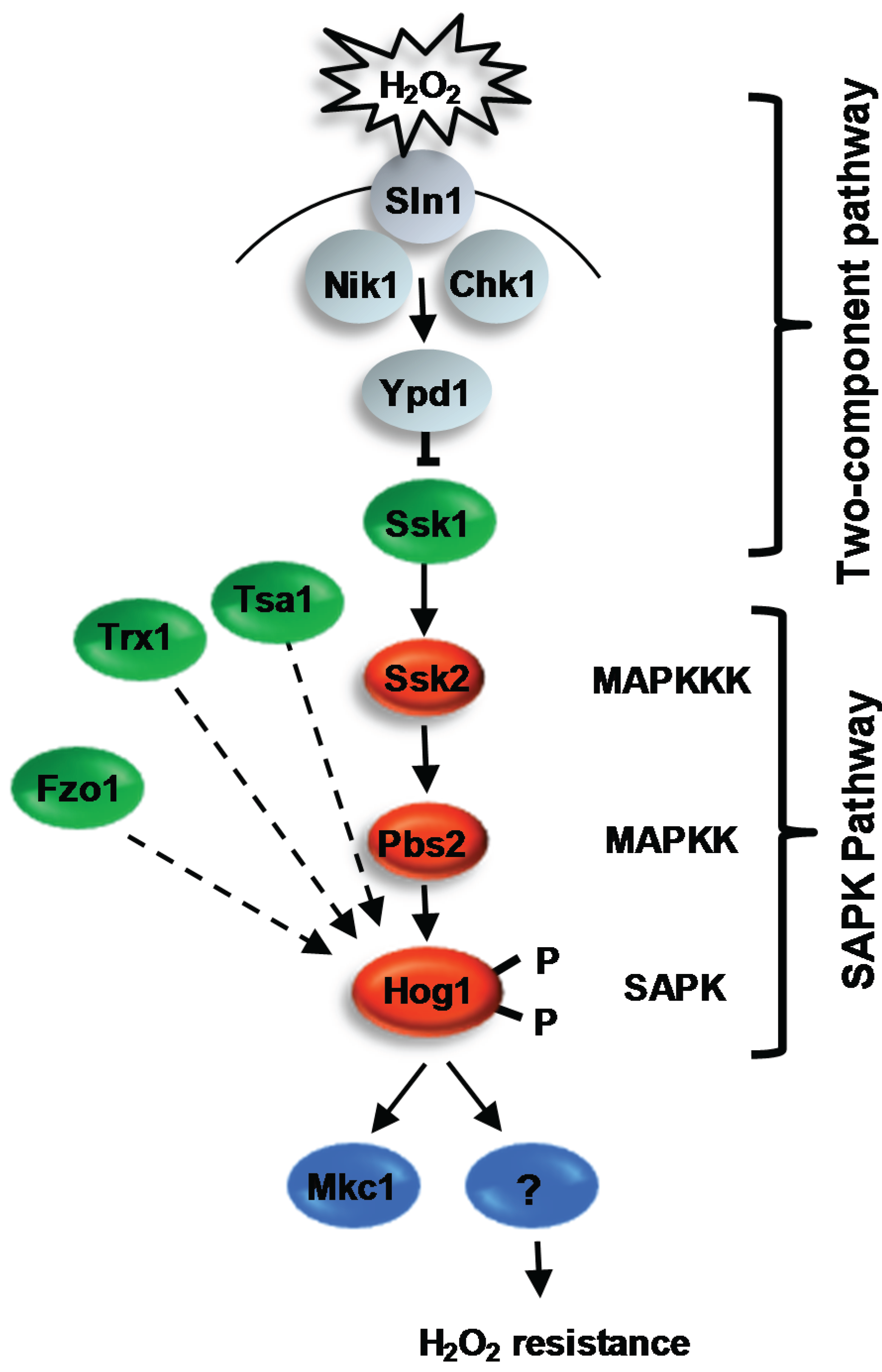
4.2.1. Regulation of Hog1 in Response to ROS

Whilst little is known regarding the cellular targets of Hog and the cellular role(s) of this kinase in promoting oxidative stress tolerance, more progress has been made in delineating how H2O2 signals are relayed to Hog1 (Figure 2). Oxidative stress-induced activation of Hog1 is entirely dependent on the Pbs2 MAPKK [93], which, in turn, is regulated by a single MAPKKK Ssk2 [94]. Furthermore, a recent study reported that deletion of a mitochondrial biogenesis factor, Fzo1, significantly impairs the H2O2-induced activation of Hog1 [95]. Thus, intriguingly, functional mitochondria may play an important role in the regulation of the Hog1 pathway in response to oxidative stress. In addition, both two-component related proteins and redox-sensitive antioxidants are necessary for the activation of the Hog1 SAPK in response to oxidative stress, and these will be described in turn.

4.2.1.1. Two-Component Mediated Regulation of Hog1

In the model yeasts, two-component signalling pathways have been shown to play an important role in the sensing and transmission of stress signals to their respective SAPK pathways. Such pathways are comprised of a histidine kinase, an intermediary phosphorelay protein and a response regulator protein. In S. cerevisiae, the Sln1 histidine kinase is inactivated in response to osmotic stress. This halts phosphorelay through the Ypd1 phosphorelay protein, leading to the rapid dephosphorylation of the Ssk1 response regulator. Dephosphorylated Ssk1 is a potent activator of the Ssk2/Ssk22 MAPKKKs in S. cerevisiae, which regulate Hog1 activation [96,97]. In C. albicans, deletion of the analogous SSK1 gene prevents Hog1 activation in response to oxidative stress, and consistent with this, ssk1Δ cells are sensitive to oxidative stress [98]. Although Ssk1 is involved in the transmission of oxidative stress signals to Hog1, the identity of the histidine kinase(s) responsible for sensing and signalling oxidative stress signals to Ssk1 in C. albicans remains elusive [99,100]. Of the three histidine kinases present in C. albicans, Chk1 would appear to be the most likely candidate for a potential peroxide-sensing histidine kinase, as this shows significant similarity to the S. pombe peroxide-sensing histidine kinases, Mak2 and Mak3 [101,102]. However, deletion of CHK1 alone or in combination with either of the genes encoding the two remaining histidine kinases, SLN1 or NIK1, does not impair H2O2-induced activation of Hog1 [100,103]. Hence, it is currently unclear as to which histidine kinase(s) senses oxidative stress and regulates phosphorelay to Ssk1. Moreover, observations that Hog1 activation is seen in cells expressing a non-phosphorylatable Ssk1 mutant [103] or in cells lacking the Ypd1 phosphorelay protein in which Ssk1 is predicted to be unphosphorylated [104] indicate that Ssk1 may relay H2O2 signals to Hog1 in a mechanism independent of two-component signalling. It is also noteworthy that a novel response regulator, named Crr1/Srr1, has been recently identified that is only present in fungi belonging to the Candida clade [105,106]. Cells lacking Crr1 or expressing a mutant lacking the predicted aspartate phosphorylation site are sensitive to H2O2 [105]. However, in contrast with Ssk1, Crr1 is not required for the H2O2-induced activation of Hog1 [105]. Thus, whilst this novel response regulator mediates the response of C. albicans to H2O2, it does so in a Hog1-independent manner. Finally, in S. cerevisiae, the transmembrane protein, Sho1, relays osmotic stress signals to the Hog1 SAPK in parallel with the Sln1-mediated two-component signalling pathway [107]. In C. albicans, the analogous Sho1 protein appears to have been reassigned to oxidative stress signalling [100]. However, it is not clear how this is mediated, as Sho1 is not required for ROS-stimulated activation of the Hog1 pathway [100].

4.2.1.2. Redox-Sensitive Antioxidant Proteins as Regulators of Hog1

It is now well recognized that redox-sensitive antioxidant proteins have important sensing and signalling roles in the cellular response to oxidative stress [108]. In C. albicans, the redox-sensitive thioredoxin peroxidase enzyme, Tsa1, is specifically required for H2O2-induced activation of Hog1 [42]. This is similar to that previously reported in S. pombe, as H2O2-induced activation of the Sty1 SAPK also requires the analogous thioredoxin peroxidase enzyme, Tpx1 [109]. In S. pombe, intermolecular disulphide bonds are formed between conserved cysteine residues in Sty1 and Tpx1 following H2O2 stress, which suggests that Tpx1 regulates Sty1 function directly. However, the mechanism of Tsa1 regulation of Hog1 in C. albicans may be different, as the conserved peroxidatic cysteine residue of Tsa1, which is essential for Tpx1 regulation of Sty1, is dispensable for Tsa1 regulation of Hog1 [42]. Furthermore, the thioredoxin enzyme, Trx1, which regulates the redox status of Tsa1, is also essential for the relay of oxidative stress signals to the Hog1 SAPK module [42]. eletion of TRX1 or mutation of the catalytic cysteine residues of Trx1 drastically impairs Hog1 phosphorylation in response to H2O2. However, it would appear that Trx1 regulates Hog1 independently of Tsa1, as the catalytic cysteine residues of Tsa1, which are reduced by Trx1, are dispensable for Hog1 activation [42]. In mammalian systems, thioredoxin functions as a repressor of the Hog1-related JNK and p38 SAPK signalling cascades [110]. The upstream Ask1 MAPKKK in the mammalian SAPK pathways is activated via cysteine oxidation, and Trx1 negatively regulates this pathway by reducing the oxidized cysteines of Ask1 [111,112]. As Trx1 is a positive regulator of the Hog1 SAPK in C. albicans, it seems unlikely that a similar mechanism is in place. It is also interesting to note that protein tyrosine phosphatases, which are negative regulators of SAPKs, are susceptible to inactivation by oxidation of their catalytic cysteine residue [113]. Whether thioredoxin regulates such phosphatases that dephosphorylate Hog1 in C. albicans, however, remains to be established.

4.2.2. Role of the Hog1 SAPK in Virulence

The stress-activated MAPK Hog1 in C. albicans is phosphorylated and accumulates in the nucleus, in response to a range of stresses likely to be encountered in the host, including ROS, osmotic stress and anti-microbial peptides [114]. Cells lacking Hog1 display impaired virulence in a wide range of infection models, including murine systemic and commensal models [114,115,116], and are more susceptible to killing by macrophages or neutrophils [117]. As Hog1 regulates a number of distinct stress responses, it is difficult to dissect whether it is the role of Hog1 in oxidative stress responses or a different aspect of Hog1 signalling that is important for virulence in these models. Importantly, however, although Hog1 signalling has also been implicated in morphogenetic regulation, mutational analysis has inferred that the importance of Hog1 in virulence is due to its role in stress protection, rather than its role in repressing the yeast to hyphal transition [115].

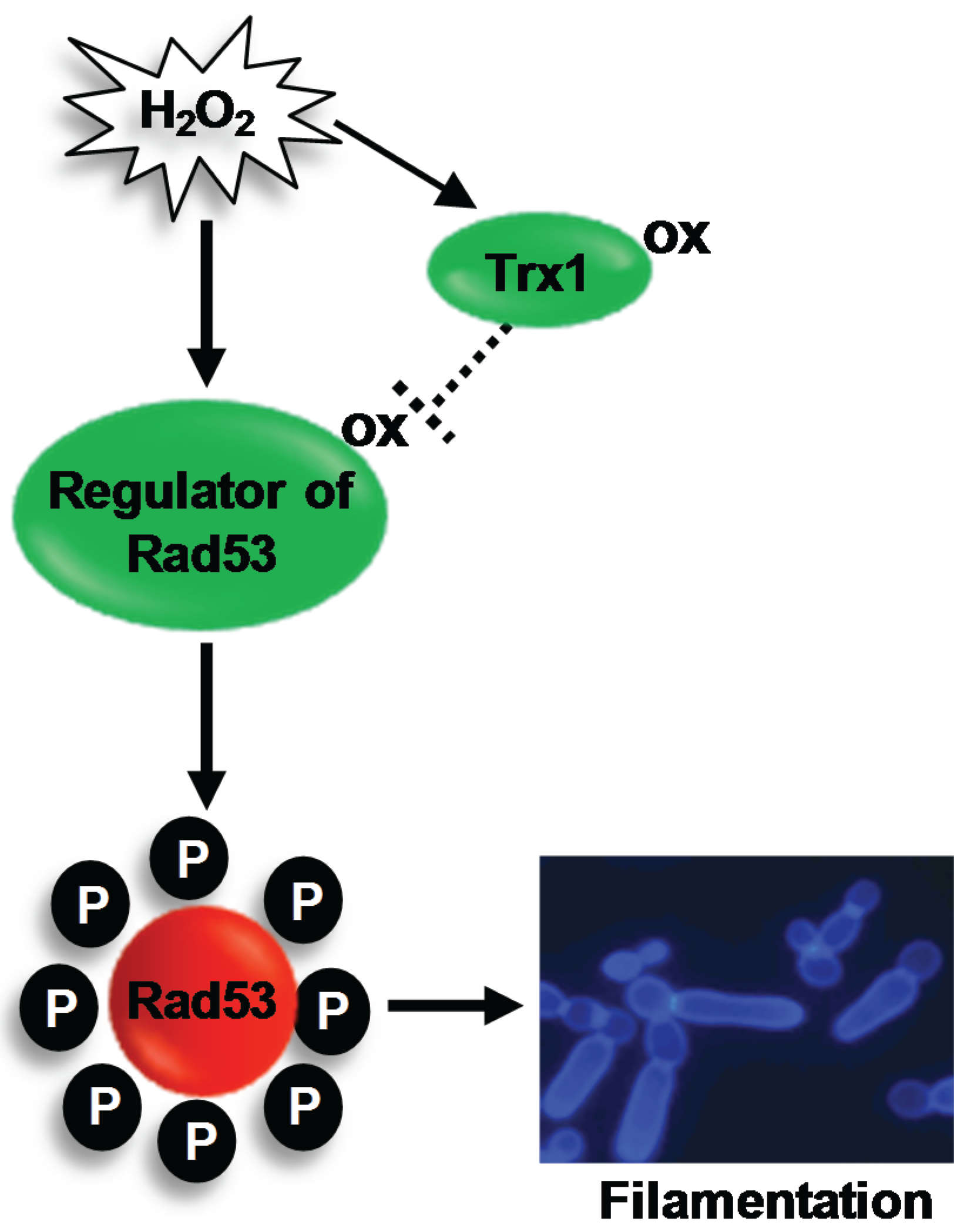
4.3. The Rad53 DNA Damage Checkpoint Kinase

Following exposure to H2O2, C. albicans forms hyperpolarised buds, which are morphologically distinct from hyphae and pseudohyphae filamentous forms (Section 3.4). Consistent with this, H2O2-induced hyperpolarized bud formation occurs independently of the key hyphal regulators, Efg1 and Cph1, and, instead, depends on the activation of the Rad53 DNA damage checkpoint pathway [42,61] (Figure 3). A wide range of genotoxic stresses, including UV, methyl methanesulfonate (MMS) and the ribonucleotide reductase inhibitor hydroxyurea have been shown to activate the Rad53 kinase in C. albicans [61], and loss of Rad53 or upstream regulators of Rad53, prevents hyperpolarised bud formation [61,118]. ROS are also genotoxic agents due to the induction of DNA damage [119], which, in turn, triggers the activation of the Rad53 DNA checkpoint pathway [120]. Indeed, treatment of C. albicans cells with H2O2 elicits the phosphorylation of Rad53, and cells lacking RAD53 fail to form hyperpolarised buds in response to H2O2 [42].
Figure 3. H2O2-induced activation of Rad53 triggers filamentation in C. albicans. The redox sensitive oxidoreductase Trx1 inhibits H2O2-induced activation of the DNA damage checkpoint kinase Rad53. This suggests that a regulator of Rad53 is activated by oxidation, and this active oxidised form is reduced by Trx1. Activation of the DNA damage checkpoint triggers the formation of hyperpolarised buds. See text for details.
Biomolecules 05 00142 g003

Regulation of Rad53

Regarding the H2O2-mediated activation of Rad53, a recent study illustrated that H2O2-induced oxidation, and inactivation, of the thioredoxin protein Trx1 is important for the activation of Rad53 and polarized cell growth [42]. Rad53 is constitutively phosphorylated in cells lacking Trx1, which display a hyperpolarized bud morphology. Conversely, ectopic expression of the thioredoxin reductase gene, TRR1, which reduces oxidized Trx1, inhibited H2O2-induced filamentation [42]. Taken together, these results illustrate that oxidation of Trx1 following H2O2 exposure is key in the activation of Rad53 that drives hyperpolarised bud formation. The finding that Trx1 inhibits Rad53 activation under non-stressed conditions may be conserved in higher eukaryotes, as ectopic expression of thioredoxin inhibits the phosphorylation of the analogous DNA damage checkpoint kinase, Chk2, in mammalian cells [121]. However, the mechanism of Trx1 regulation of Rad53 is unclear. As Trx1 functions to reduce oxidised proteins, an attractive hypothesis is that Rad53, or a regulator of this kinase, is activated by oxidation (Figure 3). In this regard, it is interesting that the human homologue (ATM) of the fungal Tel1 DNA-damage sensing kinase, which regulates Rad53, has recently been shown to be activated by oxidation [122]. Further studies are needed to determine if Tel1 is similarly regulated to mediate H2O2-induced filamentation in C. albicans.

4.4. Other Signaling Pathways that Contribute to Oxidative Stress Resistance

The cAMP/PKA signalling pathway has a negative impact on oxidative stress responses in C. albicans. For example, induction of the pathway by inactivation of the phosphodiesterase, Pde2, which degrades cAMP, results in increased sensitivity to H2O2 [123]. Related to this, farnesol treatment of C. albicans cells results in increased resistance to H2O2, due to the inhibition of the cAMP/PKA signalling pathway [124]. Such changes in resistance are possibly due to changes in the levels of anti-oxidant gene expression [124]; however, the mechanism linking cAMP/PKA to their regulation is unknown.
There is also evidence that the spindle assembly checkpoint is required for C. albicans oxidative stress resistance. Cells lacking the spindle checkpoint protein kinase, Mps1, are sensitive to H2O2 [125] and, similar to other oxidative stress sensitive mutants [56], fail to filament following phagocytosis. Related to this, the spindle assembly checkpoint protein, Mad2, is essential for C. albicans survival in macrophages [126].

5. Conclusions and Future Perspectives

In this review, we have summarized the current literature of oxidative stress responses and how they are regulated in the human fungal pathogen C. albicans. This is an important area of research, as oxidative stress adaptation is emerging as an important virulence trait in this, and other, fungal pathogens. Table 1 summarizes studies that have documented the impact of the loss of oxidative stress regulatory proteins or antioxidant enzymes on C. albicans virulence in either a murine systemic infection model or a macrophage/neutrophil phagocyte-survival infection model. From this summary, a number of observations can be made. First of all, when examined, mutants that display an impaired tolerance to oxidative stress show an impaired ability to survive phagocyte killing. This is consistent with previous observations that C. albicans mounts a robust transcriptional response to oxidative stress following phagocytosis [34,35,36,37,38]. Secondly, not all oxidative stress-sensitive C. albicans mutants display attenuated virulence in a murine systemic infection model. Perhaps most striking is the observation that the major regulator of anti-oxidant gene expression, Cap1, is dispensable for virulence in such an infection model. This is particularly intriguing, as certain genes, which are dependent on Cap1 for induction following oxidative stress, are important for virulence in systemic models. Thirdly, the role of many other oxidative stress-responsive proteins in mediating C. albicans virulence, such as the Rad53-mediated DNA damage checkpoint pathway, have yet to be explored.
Table 1. Summary of the role of oxidative stress-responsive signalling proteins and antioxidant enzymes in C. albicans virulence. The importance of proteins needed for resistance to oxidative stress in mediating C. albicans virulence in either a systemic infection model (SIM) or phagocyte infection model (PIM) is indicated; +, important for virulence; −, dispensable for virulence; nd, not determined. For further explanation, see the text.
ProteinFunctionSIMPIMReferences
Signalling Proteins
Hog1Stress-activated protein kinase++[114,115,117]
Ssk1Response regulator++[127,128]
Cap1Transcription factor+[56,81]
Ybp1Cap1 regulator+[56]
Gpx3Cap1 regulator+[56]
Skn7Transcription factornd[80]
Signalling Proteins
Pde2Phosphodiesterase+nd[123]
Mps1Spindle checkpointnd+[125]
Mad2Spindle checkpoint++[126]
Antioxidant Enzymes
Cat1Catalase++[45]
Trx1Thioredoxin+nd[42]
Tsa1Thioredoxin peroxidasend[47]
Sod1Superoxide dismutase++[43]
Sod5Superoxide dismutase++[23,44]
Grx2Glutaredoxin+nd[41]
Gpx31-33Glutathione peroxidasesnd+[46]
In addition to gaps in our knowledge regarding the relative importance of specific oxidative stress responses in mediating C. albicans virulence, there are also additional key questions that remain to be addressed. For example, what is the role of the Hog1 SAPK in mediating oxidative stress resistance in C. albicans, and how is this regulated? This is important, as, although Hog1 is an essential virulence determinant in C. albicans, the conservation with highly related SAPKs in human cells suggests that Hog1 itself may be unsuitable as a specific antifungal target. Thus, there is much interest in identifying fungal-specific SAPK regulators or substrates, as such proteins hold greater promise for future therapeutic strategies. In addition, the recent findings that C. albicans is exquisitely sensitive to combinations of stress that are encountered following phagocytosis represent a new unchartered area in the field of stress signalling. A key question for the future is how do combinations of stress imposed by the phagosome inhibit oxidative stress adaptation and survival of C. albicans? Addressing this question is critical to further our understanding of Candida-host interactions during disease progression.

Acknowledgments

We thank our friends and colleagues, in particular Al Brown, Lars Erwig, Ken Haynes, Brian Morgan and Elizabeth Veal, for stimulating discussions on oxidative stress signalling mechanisms and fungal pathogenesis. Research in the Janet Quinn lab is funded by the BBSRC BB/K016393/1, an MRC-DTP studentship, the NIHR Newcastle Biomedical Research Centre and the Wellcome Trust Strategic Award for Medical Mycology and Fungal Immunology 097377/Z/11/Z.

Author Contributions

All authors contributed to the initial draft of this review, which was edited by Janet Quinn.

Conflicts of Interest

The authors declare no conflict of interest.

References and Notes

  1. Odds, F.C. Candida and Candidosis, 2nd ed.; Bailliere-Tindall: London, UK, 1988. [Google Scholar]
  2. Calderone, R.A.; Clancy, C.J. Candida and Candidiasis; ASM Press: Washington, DC, USA, 2012. [Google Scholar]
  3. Anaissie, E.J.; McGinnis, M.R.; Pfaller, M.A. Clinical Mycology; Churchill Livingstone: London, UK, 2009. [Google Scholar]
  4. Flevari, A.; Theodorakopoulou, M.; Velegraki, A.; Armaganidis, A.; Dimopoulos, G. Treatment of invasive candidiasis in the elderly: A review. Clin. Interv. Aging 2013, 8, 1199–1208. [Google Scholar] [PubMed]
  5. Healy, C.M.; Campbell, J.R.; Zaccaria, E.; Baker, C.J. Fluconazole prophylaxis in extremely low birth weight neonates reduces invasive candidiasis mortality rates without emergence of fluconazole-resistant candida species. Pediatrics 2008, 121, 703–710. [Google Scholar] [CrossRef] [PubMed]
  6. Sobel, J.D. Vulvovaginal candidosis. Lancet 2007, 369, 1961–1971. [Google Scholar] [CrossRef] [PubMed]
  7. Lilic, D. Unravelling fungal immunity through primary immune deficiencies. Curr. Opin. Microbiol. 2012, 15, 420–426. [Google Scholar] [CrossRef] [PubMed]
  8. Morgan, J. Global trends in candidemia: Review of reports from 1995–2005. Curr. Infect. Dis. Rep. 2005, 7, 429–439. [Google Scholar] [CrossRef] [PubMed]
  9. Brown, G.D.; Denning, D.W.; Gow, N.A.; Levitz, S.M.; Netea, M.G.; White, T.C. Hidden killers: Human fungal infections. Sci. Transl. Med. 2012. [Google Scholar] [CrossRef]
  10. Koh, A.Y.; Kohler, J.R.; Coggshall, K.T.; van Rooijen, N.; Pier, G.B. Mucosal damage and neutropenia are required for Candida albicans dissemination. PLOS Pathog. 2008, 4, e35. [Google Scholar] [CrossRef] [PubMed]
  11. Seider, K.; Heyken, A.; Luttich, A.; Miramon, P.; Hube, B. Interaction of pathogenic yeasts with phagocytes: Survival, persistence and escape. Curr. Opin. Microbiol. 2010, 13, 392–400. [Google Scholar] [CrossRef] [PubMed]
  12. Babior, B.M. NADPH oxidase. Curr. Opin. Immunol. 2004, 16, 42–47. [Google Scholar] [CrossRef] [PubMed]
  13. Bedard, K.; Krause, K.H. The NOX family of ROS-generating NADPH oxidases: Physiology and pathophysiology. Physiol. Rev. 2007, 87, 245–313. [Google Scholar] [CrossRef] [PubMed]
  14. Pick, E. Role of the Rho GTPase Rac in the activation of the phagocyte NADPH oxidase: Outsourcing a key task. Small GTPases 2014, 5, e27952. [Google Scholar] [CrossRef] [PubMed]
  15. Hampton, M.B.; Kettle, A.J.; Winterbourn, C.C. Involvement of superoxide and myeloperoxidase in oxygen-dependent killing of Staphylococcus aureus by neutrophils. Infect. Immun. 1996, 64, 3512–3517. [Google Scholar] [PubMed]
  16. Reeves, E.P.; Lu, H.; Jacobs, H.L.; Messina, C.G.; Bolsover, S.; Gabella, G.; Potma, E.O.; Warley, A.; Roes, J.; Segal, A.W. Killing activity of neutrophils is mediated through activation of proteases by K+ flux. Nature 2002, 416, 291–297. [Google Scholar] [CrossRef] [PubMed]
  17. Holland, S.M. Chronic granulomatous disease. Clin. Rev. Allerg. Immunol. 2010, 38, 3–10. [Google Scholar] [CrossRef]
  18. Cohen, M.S.; Isturiz, R.E.; Malech, H.L.; Root, R.K.; Wilfert, C.M.; Gutman, L.; Buckley, R.H. Fungal infection in chronic granulomatous disease. The importance of the phagocyte in defense against fungi. Am. J. Med. 1981, 71, 59–66. [Google Scholar] [CrossRef] [PubMed]
  19. Brothers, K.M.; Gratacap, R.L.; Barker, S.E.; Newman, Z.R.; Norum, A.; Wheeler, R.T. Nadph oxidase-driven phagocyte recruitment controls Candida albicans filamentous growth and prevents mortality. PLOS Pathog. 2013, 9, e1003634. [Google Scholar] [CrossRef] [PubMed]
  20. Brown, A.J.; Haynes, K.; Quinn, J. Nitrosative and oxidative stress responses in fungal pathogenicity. Curr. Opin. Microbiol. 2009, 12, 384–391. [Google Scholar] [CrossRef] [PubMed]
  21. Fang, F.C. Antimicrobial reactive oxygen and nitrogen species: Concepts and controversies. Nat. Rev. Microbiol. 2004, 2, 820–832. [Google Scholar] [CrossRef] [PubMed]
  22. Kaloriti, D.; Jacobsen, M.; Yin, Z.; Patterson, M.; Tillmann, A.; Smith, D.A.; Cook, E.; You, T.; Grimm, M.J.; Bohovych, I.; et al. Mechanisms underlying the exquisite sensitivity of Candida albicans to combinatorial cationic and oxidative stress that enhances the potent fungicidal activity of phagocytes. MBio 2014, 5, e01334–e01314. [Google Scholar] [CrossRef] [PubMed]
  23. Frohner, I.E.; Bourgeois, C.; Yatsyk, K.; Majer, O.; Kuchler, K. Candida albicans cell surface superoxide dismutases degrade host-derived reactive oxygen species to escape innate immune surveillance. Mol. Microbiol. 2009, 71, 240–252. [Google Scholar] [CrossRef] [PubMed]
  24. Miramon, P.; Dunker, C.; Windecker, H.; Bohovych, I.M.; Brown, A.J.; Kurzai, O.; Hube, B. Cellular responses of Candida albicans to phagocytosis and the extracellular activities of neutrophils are critical to counteract carbohydrate starvation, oxidative and nitrosative stress. PLOS ONE 2012, 7, e52850. [Google Scholar] [CrossRef] [PubMed] [Green Version]
  25. Huycke, M.M.; Abrams, V.; Moore, D.R. Enterococcus faecalis produces extracellular superoxide and hydrogen peroxide that damages colonic epithelial cell DNA. Carcinogenesis 2002, 23, 529–536. [Google Scholar] [CrossRef] [PubMed]
  26. Fitzsimmons, N.; Berry, D.R. Inhibition of Candida albicans by lactobacillus acidophilus: Evidence for the involvement of a peroxidase system. Microbios 1994, 80, 125–133. [Google Scholar] [PubMed]
  27. Cruz, M.R.; Graham, C.E.; Gagliano, B.C.; Lorenz, M.C.; Garsin, D.A. Enterococcus faecalis inhibits hyphal morphogenesis and virulence of Candida albicans. Infect. Immun. 2013, 81, 189–200. [Google Scholar] [CrossRef] [PubMed]
  28. Phillips, A.J.; Sudbery, I.; Ramsdale, M. Apoptosis induced by environmental stresses and amphotericin B in Candida albicans. Proc. Natl. Acad. Sci. USA 2003, 100, 14327–14332. [Google Scholar] [CrossRef] [PubMed]
  29. Halliwell, B. Oxidative stress and cancer: Have we moved forward? Biochem. J. 2007, 401, 1–11. [Google Scholar] [CrossRef] [PubMed]
  30. Jamieson, D.J.; Stephen, D.W.; Terriere, E.C. Analysis of the adaptive oxidative stress response of Candida albicans. FEMS Microbiol. Lett. 1996, 138, 83–88. [Google Scholar] [CrossRef] [PubMed]
  31. Nikolaou, E.; Agrafioti, I.; Stumpf, M.; Quinn, J.; Stansfield, I.; Brown, A.J. Phylogenetic diversity of stress signalling pathways in fungi. BMC Evol. Biol. 2009. [Google Scholar] [CrossRef] [Green Version]
  32. Enjalbert, B.; Nantel, A.; Whiteway, M. Stress-induced gene expression in Candida albicans: Absence of a general stress response. Mol. Biol. Cell 2003, 14, 1460–1467. [Google Scholar] [CrossRef] [PubMed]
  33. Enjalbert, B.; Smith, D.A.; Cornell, M.J.; Alam, I.; Nicholls, S.; Brown, A.J.; Quinn, J. Role of the Hog1 stress-activated protein kinase in the global transcriptional response to stress in the fungal pathogen Candida albicans. Mol. Biol. Cell 2006, 17, 1018–1032. [Google Scholar] [CrossRef] [PubMed]
  34. Enjalbert, B.; MacCallum, D.M.; Odds, F.C.; Brown, A.J. Niche-specific activation of the oxidative stress response by the pathogenic fungus Candida albicans. Infect. Immun. 2007, 75, 2143–2151. [Google Scholar] [CrossRef] [PubMed]
  35. Fradin, C.; de Groot, P.; MacCallum, D.; Schaller, M.; Klis, F.; Odds, F.C.; Hube, B. Granulocytes govern the transcriptional response, morphology and proliferation of Candida albicans in human blood. Mol. Microbiol. 2005, 56, 397–415. [Google Scholar] [CrossRef] [PubMed]
  36. Fradin, C.; Kretschmar, M.; Nichterlein, T.; Gaillardin, C.; d’Enfert, C.; Hube, B. Stage-specific gene expression of Candida albicans in human blood. Mol. Microbiol. 2003, 47, 1523–1543. [Google Scholar] [CrossRef] [PubMed]
  37. Lorenz, M.C.; Bender, J.A.; Fink, G.R. Transcriptional response of Candida albicans upon internalization by macrophages. Eukaryot. Cell 2004, 3, 1076–1087. [Google Scholar] [CrossRef] [PubMed]
  38. Rubin-Bejerano, I.; Fraser, I.; Grisafi, P.; Fink, G.R. Phagocytosis by neutrophils induces an amino acid deprivation response in Saccharomyces cerevisiae and Candida albicans. Proc. Natl. Acad. Sci. USA 2003, 100, 11007–11012. [Google Scholar] [CrossRef] [PubMed]
  39. Thewes, S.; Kretschmar, M.; Park, H.; Schaller, M.; Filler, S.G.; Hube, B. In vivo and ex vivo comparative transcriptional profiling of invasive and non-invasive Candida albicans isolates identifies genes associated with tissue invasion. Mol. Microbiol. 2007, 63, 1606–1628. [Google Scholar] [CrossRef] [PubMed]
  40. Walker, L.A.; Maccallum, D.M.; Bertram, G.; Gow, N.A.; Odds, F.C.; Brown, A.J. Genome-wide analysis of Candida albicans gene expression patterns during infection of the mammalian kidney. Fungal Genet. Biol. 2009, 46, 210–219. [Google Scholar] [CrossRef] [PubMed]
  41. Chaves, G.M.; Bates, S.; Maccallum, D.M.; Odds, F.C. Candida albicans GRX2, encoding a putative glutaredoxin, is required for virulence in a murine model. Genet. Mol. Res. 2007, 6, 1051–1063. [Google Scholar] [PubMed]
  42. Da Silva Dantas, A.; Patterson, M.J.; Smith, D.A.; Maccallum, D.M.; Erwig, L.P.; Morgan, B.A.; Quinn, J. Thioredoxin regulates multiple hydrogen peroxide-induced signaling pathways in Candida albicans. Mol. Cell. Biol. 2010, 30, 4550–4563. [Google Scholar] [CrossRef] [PubMed]
  43. Hwang, C.S.; Rhie, G.E.; Oh, J.H.; Huh, W.K.; Yim, H.S.; Kang, S.O. Copper- and zinc-containing superoxide dismutase (Cu/ZnSOD) is required for the protection of Candida albicans against oxidative stresses and the expression of its full virulence. Microbiology 2002, 148, 3705–3713. [Google Scholar] [PubMed]
  44. Martchenko, M.; Alarco, A.M.; Harcus, D.; Whiteway, M. Superoxide dismutases in Candida albicans: Transcriptional regulation and functional characterization of the hyphal-induced SOD5 gene. Mol. Biol. Cell 2004, 15, 456–467. [Google Scholar] [CrossRef] [PubMed]
  45. Wysong, D.R.; Christin, L.; Sugar, A.M.; Robbins, P.W.; Diamond, R.D. Cloning and sequencing of a Candida albicans catalase gene and effects of disruption of this gene. Infect. Immun. 1998, 66, 1953–1961. [Google Scholar] [PubMed]
  46. Miramon, P.; Dunker, C.; Kasper, L.; Jacobsen, I.D.; Barz, D.; Kurzai, O.; Hube, B. A family of glutathione peroxidases contributes to oxidative stress resistance in Candida albicans. Med. Mycol. 2014, 52, 223–239. [Google Scholar] [CrossRef] [PubMed]
  47. Urban, C.; Xiong, X.; Sohn, K.; Schroppel, K.; Brunner, H.; Rupp, S. The moonlighting protein tsa1p is implicated in oxidative stress response and in cell wall biogenesis in Candida albicans. Mol. Microbiol. 2005, 57, 1318–1341. [Google Scholar] [CrossRef] [PubMed]
  48. Steinberg, B.E.; Huynh, K.K.; Grinstein, S. Phagosomal acidification: Measurement, manipulation and functional consequences. Biochem. Soc. Trans. 2007, 35, 1083–1087. [Google Scholar] [CrossRef] [PubMed]
  49. Kaloriti, D.; Tillmann, A.; Cook, E.; Jacobsen, M.; You, T.; Lenardon, M.; Ames, L.; Barahona, M.; Chandrasekaran, K.; Coghill, G.; et al. Combinatorial stresses kill pathogenic Candida species. Med. Mycol. 2012, 50, 699–709. [Google Scholar] [CrossRef] [PubMed]
  50. Schuller, C.; Brewster, J.L.; Alexander, M.R.; Gustin, M.C.; Ruis, H. The hog pathway controls osmotic regulation of transcription via the stress response element (STRE) of the Saccharomyces cerevisiae CTT1 gene. EMBO J. 1994, 13, 4382–4389. [Google Scholar] [PubMed]
  51. Heilmann, C.J.; Sorgo, A.G.; Siliakus, A.R.; Dekker, H.L.; Brul, S.; de Koster, C.G.; de Koning, L.J.; Klis, F.M. Hyphal induction in the human fungal pathogen Candida albicans reveals a characteristic wall protein profile. Microbiology 2011, 157, 2297–2307. [Google Scholar] [CrossRef] [PubMed]
  52. Gleason, J.E.; Galaleldeen, A.; Peterson, R.L.; Taylor, A.B.; Holloway, S.P.; Waninger-Saroni, J.; Cormack, B.P.; Cabelli, D.E.; Hart, P.J.; Culotta, V.C. Candida albicans SOD5 represents the prototype of an unprecedented class of Cu-only superoxide dismutases required for pathogen defense. Proc. Natl. Acad. Sci. USA 2014, 111, 5866–5871. [Google Scholar] [CrossRef] [PubMed]
  53. Festa, R.A.; Thiele, D.J. Copper at the front line of the host-pathogen battle. PLOS Pathog. 2012, 8, e1002887. [Google Scholar] [CrossRef] [PubMed]
  54. Crowe, J.D.; Sievwright, I.K.; Auld, G.C.; Moore, N.R.; Gow, N.A.; Booth, N.A. Candida albicans binds human plasminogen: Identification of eight plasminogen-binding proteins. Mol. Microbiol. 2003, 47, 1637–1651. [Google Scholar] [CrossRef] [PubMed]
  55. Lewis, L.E.; Bain, J.M.; Okai, B.; Gow, N.A.; Erwig, L.P. Live-cell video microscopy of fungal pathogen phagocytosis. J. Vis. Exp. 2013. [Google Scholar] [CrossRef]
  56. Patterson, M.J.; McKenzie, C.G.; Smith, D.A.; da Silva Dantas, A.; Sherston, S.; Veal, E.A.; Morgan, B.A.; MacCallum, D.M.; Erwig, L.P.; Quinn, J. Ybp1 and Gpx3 signaling in Candida albicans govern hydrogen peroxide-induced oxidation of the Cap1 transcription factor and macrophage escape. Antioxid. Redox Signal. 2013, 19, 2244–2260. [Google Scholar] [CrossRef] [PubMed] [Green Version]
  57. Brothers, K.M.; Newman, Z.R.; Wheeler, R.T. Live imaging of disseminated candidiasis in zebrafish reveals role of phagocyte oxidase in limiting filamentous growth. Eukaryot. Cell 2011, 10, 932–944. [Google Scholar] [CrossRef] [PubMed]
  58. Jimenez-Lopez, C.; Collette, J.R.; Brothers, K.M.; Shepardson, K.M.; Cramer, R.A.; Wheeler, R.T.; Lorenz, M.C. Candida albicans induces arginine biosynthetic genes in response to host-derived reactive oxygen species. Eukaryot. Cell 2013, 12, 91–100. [Google Scholar] [CrossRef] [PubMed]
  59. Nasution, O.; Srinivasa, K.; Kim, M.; Kim, Y.J.; Kim, W.; Jeong, W.; Choi, W. Hydrogen peroxide induces hyphal differentiation in Candida albicans. Eukaryot. Cell 2008, 7, 2008–2011. [Google Scholar] [CrossRef] [PubMed]
  60. Bachewich, C.; Nantel, A.; Whiteway, M. Cell cycle arrest during S or M phase generates polarized growth via distinct signals in Candida albicans. Mol. Microbiol. 2005, 57, 942–959. [Google Scholar] [CrossRef] [PubMed]
  61. Shi, Q.M.; Wang, Y.M.; Zheng, X.D.; Lee, R.T.; Wang, Y. Critical role of DNA checkpoints in mediating genotoxic-stress-induced filamentous growth in Candida albicans. Mol. Biol. Cell 2007, 18, 815–826. [Google Scholar] [CrossRef] [PubMed]
  62. Kusch, H.; Engelmann, S.; Albrecht, D.; Morschhauser, J.; Hecker, M. Proteomic analysis of the oxidative stress response in Candida albicans. Proteomics 2007, 7, 686–697. [Google Scholar] [CrossRef] [PubMed]
  63. Wang, Y.; Cao, Y.Y.; Jia, X.M.; Cao, Y.B.; Gao, P.H.; Fu, X.P.; Ying, K.; Chen, W.S.; Jiang, Y.Y. Cap1p is involved in multiple pathways of oxidative stress response in Candida albicans. Free Radic. Biol. Med. 2006, 40, 1201–1209. [Google Scholar] [CrossRef] [PubMed]
  64. Moye-Rowley, W.S. Regulation of the transcriptional response to oxidative stress in fungi: Similarities and differences. Eukaryot. Cell 2003, 2, 381–389. [Google Scholar] [CrossRef] [PubMed]
  65. Toone, W.M.; Morgan, B.A.; Jones, N. Redox control of AP-1-like factors in yeast and beyond. Oncogene 2001, 20, 2336–2346. [Google Scholar] [CrossRef] [PubMed]
  66. Alarco, A.M.; Raymond, M. The bzip transcription factor Cap1p is involved in multidrug resistance and oxidative stress response in Candida albicans. J. Bacteriol. 1999, 181, 700–708. [Google Scholar] [PubMed]
  67. Zhang, X.; de Micheli, M.; Coleman, S.T.; Sanglard, D.; Moye-Rowley, W.S. Analysis of the oxidative stress regulation of the Candida albicans transcription factor, Cap1p. Mol. Microbiol. 2000, 36, 618–629. [Google Scholar] [CrossRef] [PubMed]
  68. Znaidi, S.; Barker, K.S.; Weber, S.; Alarco, A.M.; Liu, T.T.; Boucher, G.; Rogers, P.D.; Raymond, M. Identification of the Candida albicans Cap1p regulon. Eukaryot. Cell 2009, 8, 806–820. [Google Scholar] [CrossRef] [PubMed]
  69. Ramirez-Zavala, B.; Mogavero, S.; Scholler, E.; Sasse, C.; Rogers, P.D.; Morschhauser, J. SAGA/ADA complex subunit Ada2 is required for Cap1- but not Mrr1-mediated upregulation of the Candida albicans multidrug efflux pump Mdr1. Antimicrob. Agents Chemother. 2014, 58, 5102–5110. [Google Scholar] [CrossRef] [PubMed]
  70. Sellam, A.; Askew, C.; Epp, E.; Lavoie, H.; Whiteway, M.; Nantel, A. Genome-wide mapping of the coactivator Ada2p yields insight into the functional roles of SAGA/ADA complex in Candida albicans. Mol. Biol. Cell 2009, 20, 2389–2400. [Google Scholar] [CrossRef] [PubMed]
  71. Delaunay, A.; Isnard, A.D.; Toledano, M.B. H2O2 sensing through oxidation of the Yap1 transcription factor. EMBO J. 2000, 19, 5157–5166. [Google Scholar] [CrossRef] [PubMed]
  72. Delaunay, A.; Pflieger, D.; Barrault, M.B.; Vinh, J.; Toledano, M.B. A thiol peroxidase is an H2O2 receptor and redox-transducer in gene activation. Cell 2002, 111, 471–481. [Google Scholar] [CrossRef] [PubMed]
  73. Okazaki, S.; Tachibana, T.; Naganuma, A.; Mano, N.; Kuge, S. Multistep disulfide bond formation in Yap1 is required for sensing and transduction of H2O2 stress signal. Mol. Cell. 2007, 27, 675–688. [Google Scholar] [CrossRef] [PubMed]
  74. Okazaki, S.; Naganuma, A.; Kuge, S. Peroxiredoxin-mediated redox regulation of the nuclear localization of Yap1, a transcription factor in budding yeast. Antioxid. Redox Signal. 2005, 7, 327–334. [Google Scholar] [CrossRef] [PubMed]
  75. Tachibana, T.; Okazaki, S.; Murayama, A.; Naganuma, A.; Nomoto, A.; Kuge, S. A major peroxiredoxin-induced activation of Yap1 transcription factor is mediated by reduction-sensitive disulfide bonds and reveals a low level of transcriptional activation. J. Biol. Chem. 2009, 284, 4464–4472. [Google Scholar] [CrossRef] [PubMed]
  76. Veal, E.A.; Ross, S.J.; Malakasi, P.; Peacock, E.; Morgan, B.A. Ybp1 is required for the hydrogen peroxide-induced oxidation of the Yap1 transcription factor. J. Biol. Chem. 2003, 278, 30896–30904. [Google Scholar] [CrossRef] [PubMed]
  77. Gulshan, K.; Thommandru, B.; Moye-Rowley, W.S. Proteolytic degradation of the Yap1 transcription factor is regulated by subcellular localization and the E3 ubiquitin ligase Not4. J. Biol. Chem. 2012, 287, 36796–36805. [Google Scholar] [CrossRef]
  78. Kitamura, K.; Taki, M.; Tanaka, N.; Yamashita, I. Fission yeast Ubr1 ubiquitin ligase influences the oxidative stress response via degradation of active Pap1 bZIP transcription factor in the nucleus. Mol. Microbiol. 2011, 80, 739–755. [Google Scholar] [CrossRef] [PubMed]
  79. Morgan, B.A.; Banks, G.R.; Toone, W.M.; Raitt, D.; Kuge, S.; Johnston, L.H. The Skn7 response regulator controls gene expression in the oxidative stress response of the budding yeast Saccharomyces cerevisiae. EMBO J. 1997, 16, 1035–1044. [Google Scholar] [CrossRef] [PubMed]
  80. Singh, P.; Chauhan, N.; Ghosh, A.; Dixon, F.; Calderone, R. Skn7 of Candida albicans: Mutant construction and phenotype analysis. Infect. Immun. 2004, 72, 2390–2394. [Google Scholar] [CrossRef] [PubMed]
  81. Jain, C.; Pastor, K.; Gonzalez, A.Y.; Lorenz, M.C.; Rao, R.P. The role of Candida albicans AP-1 protein against host derived ROS in in vivo models of infection. Virulence 2013, 4, 67–76. [Google Scholar] [CrossRef] [PubMed]
  82. Smith, D.A.; Morgan, B.A.; Quinn, J. Stress signalling to fungal stress-activated protein kinase pathways. FEMS Microbiol. Lett. 2010, 306, 1–8. [Google Scholar] [CrossRef] [PubMed]
  83. Bellon, S.; Fitzgibbon, M.J.; Fox, T.; Hsiao, H.M.; Wilson, K.P. The structure of phosphorylated p38gamma is monomeric and reveals a conserved activation-loop conformation. Structure 1999, 7, 1057–1065. [Google Scholar] [CrossRef] [PubMed]
  84. Alonso-Monge, R.; Navarro-Garcia, F.; Roman, E.; Negredo, A.I.; Eisman, B.; Nombela, C.; Pla, J. The Hog1 mitogen-activated protein kinase is essential in the oxidative stress response and chlamydospore formation in Candida albicans. Eukaryot. Cell 2003, 2, 351–361. [Google Scholar] [CrossRef] [PubMed]
  85. Smith, D.A.; Nicholls, S.; Morgan, B.A.; Brown, A.J.; Quinn, J. A conserved stress-activated protein kinase regulates a core stress response in the human pathogen Candida albicans. Mol. Biol. Cell 2004, 15, 4179–4190. [Google Scholar] [CrossRef] [PubMed]
  86. Chen, D.; Toone, W.M.; Mata, J.; Lyne, R.; Burns, G.; Kivinen, K.; Brazma, A.; Jones, N.; Bahler, J. Global transcriptional responses of fission yeast to environmental stress. Mol. Biol. Cell 2003, 14, 214–229. [Google Scholar] [CrossRef] [PubMed]
  87. Asp, E.; Nilsson, D.; Sunnerhagen, P. Fission yeast mitogen-activated protein kinase Sty1 interacts with translation factors. Eukaryot. Cell 2008, 7, 328–338. [Google Scholar] [CrossRef] [PubMed]
  88. Yin, Z.; Stead, D.; Walker, J.; Selway, L.; Smith, D.; Brown, A.J.P.; Quinn, J. A proteomic analysis of the salt, cadmium and peroxide stress responses in Candida albicans and the role of the Hog1 Sapk in regulating the stress-induced proteome. Proteomics 2009, 9, 4680–4703. [Google Scholar] [CrossRef]
  89. Alonso-Monge, R.; Carvaihlo, S.; Nombela, C.; Rial, E.; Pla, J. The Hog1 MAP kinase controls respiratory metabolism in the fungal pathogen Candida albicans. Microbiology 2009, 155, 413–423. [Google Scholar] [CrossRef] [PubMed]
  90. Navarro-Garcia, F.; Eisman, B.; Fiuza, S.M.; Nombela, C.; Pla, J. The MAP kinase Mkc1p is activated under different stress conditions in Candida albicans. Microbiology 2005, 151, 2737–2749. [Google Scholar] [CrossRef] [PubMed]
  91. Rauceo, J.M.; Blankenship, J.R.; Fanning, S.; Hamaker, J.J.; Deneault, J.S.; Smith, F.J.; Nantel, A.; Mitchell, A.P. Regulation of the Candida albicans cell wall damage response by transcription factor Sko1 and PAS kinase Psk1. Mol. Biol. Cell 2008, 19, 2741–2751. [Google Scholar] [CrossRef] [PubMed]
  92. Alonso-Monge, R.; Roman, E.; Arana, D.M.; Prieto, D.; Urrialde, V.; Nombela, C.; Pla, J. The sko1 protein represses the yeast-to-hypha transition and regulates the oxidative stress response in Candida albicans. Fungal Genet. Biol. 2010, 47, 587–601. [Google Scholar] [CrossRef] [PubMed]
  93. Arana, D.M.; Nombela, C.; Alonso-Monge, R.; Pla, J. The Pbs2 MAP kinase kinase is essential for the oxidative-stress response in the fungal pathogen Candida albicans. Microbiology 2005, 151, 1033–1049. [Google Scholar] [CrossRef] [PubMed]
  94. Cheetham, J.; Smith, D.A.; da Silva Dantas, A.; Doris, K.S.; Patterson, M.J.; Bruce, C.R.; Quinn, J. A single MAPKKK regulates the Hog1 MAPK pathway in the pathogenic fungus Candida albicans. Mol. Biol. Cell 2007, 18, 4603–4614. [Google Scholar] [CrossRef] [PubMed]
  95. Thomas, E.; Roman, E.; Claypool, S.; Manzoor, N.; Pla, J.; Panwar, S.L. Mitochondria influence Cdr1 efflux pump activity, Hog1-mediated oxidative stress pathway, iron homeostasis, and ergosterol levels in Candida albicans. Antimicrob. Agents Chemother. 2013, 57, 5580–5599. [Google Scholar] [CrossRef] [PubMed]
  96. Horie, T.; Tatebayashi, K.; Yamada, R.; Saito, H. Phosphorylated Ssk1 prevents unphosphorylated Ssk1 from activating the Ssk2 mitogen-activated protein kinase kinase kinase in the yeast high-osmolarity glycerol osmoregulatory pathway. Mol. Cell. Biol. 2008, 28, 5172–5183. [Google Scholar] [CrossRef] [PubMed]
  97. Posas, F.; Saito, H. Activation of the yeast Ssk2 map kinase kinase kinase by the Ssk1 two-component response regulator. EMBO J. 1998, 17, 1385–1394. [Google Scholar] [CrossRef] [PubMed]
  98. Chauhan, N.; Inglis, D.; Roman, E.; Pla, J.; Li, D.; Calera, J.A.; Calderone, R. Candida albicans response regulator gene Ssk1 regulates a subset of genes whose functions are associated with cell wall biosynthesis and adaptation to oxidative stress. Eukaryot. Cell 2003, 2, 1018–1024. [Google Scholar] [CrossRef] [PubMed]
  99. Li, D.; Gurkovska, V.; Sheridan, M.; Calderone, R.; Chauhan, N. Studies on the regulation of the two-component histidine kinase gene CHK1 in Candida albicans using the heterologous lacz reporter gene. Microbiology 2004, 150, 3305–3313. [Google Scholar] [CrossRef] [PubMed]
  100. Roman, E.; Nombela, C.; Pla, J. The SHO1 adaptor protein links oxidative stress to morphogenesis and cell wall biosynthesis in the fungal pathogen Candida albicans. Mol. Cell. Biol. 2005, 25, 10611–10627. [Google Scholar] [CrossRef] [PubMed]
  101. Buck, V.; Quinn, J.; Soto Pino, T.; Martin, H.; Saldanha, J.; Makino, K.; Morgan, B.A.; Millar, J.B. Peroxide sensors for the fission yeast stress-activated mitogen-activated protein kinase pathway. Mol. Biol. Cell 2001, 12, 407–419. [Google Scholar] [CrossRef] [PubMed]
  102. Quinn, J.; Malakasi, P.; Smith, D.A.; Cheetham, J.; Buck, V.; Millar, J.B.; Morgan, B.A. Two-component mediated peroxide sensing and signal transduction in fission yeast. Antioxid. Redox Signal. 2011, 15, 153–165. [Google Scholar] [CrossRef] [PubMed]
  103. Menon, V.; Li, D.; Chauhan, N.; Rajnarayanan, R.; Dubrovska, A.; West, A.H.; Calderone, R. Functional studies of the Ssk1p response regulator protein of Candida albicans as determined by phenotypic analysis of receiver domain point mutants. Mol. Microbiol. 2006, 62, 997–1013. [Google Scholar] [PubMed]
  104. Mavrianos, J.; Desai, C.; Chauhan, N. Two-component histidine phosphotransfer protein Ypd1 is not essential for viability in Candida albicans. Eukaryot. Cell 2014, 13, 452–460. [Google Scholar] [CrossRef] [PubMed]
  105. Bruce, C.R.; Smith, D.A.; Rodgers, D.; da Silva Dantas, A.; MacCallum, D.M.; Morgan, B.A.; Quinn, J. Identification of a novel response regulator, Crr1, that is required for hydrogen peroxide resistance in Candida albicans. PLOS ONE 2011, 6, e27979. [Google Scholar] [CrossRef] [PubMed] [Green Version]
  106. Desai, C.; Mavrianos, J.; Chauhan, N. Candida albicans Srr1, a putative two-component response regulator gene, is required for stress adaptation, morphogenesis, and virulence. Eukaryot. Cell 2011, 10, 1370–1374. [Google Scholar] [CrossRef] [PubMed]
  107. O’Rourke, S.M.; Herskowitz, I. A third osmosensing branch in Saccharomyces cerevisiae requires the Msb2 protein and functions in parallel with the Sho1 branch. Mol. Cell. Biol. 2002, 22, 4739–4749. [Google Scholar] [CrossRef] [PubMed]
  108. Veal, E.A.; Day, A.M.; Morgan, B.A. Hydrogen peroxide sensing and signaling. Mol. Cell 2007, 26, 1–14. [Google Scholar] [CrossRef] [PubMed]
  109. Veal, E.A.; Findlay, V.J.; Day, A.M.; Bozonet, S.M.; Evans, J.M.; Quinn, J.; Morgan, B.A. A 2-cys peroxiredoxin regulates peroxide-induced oxidation and activation of a stress-activated MAP kinase. Mol. Cell 2004, 15, 129–139. [Google Scholar] [CrossRef] [PubMed]
  110. Saitoh, M.; Nishitoh, H.; Fujii, M.; Takeda, K.; Tobiume, K.; Sawada, Y.; Kawabata, M.; Miyazono, K.; Ichijo, H. Mammalian thioredoxin is a direct inhibitor of apoptosis signal-regulating kinase (ASK) 1. EMBO J. 1998, 17, 2596–2606. [Google Scholar] [CrossRef] [PubMed]
  111. Nadeau, P.J.; Charette, S.J.; Landry, J. Redox reaction at ASK1-Cys250 is essential for activation of jnk and induction of apoptosis. Mol. Biol. Cell 2009, 20, 3628–3637. [Google Scholar] [CrossRef] [PubMed]
  112. Nadeau, P.J.; Charette, S.J.; Toledano, M.B.; Landry, J. Disulfide bond-mediated multimerization of ASK1 and its reduction by thioredoxin-1 regulate H2O2-induced c-Jun NH2-terminal kinase activation and apoptosis. Mol. Biol. Cell 2007, 18, 3903–3913. [Google Scholar] [CrossRef] [PubMed]
  113. Brandes, N.; Schmitt, S.; Jakob, U. Thiol-based redox switches in eukaryotic proteins. Antioxid. Redox Signal. 2009, 11, 997–1014. [Google Scholar] [CrossRef] [PubMed]
  114. Alonso-Monge, R.; Navarro-Garcia, F.; Molero, G.; Diez-Orejas, R.; Gustin, M.; Pla, J.; Sanchez, M.; Nombela, C. Role of the mitogen-activated protein kinase Hog1p in morphogenesis and virulence of Candida albicans. J. Bacteriol. 1999, 181, 3058–3068. [Google Scholar] [PubMed]
  115. Cheetham, J.; MacCallum, D.M.; Doris, K.S.; da Silva Dantas, A.; Scorfield, S.; Odds, F.; Smith, D.A.; Quinn, J. MAPKKK-independent regulation of the Hog1 stress-activated protein kinase in Candida albicans. J. Biol. Chem. 2011, 286, 42002–42016. [Google Scholar] [CrossRef] [PubMed]
  116. Prieto, D.; Roman, E.; Correia, I.; Pla, J. The Hog pathway is critical for the colonization of the mouse gastrointestinal tract by Candida albicans. PLOS ONE 2014, 9, e87128. [Google Scholar] [CrossRef] [PubMed]
  117. Arana, D.M.; Alonso-Monge, R.; Du, C.; Calderone, R.; Pla, J. Differential susceptibility of mitogen-activated protein kinase pathway mutants to oxidative-mediated killing by phagocytes in the fungal pathogen Candida albicans. Cell. Microbiol. 2007, 9, 1647–1659. [Google Scholar] [CrossRef] [PubMed]
  118. Loll-Krippleber, R.; d’Enfert, C.; Feri, A.; Diogo, D.; Perin, A.; Marcet-Houben, M.; Bougnoux, M.E.; Legrand, M. A study of the DNA damage checkpoint in Candida albicans: Uncoupling of the functions of Rad53 in DNA repair, cell cycle regulation and genotoxic stress-induced polarized growth. Mol. Microbiol. 2014, 91, 452–471. [Google Scholar] [CrossRef] [PubMed]
  119. Shockley, A.H.; Doo, D.W.; Rodriguez, G.P.; Crouse, G.F. Oxidative damage and mutagenesis in Saccharomyces cerevisiae: Genetic studies of pathways affecting replication fidelity of 8-oxoguanine. Genetics 2013, 195, 359–367. [Google Scholar] [CrossRef] [PubMed]
  120. Leroy, C.; Mann, C.; Marsolier, M.C. Silent repair accounts for cell cycle specificity in the signaling of oxidative DNA lesions. EMBO J. 2001, 20, 2896–2906. [Google Scholar] [CrossRef] [PubMed]
  121. Muniyappa, H.; Song, S.; Mathews, C.K.; Das, K.C. Reactive oxygen species-independent oxidation of thioredoxin in hypoxia: Inactivation of ribonucleotide reductase and redox-mediated checkpoint control. J. Biol. Chem. 2009, 284, 17069–17081. [Google Scholar] [CrossRef] [PubMed]
  122. Guo, Z.; Kozlov, S.; Lavin, M.F.; Person, M.D.; Paull, T.T. ATM activation by oxidative stress. Science 2010, 330, 517–521. [Google Scholar] [CrossRef] [PubMed]
  123. Wilson, D.; Tutulan-Cunita, A.; Jung, W.; Hauser, N.C.; Hernandez, R.; Williamson, T.; Piekarska, K.; Rupp, S.; Young, T.; Stateva, L. Deletion of the high-affinity camp phosphodiesterase encoded by Pde2 affects stress responses and virulence in Candida albicans. Mol. Microbiol. 2007, 65, 841–856. [Google Scholar] [CrossRef] [PubMed]
  124. Deveau, A.; Piispanen, A.E.; Jackson, A.A.; Hogan, D.A. Farnesol induces hydrogen peroxide resistance in Candida albicans yeast by inhibiting the Ras-cyclic AMP signaling pathway. Eukaryot. Cell 2010, 9, 569–577. [Google Scholar] [CrossRef] [PubMed]
  125. Kamthan, M.; Nalla, V.K.; Ruhela, D.; Kamthan, A.; Maiti, P.; Datta, A. Characterization of a putative spindle assembly checkpoint kinase Mps1, suggests its involvement in cell division, morphogenesis and oxidative stress tolerance in Candida albicans. PLOS ONE 2014, 9, e101517. [Google Scholar] [CrossRef] [PubMed]
  126. Bai, C.; Ramanan, N.; Wang, Y.M.; Wang, Y. Spindle assembly checkpoint component CaMad2p is indispensable for Candida albicans survival and virulence in mice. Mol. Microbiol. 2002, 45, 31–44. [Google Scholar] [CrossRef] [PubMed]
  127. Du, C.; Calderone, R.; Richert, J.; Li, D. Deletion of the SSK1 response regulator gene in Candida albicans contributes to enhanced killing by human polymorphonuclear neutrophils. Infect. Immun. 2005, 73, 865–871. [Google Scholar] [CrossRef] [PubMed]
  128. Calera, J.A.; Zhao, X.J.; Calderone, R. Defective hyphal development and avirulence caused by a deletion of the SSK1 response regulator gene in Candida albicans. Infect. Immun. 2000, 68, 518–525. [Google Scholar] [CrossRef] [PubMed]

