Article Text

Statistics from Altmetric.com
Signalling lymphocytic activation molecule (SLAM, CD150), originally identified as a lymphocyte activation molecule, is now known to be expressed also on mature dendritic cells and on activated monocytes. Importantly, SLAM is distinct from other monocyte activation markers because its expression on monocytes is readily induced in vitro by bacteria derived ligands of Toll-like receptors (TLRs), and not by single stimulation with inflammatory cytokines.1
This study analysed SLAM expression on monocytes and macrophages in two distinct chronic inflammatory pathologies. In Crohn’s disease, the chronic inflammation in the gut is thought to result from inappropriate and ongoing activation of the mucosal immune system driven by the presence of normal luminal flora. The bacterial product flagellin has been suggested to be a dominant antigen in Crohn’s disease.2 Flagellin is the ligand for TLR5 and induces SLAM on monocytes.1 In contrast to Crohn’s disease, in multiple sclerosis (MS), the chronic inflammation in the central nervous system is commonly assumed not to be linked to bacterial infections, but rather is driven by autoreactive T cells that recognise self antigens.3
Frozen tissue sections from two normal colons, two normal ileums, colon specimens from two patients with Crohn’s disease, two lesions from one patient with MS (case 2 in Babbe and colleagues4), and three normal brains, were stained with the following monoclonal antibodies: anti-CD14–biotin (BMA Biomedicals, Augst, Switzerland), anti-SLAM (Kamiya Biomedical, Seattle, Washington, USA), and anti-CD68 (Dako, Hamburg, Germany).
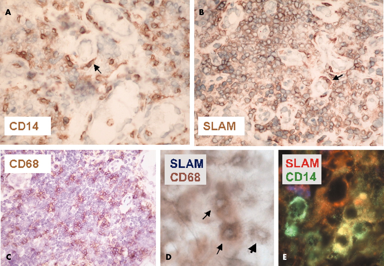
Considerable numbers of CD68+ macrophages and to a lesser extent CD14+ cells were present in the wall of the normal gut and in Peyer’s patches. SLAM+ cells were detected only in Peyer’s patches, and not in other parts of the normal tissue (not shown). Numerous CD14+ monocytes, CD68+ macrophages, and SLAM+ cells were detected throughout the inflamed gut of patients with Crohn’s disease (fig 1A–C). Some SLAM+ cells were round, but many had processes, indicating that they belonged to the monocyte/macrophage lineage (fig 1B). Double staining demonstrated SLAM expression on CD14+ monocytes and on CD68+ macrophages (fig 1D, E).

Expression of CD14, CD68, and SLAM in Crohn’s disease. Antibodies to (A) CD14, (B) SLAM, and (C) CD68 showed extensive accumulation of inflammatory cells in the mucosa of the gut. Elongated cells with processes were seen after staining for both CD14 and SLAM (arrows in A and B). Cell nuclei were counterstained with haematoxylin in A–C. (D) Immunohistochemical staining showing blue labelling of SLAM expressing cells and brown labelling of CD68 expressing cells. Two CD68+ cells coexpressing SLAM (arrow) and one cell expressing SLAM only (arrowhead) are labelled. (E) Double staining with anti-CD14 and anti-SLAM revealed cells that were positive for CD14 only (green), positive for SLAM only (red), and double positive (yellow). Original magnification: ×400 (A–C); ×1000 (D, E).
In normal brain specimens very few CD14+, CD68+, and SLAM+ cells were detected (not shown). The MS lesion was characterised by an active plaque and composed mainly of activated monocytes and macrophages (fig 2A, C, D). The CD68+ cells extended into the parenchyma and together with CD14+ cells surrounded the inflamed blood vessels, where only very few SLAM positive cells were detected (fig 2B, E). Thus, most, if not all, CD68+ and CD14+ cells did not express SLAM in this MS tissue lesion. Further studies taking into account the pathological heterogeneity of MS5 are required to elaborate this.

Expression of CD14, CD68, and SLAM in multiple sclerosis (MS). Staining for (A, D) CD14, (B, E) SLAM, and (C) CD68 is shown in an active MS lesion. The sharply demarcated lesion edge is visible (arrow in A). (A, C, E) Numerous CD14+ and CD68+ cells were densely packed around the blood vessels. (C) CD68 expressing cells also infiltrated the parenchyma. (B, E) A few SLAM positive cells can be seen around the blood vessels, but they do not extend into the parenchyma. In all sections nuclei were counterstained with haematoxylin. Original magnification: ×100 (A); ×400 (B–E).
Thus, our study demonstrates for the first time the presence of SLAM+ monocytes and macrophages in a chronic inflammatory disease, Crohn’s disease. Furthermore, we show that this is not a feature common to all types of inflammation, because monocytes and macrophages did not express SLAM in the analysed MS lesions. The expression of SLAM on monocytes and macrophages in Crohn’s disease supports the concept that the inflammation in this disease is driven by bacteria derived TLR ligands.
Acknowledgments
Supported by the DFG (SFB 571). The Institute for Clinical Neuroimmunology is supported by the Hermann and Lilly Schilling Foundation. We thank Professor G Niedobitek (University of Erlangen, Germany), Dr M Heiss, Dr H Hartl, and I Paripovic (University of Munich, Germany) for their help, Ms J Benson, Drs A Flügel, and A Holz for helpful comments on the manuscript.
Footnotes
-
The first two authors contributed equally to this work.